Review
. 2019 Aug;16(8):665-680.
doi: 10.1080/14789450.2019.1645604. Epub 2019 Jul 25.

Glycosylation and its implications in breast cancer

Affiliations
Review

Glycosylation and its implications in breast cancer

Danielle A Scott et al. Expert Rev Proteomics. 2019 Aug.

Abstract

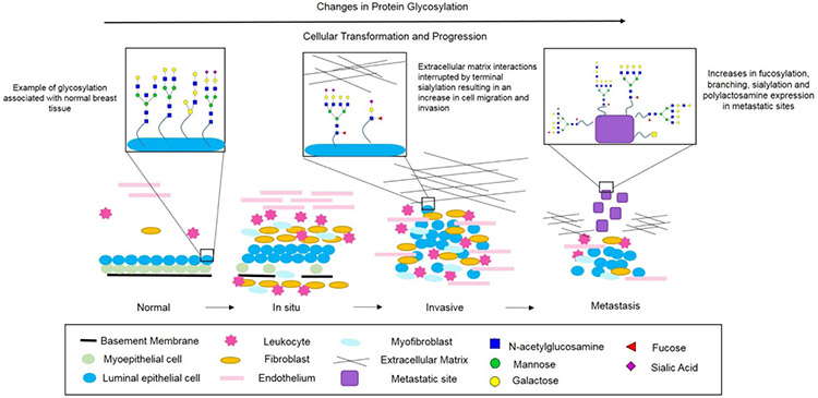
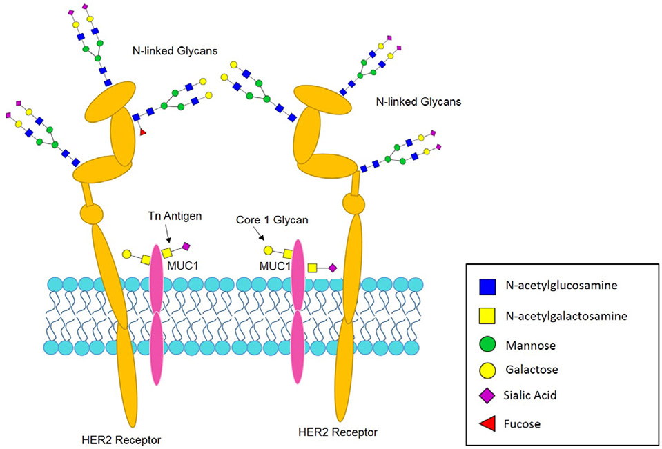
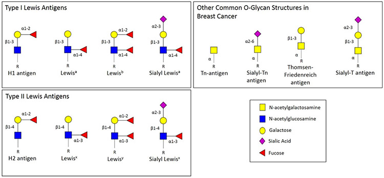
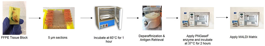
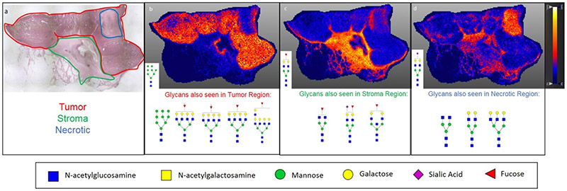
Introduction: For decades, the role of glycans and glycoproteins in the progression of breast cancer and other cancers have been evaluated. Through extensive studies focused on elucidating the biological functions of glycosylation, researchers have been able to implicate alterations in these functions to tumor formation and metastasis. Areas covered: In this review, we summarize how changes in glycosylation are associated with tumorigenesis, with emphasis on breast cancers. An overview of the changes in N-linked and O-linked glycans associated with breast cancer tumors and biofluids are described. Recent advances in glycomics are emphasized in the context of continuing to decipher the glycosylation changes associated with breast cancer progression. Expert opinion: While changes in glycosylation have been studied in breast cancer for many years, the clinical relevance of these studies has been limited. This reflects the inherent biological and clinical heterogeneity of breast cancers. Glycomics analysis lags behind the advances in genomics and proteomics, but new approaches are emerging. A summary of known glycosylation changes associated with breast cancer is necessary to implement new findings in the context of clinical outcomes and therapeutic strategies. A better understanding of the dynamics of tumor and immune glycosylation is critical to improving emerging immunotherapeutic treatments.

Keywords: Biomarkers; breast cancer; glycan; glycosylation; mass spectrometry.


Conflict of interest statement

Declaration of interest

The authors have no relevant affiliations or financial involvement with any organization or entity with a financial interest in or financial conflict with the subject matter or materials discussed in the manuscript. This includes employment, consultancies, honoraria, stock ownership or options, expert testimony, grants or patents received or pending, or royalties.

Figures

Figure 1:
Figure 1:
Schematic Overview of Normal, in situ, Invasive and Metastatic Breast Carcinoma Progression. Normal breast ducts contain a basement membrane with a layer of myoepithelial cells and a layer of luminal epithelial cells with various endothelial cells, fibroblasts and leukocytes in the stroma. Glycans on normal breast epithelium are basic bi- and tri-antennary structures typically lacking expression of core-fucosylated structures. As in situ carcinoma develops, the basement membrane begins to degrade and the number of myoepithelial cells decreases. Additionally, the number of stromal immune cells increases. A cancer is considered invasive when the basement membrane is no longer present, allowing the tumor cells to invade surrounding tissues, enter the blood stream and eventually form distant metastatic sites. As a cancer becomes invasive and metastatic, N-glycans expressed display increases in branching, sialylation and fucosylation. Additionally, O-glycans decrease in chain length and the expression of Tn antigen, Thomsen-Friedenreich antigen, sialyl-T antigen and sialyl-Tn antigen increases.
Figure 2:
Figure 2:
HER2 Receptor and MUC1 as Example Glycoproteins. A schematic diagram indicating the presence of N- and O- linked glycans on common cell surface glycoproteins found in breast cancer.
Figure 3:
Figure 3:
Lewis Antigens Terminal oligosaccharide structures used to classify particular antigens based on the presence of an alpha 1-4 linked fucose (Lewis A, B) or an alpha 1-3 linked fucose (Lewis X, Y) to the GlcNAc monosaccharide. Additionally, sialylated forms of Lewis A and Lewis X structures are also shown, in addition to other common O-glycan structures in breast cancer such as Tn-antigen, sialyl-Tn antigen, Thomsen-Friedenreich antigen and sialyl-T antigen.
Figure 4:
Figure 4:
Workflow for Preparation of FFPE Tissues for Glycan Analysis by MALDI IMS. FFPE tissue blocks are sliced into 5 ?m sections and placed onto glass slides. Once sectioned slides are prepared, they are placed at 60°C for 1 hour. Next, slides are placed through a series of deparaffinization washes and antigen retrieval. PNGaseF enzyme is then applied in a fine mist to the slides using a HTX TMSprayer to cleave glycans on the slides. Slides are then incubated at 37°C for 2 hours. After incubation, a MALDI compatible matrix is applied. At the conclusion of this process, slides can be stored under vacuum until ready for MALDI IMS analysis.
Figure 5:
Figure 5:
Linking Histopathology with MALDI IMS Glycan Images. a) H&E stain of an invasive ductal carcinoma at 2X magnification with pathologist annotation of tumor (red), stroma (green), and necrotic (blue) regions. b) Distribution of Hex8HexNAc2 + 1Na (m/z 1743.5956) showing strong correlation with the tumor region of the tissue. c) Distribution of Hex5dHex1HexNAc2 + 1Na (m/z 1809.6838) throughout stroma region of the tissue. d) Distribution of Hex5HexNAc4NeuAc1 + 1Na throughout necrotic and stromal region of the tissue. Additionally, other glycan structures seen correlating with each region in b-d are shown below the respective image. Images were created using SCiLS Lab 2017a and previously described in Scott et. al. 2018.

Similar articles

Cited by

References

    1. Aub JC, Tieslau C, Lankester A. REACTIONS OF NORMAL AND TUMOR CELL SURFACES TO ENZYMES. I. WHEAT-GERM LIPASE AND ASSOCIATED MUCOPOLYSACCHARIDES. Proceedings of the National Academy of Sciences of the United States of America. 1963. October;50:613–9. PubMed PMID: 14077487; PubMed Central PMCID: PMCPMC221235. eng. - PMC - PubMed
    1. Barton JG, Bois JP, Sarr MG, et al. Predictive and prognostic value of CA 19-9 in resected pancreatic adenocarcinoma. Journal of gastrointestinal surgery : official journal of the Society for Surgery of the Alimentary Tract. 2009. November;13(11):2050–8. doi: 10.1007/s11605-009-0849-z. PubMed PMID: 19756875; eng. - DOI - PubMed
    1. Drake RR. Glycosylation and Cancer: Moving Glycomics to the Forefront. Advances in cancer research. 2015;126:1–10. - PubMed
    1. Kirwan A, Utratna M, O’Dwyer ME, et al. Glycosylation-Based Serum Biomarkers for Cancer Diagnostics and Prognostics. BioMed Research International. 2015. 10/05 04/02/received 05/28/revised 05/31/accepted;2015:490531. doi: 10.1155/2015/490531. PubMed PMID: PMC4609776. - DOI - PMC - PubMed
    1. Ludwig JA, Weinstein JN. Biomarkers in cancer staging, prognosis and treatment selection. Nat Rev Cancer. 2005. November;5(11):845–56. doi: 10.1038/nrc1739. PubMed PMID: 16239904; eng. - DOI - PubMed

Publication types