Review
. 2019 Jul 16;51(1):27-41.
doi: 10.1016/j.immuni.2019.06.025.

Inflammation and Cancer: Triggers, Mechanisms, and Consequences

Affiliations
Review

Inflammation and Cancer: Triggers, Mechanisms, and Consequences

Florian R Greten et al. Immunity. .

Abstract

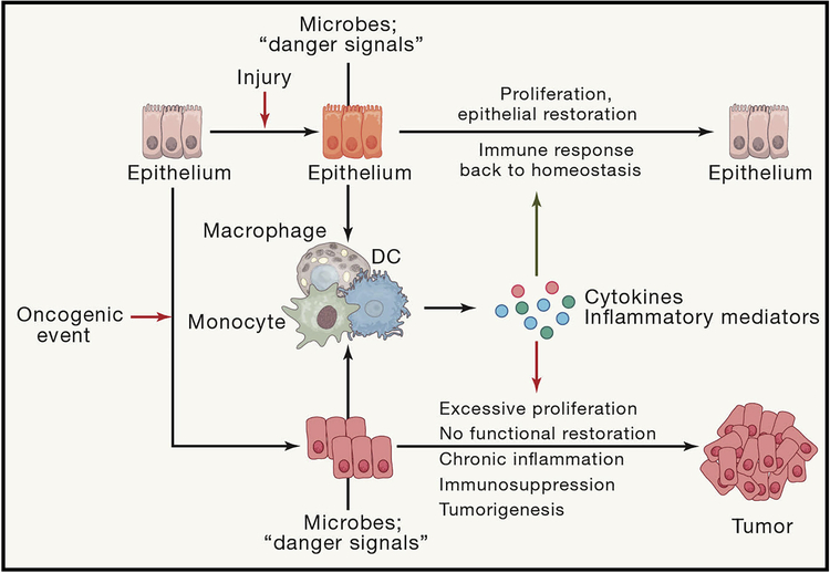
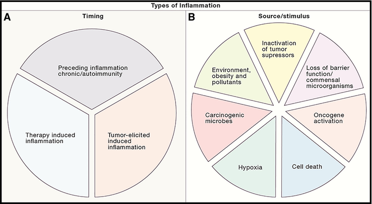
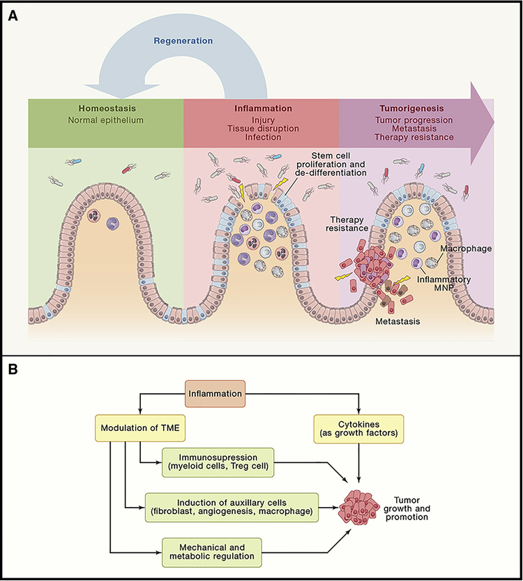
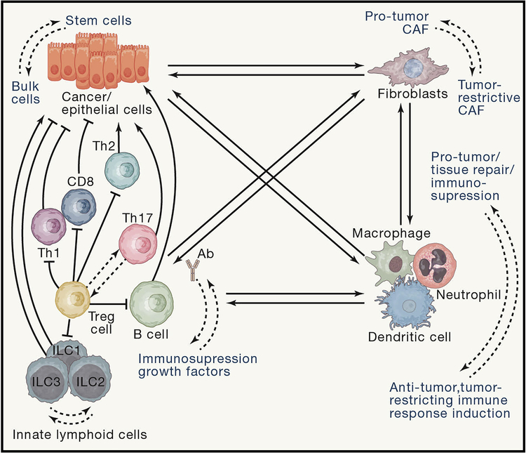
Inflammation predisposes to the development of cancer and promotes all stages of tumorigenesis. Cancer cells, as well as surrounding stromal and inflammatory cells, engage in well-orchestrated reciprocal interactions to form an inflammatory tumor microenvironment (TME). Cells within the TME are highly plastic, continuously changing their phenotypic and functional characteristics. Here, we review the origins of inflammation in tumors, and the mechanisms whereby inflammation drives tumor initiation, growth, progression, and metastasis. We discuss how tumor-promoting inflammation closely resembles inflammatory processes typically found during development, immunity, maintenance of tissue homeostasis, or tissue repair and illuminate the distinctions between tissue-protective and pro-tumorigenic inflammation, including spatiotemporal considerations. Defining the cornerstone rules of engagement governing molecular and cellular mechanisms of tumor-promoting inflammation will be essential for further development of anti-cancer therapies.

Keywords: cancer; cell plasticity; cytokine; inflammation; mechanisms; metastasis; tumor microenvironment; tumor progression.


Conflict of interest statement

Conflict of interest: The authors declare no competing financial interests.

Figures

Figure 1:
Figure 1:. Evolutionary and functional differences and similarities between inflammation in cancer and inflammation during infection and tissue regeneration
Insults to epithelial tissues caused by injury or infection cause activation of myeloid cells which start to produce inflammatory cytokines to activate innate and adaptive sterilizing immunity to get rid of the pathogen and to activate epithelial cell proliferation to close down the barrier dysfunction which allowed translocation of pathogen or to repair inflicted injury. After this concerted effort insulted epithelial tissue comes back to normal state of homeostasis. However if initial disturbance of epithelial homeostasis is caused by an oncogenic event, the sterilizing immunity will not remove the intial insult and the enhanced inflammation and cytokine-driven proliferation will facilitate tumor growth rather than restoring normal epithelial homeostasis.
Figure 2:
Figure 2:. Types of inflammation in cancer: different timing and different inducers
(A) Cancer-associated inflammation can be induced at different time points of tumor development. It can precede carcinogenesis in form of autoimmunity or infection, can be induced by malignant cells or can be triggered by anti-cancer therapy. (B) Various cell intrinsic, host dependent or environmental factors can cause tumor-associated inflammation in different tumor types.
Figure 3:
Figure 3:. Pro-tumorigenic actions of inflammation in progression, metastasis and growth
(A) Injury, infection or functional tissue disruption(as during malignant transformation) leads to activation of inflammatory responses which can drive the expansion and proliferation of stem cell pool, the process normally designed to restitute and normalize epithelium and its barrier function, i.e. causing normal “Regeneration” and a path to normal epithelium. However, if stem cells already harbor oncogenic mutations, and are de facto cancer stem cells, expansion of stem cell pool leads to enhanced metastasis and therapy resistance. (B) Inflammatory entities, such as cytokines and growth factors, release by immune cells within the TME can have a direct effect on pre-malignant and cancer cells by increasing their proliferation and resistance to cell death and stresses, thereby directly promoting tumor growth and progression. can be caused at different time points of cancer development. In addition, inflamamtory signals can shape TME to induce immunosupression via action of Tregs, immature myeloid cells and other supressive players; to enhance recruitment, proliferation and distinct functions of other pro-tumorigenic auxillary cells within the TME (such as fibroblasts, myeloid cells and endothelium of new blood vessels); and to alter mechanicaal and metabolic functions of TME. Altogether, these inflammation-driven changes also significantly contribute to tumor growths and progression.
Figure 4:
Figure 4:. Increased cell plasticity within the tumor microenvironment (TME)
An intricate reciprocal interplay between all cells (cancer, stromal and immune) in the TME shapes polarization of immune cells activation states ( for myeloid cells and lymphocyes) as well as of cancer-associated fibroblasts and states of differentiation of epithelial cells. These different cells types instruct reversible phenotypic and functional perturbations in neighboring cells types, executing precise multicellular responses to the exact needs of tissue (transformed tissue) by tailoring plastic changes in existing and available cell types, rather than demanding the existence of multiple new “rigid” cellular types “one new distinct cell types for each new condition”. These plastic cellular changes may have distinct, sometimes opposing, roles in the TME with regard to their net effect on tumor growth, progression or metastasis.

References

    1. Aceto N, Bardia A, Miyamoto DT, Donaldson MC, Wittner BS, Spencer JA, Yu M, Pely A, Engstrom A, Zhu H, et al. (2014). Circulating tumor cell clusters are oligoclonal precursors of breast cancer metastasis. Cell 158, 1110–1122. - PMC - PubMed
    1. Affara NI, Ruffell B, Medler TR, Gunderson AJ, Johansson M, Bornstein S, Bergsland E, Steinhoff M, Li Y, Gong Q, et al. (2014). B cells regulate macrophage phenotype and response to chemotherapy in squamous carcinomas. Cancer Cell 25, 809–821. - PMC - PubMed
    1. Akkari L, Gocheva V, Kester JC, Hunter KE, Quick ML, Sevenich L, Wang HW, Peters C, Tang LH, Klimstra DS, et al. (2014). Distinct functions of macrophage-derived and cancer cell-derived cathepsin Z combine to promote tumor malignancy via interactions with the extracellular matrix. Genes Dev 28, 2134–2150. - PMC - PubMed
    1. Albrengues J, Shields MA, Ng D, Park CG, Ambrico A, Poindexter ME, Upadhyay P, Uyeminami DL, Pommier A, Kuttner V, et al. (2018). Neutrophil extracellular traps produced during inflammation awaken dormant cancer cells in mice. Science 361. - PMC - PubMed
    1. Alexander J, and Cukierman E (2016). Stromal dynamic reciprocity in cancer: intricacies of fibroblastic-ECM interactions. Current opinion in cell biology 42, 80–93. - PMC - PubMed

Publication types