. 2019 Jul 16;28(3):625-639.e6.
doi: 10.1016/j.celrep.2019.06.033.

Spinal Neuropeptide Y1 Receptor-Expressing Neurons Form an Essential Excitatory Pathway for Mechanical Itch

Affiliations

Spinal Neuropeptide Y1 Receptor-Expressing Neurons Form an Essential Excitatory Pathway for Mechanical Itch

David Acton et al. Cell Rep. .

Abstract

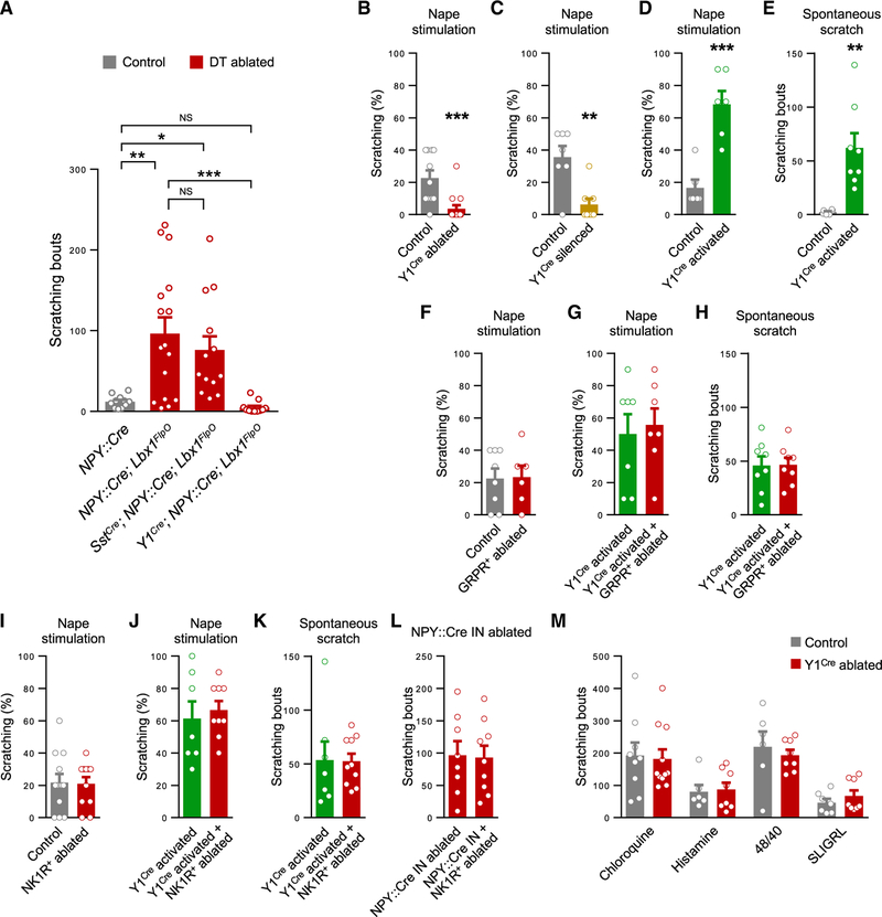
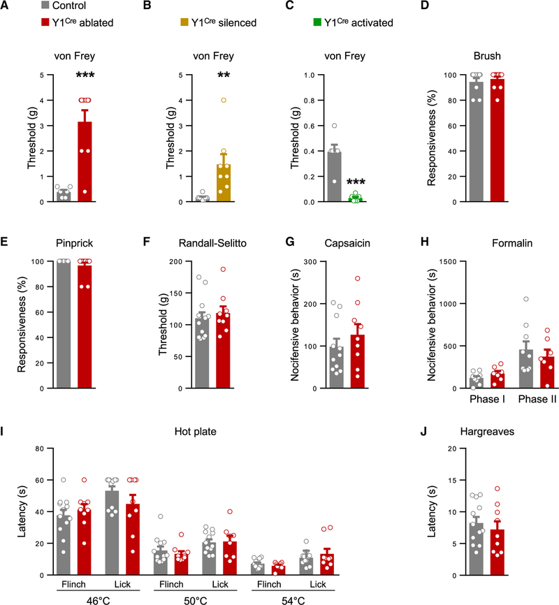
Acute itch can be generated by either chemical or mechanical stimuli, which activate separate pathways in the periphery and spinal cord. While substantial progress has been made in mapping the transmission pathway for chemical itch, the central pathway for mechanical itch remains obscure. Using complementary genetic and pharmacological manipulations, we show that excitatory neurons marked by the expression of the neuropeptide Y1 receptor (Y1Cre neurons) form an essential pathway in the dorsal spinal cord for the transmission of mechanical but not chemical itch. Ablating or silencing the Y1Cre neurons abrogates mechanical itch, while chemogenetic activation induces scratching. Moreover, using Y1 conditional knockout mice, we demonstrate that endogenous neuropeptide Y (NPY) acts via dorsal-horn Y1-expressing neurons to suppress light punctate touch and mechanical itch stimuli. NPY-Y1 signaling thus regulates the transmission of innocuous tactile information by establishing biologically relevant thresholds for touch discrimination and mechanical itch reflexes.

Keywords: NPY1R signaling; Y1 excitatory neurons; chemical itch; dorsal spinal cord; inhibition; mechanical itch transmission; neuropeptide Y; pain; somatosensory processing; touch.


Conflict of interest statement

DECLARATION OF INTERESTS

The authors declare no competing interests.

Figures

Figure 1.
Figure 1.. Y1Cre Marks a Population of Excitatory Neurons Concentrated in Laminae II-III
(A) Transverse section through the lumbar spinal cord of a P21 mouse showing Y1 mRNA expression in the dorsal horn. (B) Section from a P42 Y1Cre; Ai14lsl-tdTom mouse showing laminar distribution of Y1-tdTomato neurons. (C) Co-expression of tdTomato and Y1 mRNA in the dorsal horn of a P21 Y1Cre; Ai14lsl-tdTom mouse. (D) Summary of Y1 mRNA expression (n = 3 mice). (E and F) Transverse sections through the lumbar spinal cord of a P10 Y1Cre; Ai14lsl-tdTom mouse stained with antibodies against Lmx1b (E) and Pax2 (F). (G) Quantification of co-expression of Y1-tdTomato with antibody-labeled Lmx1b and Pax2 in P10 Y1Cre; Ai14lsl-tdTom mice (n = 5 mice). Roman numerals denote Rexed’s laminae. Scale bars: 50 μm (A, B, E, and F) and 10 μm (C). Data: mean ± SEM. See also Figures S1–S3.
Figure 2.
Figure 2.. Y1Cre Neurons Receive Extensive LTMR Input
(A and B) Examples of Y1Cre neurons in lamina IIi from lumbarspinal cord sectionsof P42 Y1Cre; Ai14lsl-tdTom mice injected with CTb into: the hairy skin of the thigh (A) and the glabrous skin of the hindpaw (B). Immunolabeled CTb+ contacts (blue) displayed vGluT1 immunoreactivity (green, arrowheads). (C) Section through the lumbar dorsal horn of a P10 Y1Cre; Lbx1FlpO; R26ds-HTB mouse injected with EnvA G-deleted rabies-mCherry virus. Arrowheads indicate infected Y1Cre neurons. mCherry+/GFP cells represent transsynaptically labeled presynaptic neurons. (D) Summary of antibody-labeled myelinated sensory afferent subtypes that are presynaptic to the Y1Cre neurons, expressed as a percentage of mCherry+ neurons (n = 4 mice). (E–J) Sections from P10 Y1Cre; Lbx1FlpO; R26ds-HTB lumbar DRGs showing presynaptically labeled sensory neurons(red) thatexpress c-Ret (E), TrkC but not parvalbumin(PV; F), TrkB(G), calcitonin gene-related peptide (CGRP) and neurofilament 200 (NF; H), and calbindin (CB; I), but not PV or calretinin (CR; J). Arrowheads indicate co-labeled sensory afferents. CB, calbindin; CR, calretinin; NF, neurofilament; PV, parvalbumin. Scale bars: 5 μm (B) and 100 μm (C and E–J). Data: mean ± SEM. See also Figure S4.
Figure 3.
Figure 3.. Y1Cre Neurons within the Dorsal Horn Transmit Mechanical but Not Chemical Itch
(A) Ablation of dorsal horn NPY::Cre INs increases spontaneous scratching in NPY::Cre; Lbx1FlpO; Tauds-DTR; Ai65ds-tdTom mice (n = 16) compared with DT-treated NPY::Cre; Tauds-DTR; Ai65ds-tdTom controls that lack DT-receptor expression (n = 11). Ablation of the Y1Cre and NPY::Cre IN populations in Y1Cre; NPY::Cre; Lbx1FlpO; Tauds-DTR; Ai65ds-tdTom mice abolishes scratching (n = 11). Scratching is unaffected when Sst+ neurons are ablated together with the NPY::Cre INs in SstCre; NPY::Cre; Lbx1FlpO; Tauds-DTR; Ai65ds-tdTom mice (n = 13). One-way ANOVA and Bonferroni post hoc tests were used to assess statistical differences. (B and C) Reduced scratching in response to stimulation of the nape with a 0.16-g von Frey hair in Y1Cre; Lbx1FlpO; Tauds-DTR; Ai65ds-tdTom mice treated with DT (n = 14) compared with saline-treated controls (n = 11; B), and in Y1Cre; Lbx1FlpO; R26ds-hM4D mice treated with clozapine N-oxide (CNO; n = 8) compared with Y1Cre; R26ds-hM4D controls, which lack FlpO-dependent hM4D expression (n = 8; C). (D) Enhanced scratching in response to stimulation of the nape in CNO-treated Y1Cre; Lbx1FlpO; R26ds-hM3D mice (n = 6) compared with Y1Cre; R26ds-hM3D controls (n = 6). (E) Spontaneous scratching in CNO-treated Y1Cre; Lbx1FlpO; R26ds-hM3D mice (n = 8) is enhanced over a 30-min period compared with control mice (n = 6). (F) Scratching responses are unchanged in wild-type mice treated with bombesin-saporin (BOM-SAP; n = 6) to ablate GRPR+ neurons compared with controls injected with saporin (SAP; n = 8). (G and H) Ablation of GRPR+ neurons in Y1Cre; Lbx1FlpO; R26ds-hM3D mice does not alter evoked scratching (G; BOM-SAP, n = 8; SAP, n = 7) or spontaneous scratching (H; BOM-SAP, n = 7; SAP, n = 7) following CNO injection. (I) Scratching responses are unchanged in wild-type mice treated with [Sar9, Met(O2)11]-substance P-SAP (SSP-SAP; n = 10) to ablate NK1R+ neurons compared to SAP-injected controls (n = 11). (J and K) In Y1Cre; Lbx1FlpO; R26ds-hM3D mice, ablation of NK1R+ neurons does not alter evoked (J; SSP-SAP, n = 9; SAP, n = 7) or spontaneous (K; SSP-SAP, n = 10; SAP, n = 7) scratching following CNO injection. (L) Unchanged scratching in NPY::Cre; Lbx1FlpO; Tauds-DTR; Ai65ds-tdTom mice 2 weeks after NK1R+ neuron ablation and 1 week following DT administration (SSP-SAP, n = 9; SAP, n = 8). (M) Unchanged scratching over a 30-min period in Y1Cre neuron-ablated mice following injection of chloroquine (control, n = 9; ablated, n = 11), histamine (control, n = 6; ablated, n = 8), compound 48/80 (control, n = 6; ablated, n = 8), and SLIGRL (control, n = 7; ablated, n = 8). *p < 0.05, **p < 0.01, ***p < 0.001, NS, not significant. Data: mean ± SEM. See also Figures S4–S6.
Figure 4.
Figure 4.. Y1Cre Neurons Selectively Transmit Light Touch Information
(A and B) Sensitivity to von Frey hair stimulation of the hindpaw glabrous skin is reduced following Y1Cre neuron ablation (A; control, n = 6; ablated, n = 9) or silencing (B; control, n = 8; silenced, n = 8). (C) Glabrous skin sensitivity to von Frey hair stimulation is elevated following Y1Cre neuron activation (control, n = 6; activated, n = 6). (D–F) Responses to dynamic touch (D; control, n = 11; ablated, n = 13), pinprick (E; control, n = 11; ablated, n = 13), and Randall-Selitto (F; control, n = 12; ablated, n = 9) are unchanged after Y1Cre neuron ablation. (G and H) Chemical pain responses following hindpaw injection of capsaicin (G; control, n = 11; ablated, n = 9) or formalin (H; control, n = 9; ablated, n = 7) are not altered following Y1Cre neuron ablation. (I and J) Y1Cre neuron-ablated mice show normal heat responses as assessed by the hot plate (I; control, n = 12; ablated, n = 9) and Hargreaves (J; control, n = 12; ablated, n = 9) assays. **p < 0.01, ***p < 0.001. Data: mean ± SEM.
Figure 5.
Figure 5.. Y1Cre Neurons Receive VGAT+ and NPY+ Synaptic Contacts from NPY::Cre Ins
(A and B) Examples of GFP-labeled cells from the lumbar spinal cord of a P60 Y1::EGFP; NPY::Cre transgenic mouse. Presynaptic contacts (red) from NPY::Cre INs onto Y1/EGFP+ cells were visualized with a Cre-dependent AAV2/1-hSyn-DIO-SypHTom virus. Putative synaptic boutons marked by VGAT (blue; A) and NPY (blue; B) immunoreactivity are indicated by arrowheads. (C) Examples of synaptic puncta labeled with antibodies against NPY (green) and gephyrin (blue) on a Y1-tdTomato+ cell body in the lumbar spinal cord of a P42 Y1Cre; Ai14lsl-tdTom mouse (arrowheads). (D) Schematic illustrating experimental conditions used to assess synaptic connectivity between NPY::Cre INs and Y1Cre neurons. (E) ReaChR-mediated activation of NPY::Cre IN generated monosynaptic outward currents in lamina IIi Y1-EGFP+ neurons in P14–21 Y1::EGFP; NPY::Cre; Lbx1FlpO; R26ds-ReaChR spinal cord slices in the presence of kynurenic acid (KA; 1.5 mM) at a holding potential of —30 mV. These currents were abolished following application of 1 μM strychnine and 60 μM picrotoxin (n = 7 cells from 5 mice). ***p < 0.001. The statistical difference was determined by the two-tailed, paired t test. (F) Traces recorded from a Y1-EGFP+ neuron showing a monosynaptic inhibitory current elicited by NPY::Cre activation (purple trace) and no response following bath application of strychnine and picrotoxin (black trace). Scale bars: 5 μm.
Figure 6.
Figure 6.. NPY-Y1 Receptor Signaling within the Dorsal Horn Gates Mechanical Itch and Light Punctate Touch
(A and B) Mice lacking the Y1 receptor in dorsal horn neurons exhibit pronounced spontaneous scratching (A; Lbx1Cre; Y1f/f, n = 10; Y1f/f control, n = 12) and hypersensitivity to light punctate mechanical stimulation of the nape (B; Lbx1Cre; Y1f/f,n = 7; Y1f/f control, n = 9). (C) The Y1 antagonist BIBP 3226 (1 mg kg−1, intraperitoneally [i.p.]) increases spontaneous scratching in control (n = 8) but not conditional knockout mice (n = 12). Two-tailed, paired t tests were used to assess statistical differences. (D) BIBP 3226 causes hypersensitivity to nape stimulation (n = 12; vehicle, n = 12). (E and F) Lbx1Cre; Y1f/f mice have reduced hindpaw von Frey thresholds (E; Lbx1Cre; Y1f/f,n= 8; Y1f/f control, n = 8) but responses to dynamic touch are unaltered (F; Lbx1Cre; Y1f/f,n= 8; Y1f/f control, n = 9). (G–I) Deletion of Y1 from dorsal horn neurons does not alter scratching frequency in response to chloroquine (Lbx1Cre; Y1f/f, n = 11; Y1f/f control, n = 9) or scratching frequency (G), duration (H), or rate (duration/frequency; I) in response to compound 48/80 (Lbx1Cre; Y1f/f, n = 14; Y1f/f control, n = 10). (J–N) Deletion of Y1 from dorsal horn neurons does not affect sensitivity to acute mechanical pain as assessed by pinprick (J; Lbx1Cre; Y1f/f,n = 9; Y1f/f control, n = 9) or the Randall-Selitto test (K; Lbx1Cre; Y1f/f,n= 8; Y1f/f control, n = 6), chemical nociception (L; Lbx1Cre; Y1f/f,n= 8; Y1f/f control, n = 8), or thermal pain as assessed by the hot plate (M; Lbx1Cre; Y1f/f,n = 9; Y1f/f control, n = 9) or Hargreaves tests (N; Lbx1Cre; Y1f/f,n = 8; Y1f/f control, n = 6). *p < 0.05 and ***p < 0.001. NS, not significant. Data: mean ± SEM.
Figure 7.
Figure 7.. NPY-Y1 Receptor Signaling Determines Sensitivity to Mechanical Itch Stimuli
(A) Spontaneous scratching in NPY::Cre; Lbx1FlpO; Tauds-DTR; Ai65ds-tdTom mice 1 week after DT treatment was reduced following injection of NPY (100 μg kg−1, i.p.; n = 9) or the selective Y1 receptor agonist [Leu31, Pro34]-NPY (100 μg kg−1, i.p.; n = 6) when compared with vehicle. (B) Evoked scratching is reduced when Y1f/f mice (n = 8) but not Lbx1Cre; Y1f/f mice (n = 9) are injected with [Leu31, Pro34]-NPY (100 μg kg−1, i.p., n = 8; vehicle, n = 8). (C and D) hM3D-mediated activation of the NPY::Cre INs reduces scratching in response to mechanical stimulation of the nape compared with controls (NPY::Cre; Lbx1FlpO; R26ds-hM3D, n = 13; NPY::Cre; R26ds-hM3D controls, n = 11; C) but not when Y1 receptors are inhibited by BIBP 3226 (n = 7; controls, n = 9; D). (E) Unchanged chloroquine-induced scratching following activation of the NPY::Cre INs (n = 12; controls, n = 12). (F–H) Activation of NPY::Cre INs reduces sensitivity to von Frey hair (F), brush (G), and pinprick (H) stimulation of the plantar hindpaw (n = 13; controls, n = 11). (I–K) Activation of NPY::Cre INs does not alter sensitivity to von Frey hair (I), brush (J), and pinprick (K) stimulation following Y1 receptor blockade by BIBP 3226 (n = 9; BIBP 3226-injected controls, n = 7). (L) Schematic showing proposed dorsal horn circuitry for mechanical and chemical itch. Dyn, dynorphin; Nppb, natriuretic peptide B; NPRA, natriuretic peptide receptor A; PN, projection neuron; Sst2A, Sst 2A receptor. Italics indicate neurotransmitters/neuropeptides. Two-tailed, paired (A and B) or unpaired (C–K) t tests were used to assess statistical differences. *p < 0.05, **p < 0.01, ***p < 0.001. NS, not significant. Data: mean ± SEM (B–L). See also Figure S7.

References

    1. Abraira VE, and Ginty DD (2013). The sensory neurons of touch. Neuron 79, 618–639. - PMC - PubMed
    1. Abraira VE, Kuehn ED, Chirila AM, Springel MW, Toliver AA, Zimmerman AL, Orefice LL, Boyle KA, Bai L, Song BJ, et al. (2017). The Cellular and Synaptic Architecture of the Mechanosensory Dorsal Horn. Cell 168, 295–310.e19. - PMC - PubMed
    1. Akiyama T, Iodi Carstens M, and Carstens E (2011). Transmitters and pathways mediating inhibition of spinal itch-signaling neurons by scratching and other counterstimuli. PLoS ONE 6, e22665. - PMC - PubMed
    1. Akiyama T, Carstens MI, Ikoma A, Cevikbas F, Steinhoff M, and Carstens E (2012). Mouse model of touch-evoked itch (alloknesis). J. Invest. Dermatol 132, 1886–1891. - PMC - PubMed
    1. Akiyama T, Tominaga M, Davoodi A, Nagamine M, Blansit K, Horwitz A, Carstens MI, and Carstens E (2013). Roles for substance P and gastrin-releasing peptide as neurotransmitters released by primary afferent pruriceptors. J. Neurophysiol 109, 742–748. - PMC - PubMed

Publication types

MeSH terms