. 2019 Jul 16;28(3):640-654.e6.
doi: 10.1016/j.celrep.2019.06.053.

Prefrontal Pathways Provide Top-Down Control of Memory for Sequences of Events

Affiliations

Prefrontal Pathways Provide Top-Down Control of Memory for Sequences of Events

Maanasa Jayachandran et al. Cell Rep. .

Abstract

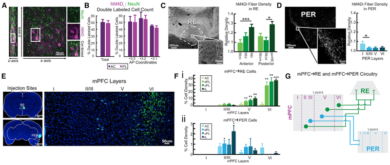
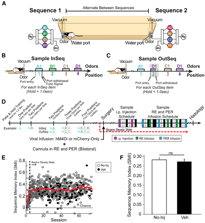
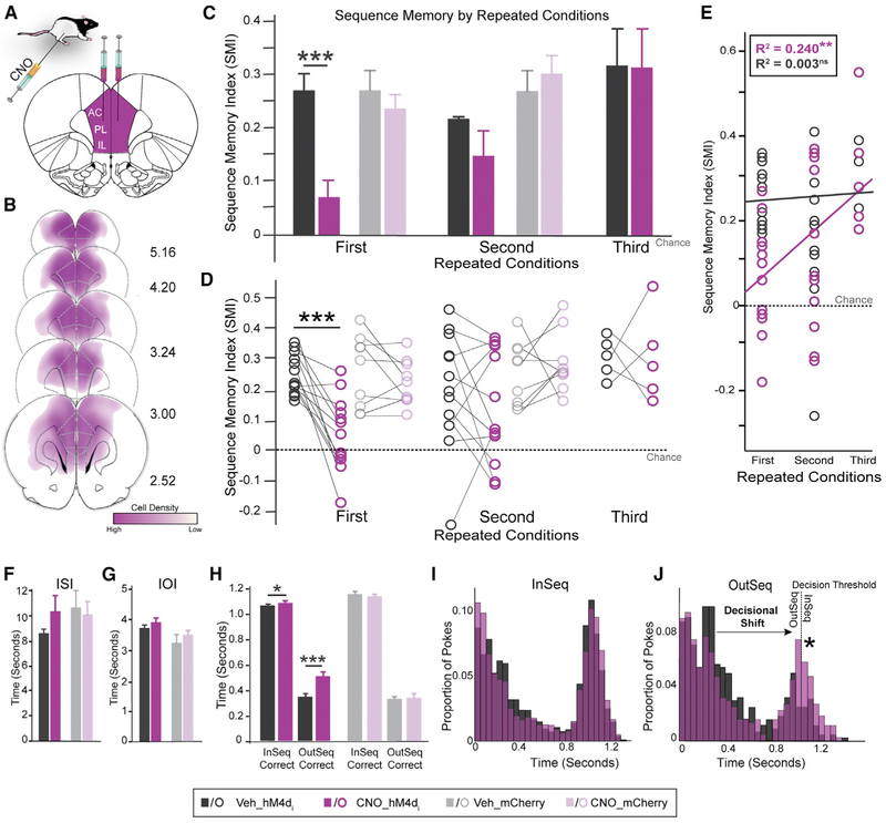
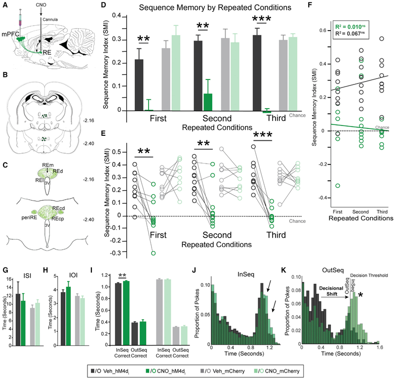
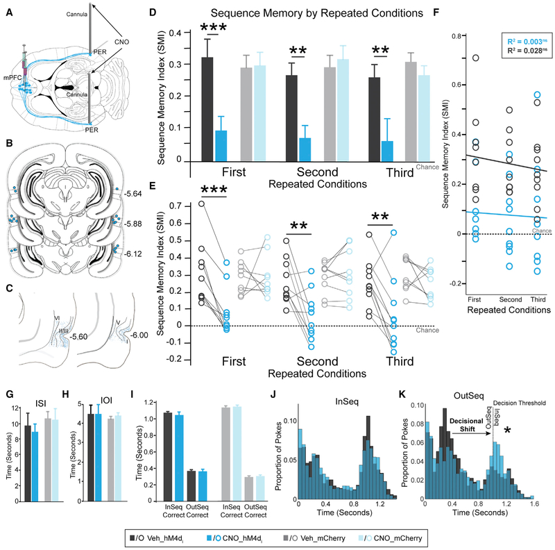
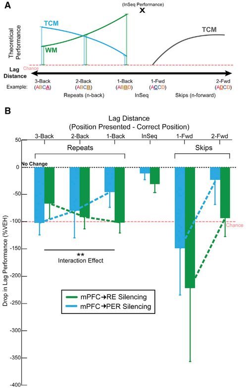
We remember our lives as sequences of events, but it is unclear how these memories are controlled during retrieval. In rats, the medial prefrontal cortex (mPFC) is positioned to influence sequence memory through extensive top-down inputs to regions heavily interconnected with the hippocampus, notably the nucleus reuniens of the thalamus (RE) and perirhinal cortex (PER). Here, we used an hM4Di synaptic-silencing approach to test our hypothesis that specific mPFC→RE and mPFC→PER projections regulate sequence memory retrieval. First, we found non-overlapping populations of mPFC cells project to RE and PER. Second, suppressing mPFC activity impaired sequence memory. Third, inhibiting mPFC→RE and mPFC→PER pathways effectively abolished sequence memory. Finally, a sequential lag analysis showed that the mPFC→RE pathway contributes to a working memory retrieval strategy, whereas the mPFC→PER pathway supports a temporal context memory retrieval strategy. These findings demonstrate that mPFC→RE and mPFC→PER pathways serve as top-down mechanisms that control distinct sequence memory retrieval strategies.

Keywords: DREADDs; cognitive control; episodic memory; hippocampus; memory retrieval; nucleus reuniens; perirhinal cortex; temporal context; thalamus; working memory.


Conflict of interest statement

DECLARATION OF INTERESTS

The authors declare no competing interests.

Figures

Figure 1.
Figure 1.. hM4Di Expression and Retrograde Labeling of RE and PER in mPFC
(A) hM4Di-mCherry (purple) and NeuN (neuron-specific; green) cells in mPFC. (B) h4MDi expression rates in the prelimbic cortex (PL) and anterior cingulate cortex (AC). (C) Axonal mPFC hM4Di expression in RE. hM4Di fiber density in RE across six subdivisions. (D) Axonal mPFC hM4Di expression in PER across layers. (E) RE (green) and PER (cyan; color was altered for consistency purposes) injections sites with CTB conjugated with Alexa Fluor 488 and 594. mPFC projects to RE and PER from separate cell populations. (F) Cell density of mPFC→RE (i) and mPFC→PER (ii) across subregions and layers. (G) Conceptual model of mPFC neurons with projections to RE and PER. mPFC provides direct excitatory inputs to RE and PER. mPFC→RE and mPFC→PER pathways originate from two distinct cell populations and project from specific cell layers in mPFC. mPFC→RE projects more toward the lateral parts of RE and mPFC→PER projects to layer I of PER. All data are represented as mean ± SEM. *p < 0.05; **p < 0.01; ***p < 0.001.
Figure 2.
Figure 2.. Sequence Memory Task
(A) A linear track was used with odor ports at each end where two separate four-odor sequences (A1, B1, C1, D1, or A2, B2, C2, D2) were presented. (B and C) Rats had to correctly identify the odor as either InSeq (70% of the time; B) or OutSeq (30% of the time; C). (D) After reaching steady-state performance, we focused on two experimental blocks: (1) i.p. injection suppressing mPFC neurons and (2) intracerebral infusions targeting mPFC terminals in RE or PER. The boxes represent a sample schedule. Veh days are denoted in black, and No-Inj days are in white. (E) We used the sequence memory index (SMI) as a summary measure. The red line represents the mean SMI of both No-Inj and Veh sessions (sliding window of 10 sessions). (F) SMI was not significantly different between the No-Inj and Veh sessions. All data are represented as mean ± SEM. ns, not significant.
Figure 3.
Figure 3.. mPFC Cortex Is Needed for Sequence Memory
(A) AAV9.hM4Di was injected bilaterally into mPFC. (B) Representation of AAV9.hM4Di viral spread in mPFC for all rats (n = 13). (C) Performance differed between hM4Di+ animals injected with Veh and CNO in the first repeated condition, but not the second and third repeated conditions. No differences between Veh and CNO were detected in the mCherry-only group. (D) Individual rat performance for each repeated condition in both groups. (E) In the hM4Di+ group, there was a positive linear relationship across repeated conditions after CNO injections, but not after Veh injections. (F) ISI was not significantly different between the Veh and CNO conditions in either group. (G) IOI was not significantly different between the Veh and CNO conditions in either group. (H) In the hM4Di+ group, nose-poke time was significantly different between the Veh and CNO conditions for both InSeqcorrect and OutSeqcorrect trials. No differences between Veh and CNO were detected in the mCherry-only group. (I) hM4Di+ group poke times show only subtle shifts in behavior. (J) hM4Di+ group poke times show a decisional shift between the Veh and CNO conditions with more OutSeq trials incorrectly identified as InSeq. All data are represented as mean ± SEM. *p < 0.05; **p < 0.01; ***p < 0.001; ns, not significant.
Figure 4.
Figure 4.. Synaptic Silencing of mPFC→RE Pathway Abolished Sequence Memory
(A) Guide cannulas targeted RE. (B) Locations of RE infusion cannulas in all rats (n = 10). (C) Schematic representation of mPFC hM4Di fiber density in RE. (D) In the hM4Di+ group, SMI was significantly different between Veh and CNO conditions in all three repeated conditions. There were no effects in the mCherry-only group. (E) Individual rat performances for each repeated condition in both groups. (F) In the hM4Di+ group, there was no significant relationship between repeated conditions and infusions. (G) ISI was not significantly different between RE Veh and CNO infusions in either group. (H) IOI was not significantly different between RE Veh and CNO infusions in either group. (I) In the hM4Di+ group, InSeqcorrect nose-poke times were significantly different between Veh and CNO. OutSeqcorrect nose-poke times, however, did not differ significantly between the Veh and CNO conditions. No differences between Veh and CNO were detected in the mCherry-only group. (J) hM4Di+ group poke times were relatively similar. (K) hM4Di+ group nose-poke times show a decisional shift (indicated by a star) between the Veh and CNO conditions toward more OutSeq odors incorrectly identified as InSeq. All data are represented as mean ± SEM. *p < 0.05; **p < 0.01; ***p < 0.001; ns, not significant.
Figure 5.
Figure 5.. Synaptic Silencing of mPFC→PER Pathway Abolished Sequence Memory
(A) Guide cannulas into the PER. (B) Locations of PER infusion cannulas in all rats (n = 10). (C) Schematic representation of mPFC hM4Di fiber density in PER (restricted to region of interest). (D) In the hM4Di+ group, SMI significantly differed between the Veh and CNO conditions for all three repeated conditions. No differences between Veh and CNO were detected in the mCherry-only group. (E) Individual rat performance in each repeated condition for both groups. (F) In the hM4Di+ group, there was no significant relationship between repeated conditions and infusions. (G) ISI was not significantly different between PER Veh and CNO conditions for either group. (H) IOI was not significantly different between PER Veh and CNO infusions for either group. (I) InSeqcorrect and OutSeqcorrect nose-poke times were not significantly different between Veh and CNO conditions in the hM4Di+ and mCherry-only groups. (J) hM4Di+ group nose-poke times show no obvious shifts in nose-poking behavior. (K) hM4Di+ group nose-poke times show a decisional shift (indicated by a star) where the rats incorrectly identified OutSeq odors as InSeq. All data are represented as mean ± SEM. *p < 0.05; **p < 0.01; ***p < 0.001; ns, not significant.
Figure 6.
Figure 6.. The mPFC→RE Pathway Supports a Working Memory Retrieval Strategy, while mPFC→PER Pathway Supports a Temporal Context Memory Retrieval Strategy
(A) If rats were using a working memory strategy, then repeated items would be easiest at short lags. By contrast, if rats were using a temporal context memory strategy, then repeated items would be the easiest to detect with longer lags. (B) mPFC→RE silencing impaired performance most on shorter distances, consistent with a loss of working memory, while mPFC→PER silencing impaired performance most at longer distance, consistent with a loss of temporal context memory. All data are represented as mean ± SEM. **p < 0.01.

References

    1. Aggleton JP, Albasser MM, Aggleton DJ, Poirier GL, and Pearce JM (2010). Lesions of the rat perirhinal cortex spare the acquisition of a complex configural visual discrimination yet impair object recognition. Behav. Neurosci. 124, 55–68. - PMC - PubMed
    1. Agster KL, Fortin NJ, and Eichenbaum H (2002). The hippocampus and disambiguation of overlapping sequences. J. Neurosci. 22, 5760–5768. - PMC - PubMed
    1. Allen TA, and Fortin NJ (2013). The evolution of episodic memory. Proc. Natl. Acad. Sci. USA 110 (Suppl 2), 10379–10386. - PMC - PubMed
    1. Allen TA, Furtak SC, and Brown TH (2007). Single-unit responses to 22 kHz ultrasonic vocalizations in rat perirhinal cortex. Behav. Brain Res. 182, 327–336. - PMC - PubMed
    1. Allen TA, Morris AM, Mattfeld AT, Stark CE, and Fortin NJ (2014). A sequence of events model ofepisodic memory shows parallels in rats and humans. Hippocampus 24, 1178–1188. - PMC - PubMed

Publication types

MeSH terms