Review
. 2019 Sep:147:104349.
doi: 10.1016/j.phrs.2019.104349. Epub 2019 Jul 14.

Inflammation within the neurovascular unit: Focus on microglia for stroke injury and recovery

Affiliations
Review

Inflammation within the neurovascular unit: Focus on microglia for stroke injury and recovery

Wael Eldahshan et al. Pharmacol Res. 2019 Sep.

Abstract

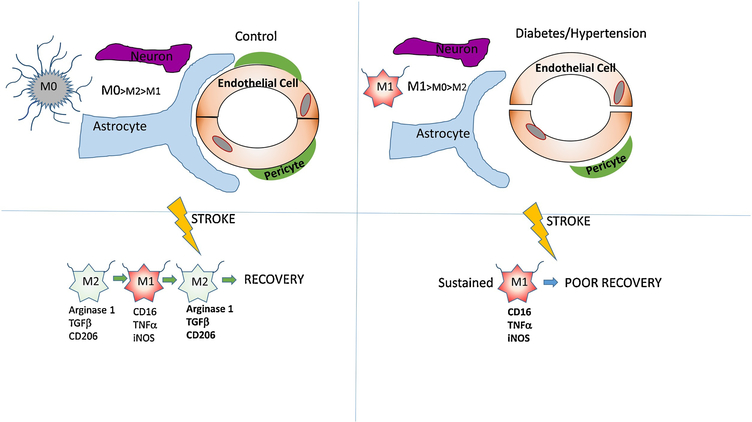
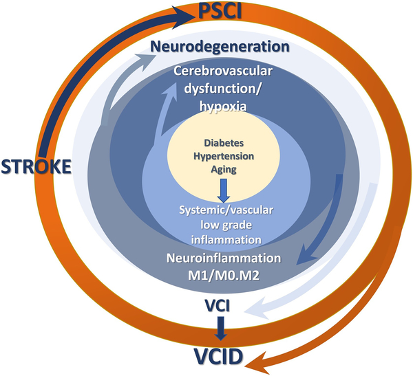
Neuroinflammation underlies the etiology of multiple neurodegenerative diseases and stroke. Our understanding of neuroinflammation has evolved in the last few years and major players have been identified. Microglia, the brain resident macrophages, are considered sentinels at the forefront of the neuroinflammatory response to different brain insults. Interestingly, microglia perform other physiological functions in addition to their role in neuroinflammation. Therefore, an updated approach in which modulation, rather than complete elimination of microglia is necessary. In this review, the emerging roles of microglia and their interaction with different components of the neurovascular unit are discussed. In addition, recent data on sex differences in microglial physiology and in the context of stroke will be presented. Finally, the multiplicity of roles assumed by microglia in the pathophysiology of ischemic stroke, and in the presence of co-morbidities such as hypertension and diabetes are summarized.

Keywords: Co-morbidities; Diabetes; Hypertension; Microglia; Neuroinflammation; Neurovascualar Unit; Sex-differences; Stroke.


Figures

Figure 1.
Figure 1.
A schematic model of the interaction between microglia and different components of the neurovascular unit in the absence and presence of comorbidities. In the left panel, microglia are in a resting state (M0) characterized by a higher number of process endpoints per cell that constantly probe the environment. After stroke, microglia transiently shift to the (M2) phenotype before shifting to (M1) pro-inflammatory phenotype. In the right panel, microglia are already primed for a pro-inflammatory phenotype in the presence of comorbidities and exhibit lower number of process endpoints per cell. After stroke, microglia shift directly to the (M1) phenotype and exhibit poorer recovery compared to microglia from animals with no comorbidities.
Figure 2.
Figure 2.
A schematic model of the role of microglia phenotypes in the vicious neuroinflammatory loop initiated by comorbid conditions. Hypoxic conditions resulting from cerebrovascular dysfunction and altered CBF causes an imbalance of pro- (M1) and anti-inflammatory (M2) microglia leading to neurodegeneration and vascular cognitive impairment (VCI). An ischemic stroke overlaid on this pathology and amplifies neurodegeneration via sustained M2 activation and causes poststroke cognitive impairment (PSCI), collectively contributing vascular contributions to cognitive impairment and dementia (VCID).
Figure 3.
Figure 3.
The impact of stroke and diabetes on microglial morphology and density. A. Heatmaps generated by IBA-1 staining after embolic MCAO show that microglial density is significantly higher in stroked animals in the ipsilateral side in the presence of diabetes. B. Cell swelling, number of protrusions from microglia cell body (red lines), number of endpoints at the tips of microglia processes (blue circles) and process length (grey lines) calculated from 40x images show that microglia of stroked diabetic animals exhibit larger cell body size and lower number of process endpoints per cell compared to control animals. (Adapted with permission from Ward, B. et al AJP: Heart and Circ Physiol.2018 (89)).

References

    1. DiSabato DJ, Quan N, Godbout JP. Neuroinflammation: the devil is in the details. J Neurochem. 2016;139 Suppl 2:136–153. - PMC - PubMed
    1. Lambertsen KL, Finsen B, Clausen BH. Post-stroke inflammation-target or tool for therapy? Acta Neuropathol. 2018. - PMC - PubMed
    1. Marcet P, Santos N, Borlongan CV. When friend turns foe: central and peripheral neuroinflammation in central nervous system injury. Neuroimmunol Neuroinflamm. 2017;4:82–92. - PMC - PubMed
    1. Rajkovic O, Potjewyd G, Pinteaux E. Regenerative Medicine Therapies for Targeting Neuroinflammation After Stroke. Front Neurol. 2018;9:734. - PMC - PubMed
    1. Gelderblom M, Leypoldt F, Steinbach K, Behrens D, Choe CU, Siler DA, Arumugam TV, Orthey E, Gerloff C, Tolosa E, Magnus T. Temporal and spatial dynamics of cerebral immune cell accumulation in stroke. Stroke. 2009;40(5):1849–1857. - PubMed

Publication types