Review
. 2019 Jul 16;10(7):474.
doi: 10.3390/mi10070474.

In Vivo Tracking of Tissue Engineered Constructs

Affiliations
Review

In Vivo Tracking of Tissue Engineered Constructs

Carmen J Gil et al. Micromachines (Basel). .

Abstract

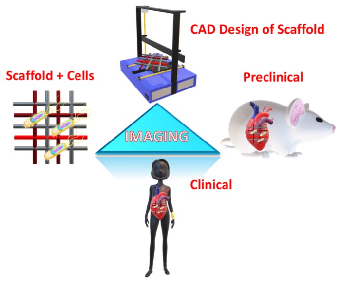
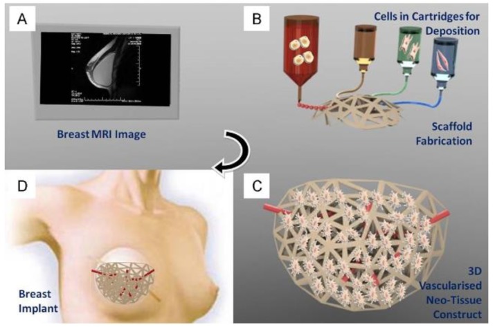
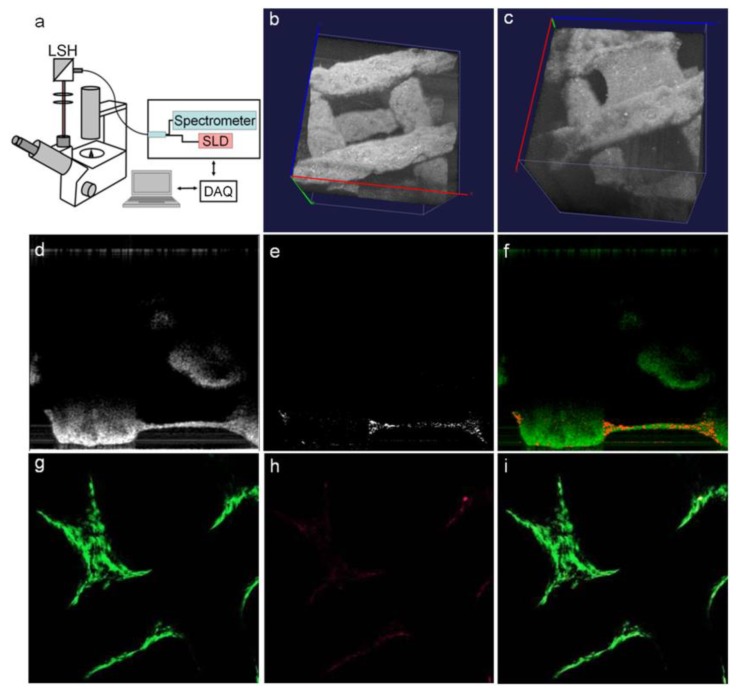
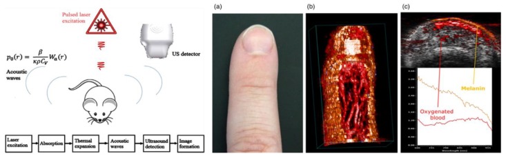
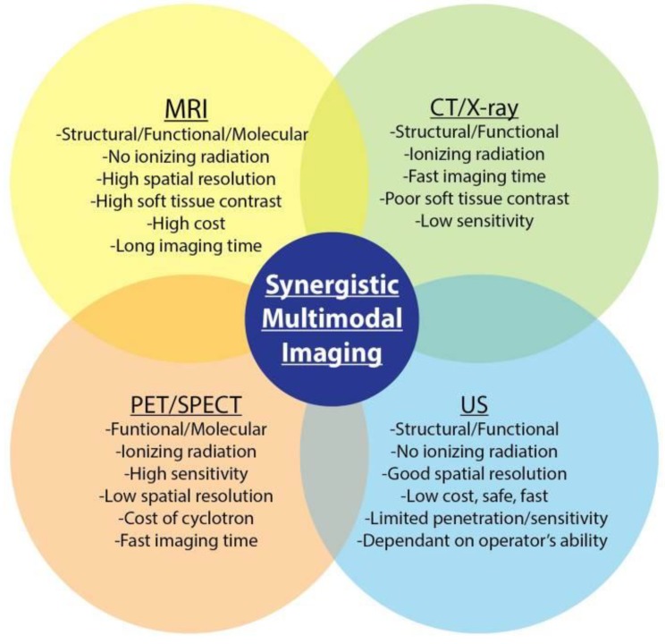
To date, the fields of biomaterials science and tissue engineering have shown great promise in creating bioartificial tissues and organs for use in a variety of regenerative medicine applications. With the emergence of new technologies such as additive biomanufacturing and 3D bioprinting, increasingly complex tissue constructs are being fabricated to fulfill the desired patient-specific requirements. Fundamental to the further advancement of this field is the design and development of imaging modalities that can enable visualization of the bioengineered constructs following implantation, at adequate spatial and temporal resolution and high penetration depths. These in vivo tracking techniques should introduce minimum toxicity, disruption, and destruction to treated tissues, while generating clinically relevant signal-to-noise ratios. This article reviews the imaging techniques that are currently being adopted in both research and clinical studies to track tissue engineering scaffolds in vivo, with special attention to 3D bioprinted tissue constructs.

Keywords: 3D bioprinting; additive manufacturing; bioluminescence; computed tomography (CT); fluorescence spectroscopy; in vivo imaging; magnetic resonant imaging (MRI); magnetic-particle imaging; multimodal imaging; optical coherence tomography; photoacoustic imaging; scaffold tracking; tissue engineering; ultrasound.


Conflict of interest statement

The authors declare no conflict of interest.

Figures

Figure 1
Figure 1
Schematic illustration for the role of imaging in tissue engineering (TE) applications at different levels: scaffold design using computer-aided design (CAD), cellular scaffolds in in vitro applications, preclinical application through implantation in animal models, and clinical application in humans.
Figure 2
Figure 2
Application of patient’s MRI data to generate a bioprinted scaffold for organ regeneration, disease treatment, or drug delivery. MR images (A) of the target organ/tissue will be acquired and processed to create a 3D STL file. The model will be 3D bioprinted using various inks and scaffolds (B), cultured in vitro to establish the new tissue structure and vasculature (C), followed by implantation in vivo to repair/regenerate target tissue/organ (D). Reproduced with permission from Ref. [19].
Figure 3
Figure 3
MR imaging of bioengineered collagen constructs used as cardiac patch to repair ischemic heart tissue. Patches were loaded with 1.5, 3.0, and 6.0 μg/mL of iron oxide nanoparticles and imaged via MRI both in vitro (A,B) and in vivo (C), in a mouse model. Manganese-enhanced MRI visualized the patch grafted onto healthy myocardial tissue in different groups including no treatment (control) (i), empty patch (ii), nanoparticle-loaded patch (iii), and loaded-empty-loaded sandwich patch (iv). Reproduced with permission from Ref. [17].
Figure 4
Figure 4
Hardware setup used in magnetic particle imaging (MPI) scans using SPIONs. (A) The Berkeley field-free-line MPI preclinical scanner. (B) To form a projection image, the magnetic field (FFL) rasters across a trajectory as shown, imaging the in vivo distribution of SPIONs in a rat. Multiple such projections can reconstruct a 3D MPI image similar to CT. Reproduced with permission from Ref. [48].
Figure 5
Figure 5
Example angiography (MR) outlining via contrasting the heart chambers and attendant vasculature. Reproduced with permission from Ref. [64].
Figure 6
Figure 6
Cellular tracking using gold nanoparticles as a contrast agent and imaged with CT. Reproduced with permission from Ref. [77].
Figure 7
Figure 7
(A,B) Experimental setup of ultrasound used to image different areas of a scaffold made of PEG hydrogel. (C) Example of output image from the system. Reproduced with permission from Ref. [96].
Figure 8
Figure 8
A novel 3D bioprinting approach with hydrogel bioinks functionalized with luminescent nanoparticles. (a) Cells and/or nanoparticles, containing the O2-sensitive luminescent indicator PtTFPP compound and an inert fluorescent coumarin dye, were incorporated into an alginate-based bioink for bioprinting. (b) Experimental setup used to image O2 distribution in bioprinted hydrogel constructs.
Figure 9
Figure 9
Fluorescence contrast of tumor growth when the mouse is injected with upconversion nanoparticles. Reproduced with permission from Ref. [115].
Figure 10
Figure 10
(ai) OCT design and the tracking of cells in a 3D bioprinted scaffold seeded with cells. Reproduced with permission from Ref. [127].
Figure 11
Figure 11
Overview of the physics and processing involved in photoacoustic imaging (PAI) (left), and a sample of 2D and 3D vasculatures acquired via PAI from hemoglobin and melanin emissions without contrast agents (right, ac). Reproduced with permission from Ref. [132].
Figure 12
Figure 12
Overview of multimodal imaging applications and their advantages and disadvantages. Reproduced with permission from Ref. [143].

References

    1. Zhang Y.S., Yao J. Imaging Biomaterial-Tissue Interactions. Trends Biotechnol. 2015;40:1291–1296. doi: 10.1016/j.tibtech.2017.09.004. - DOI - PMC - PubMed
    1. Khan F., Tanaka M. Designing Smart Biomaterials for Tissue Engineering. Int. J. Mol. Sci. 2018;19:17. doi: 10.3390/ijms19010017. - DOI - PMC - PubMed
    1. Lee E.J., Kasper F.K., Mikos A.G. Biomaterials for Tissue Engineering. Ann. Biomed. Eng. 2014;42:323–337. doi: 10.1007/s10439-013-0859-6. - DOI - PMC - PubMed
    1. Zhang X., Zhang Y. Tissue Engineering Applications of Three-Dimensional Bioprinting. Cell Biochem. Biophys. 2015;72:777–782. doi: 10.1007/s12013-015-0531-x. - DOI - PubMed
    1. Hunsberger J., Harrysson O., Shirwaiker R., Starly B., Wysk R., Cohen P., Allickson J., Yoo J., Atala A. Manufacturing Road Map for Tissue Engineering and Regenerative Medicine Technologies. Stem Cells Transl. Med. 2015;4:130–135. doi: 10.5966/sctm.2014-0254. - DOI - PMC - PubMed

LinkOut - more resources