. 2019 Jul 16;8(7):1039.
doi: 10.3390/jcm8071039.

Electrospun Polyethylene Terephthalate Nanofibers Loaded with Silver Nanoparticles: Novel Approach in Anti-Infective Therapy

Affiliations

Electrospun Polyethylene Terephthalate Nanofibers Loaded with Silver Nanoparticles: Novel Approach in Anti-Infective Therapy

Alexandru Mihai Grumezescu et al. J Clin Med. .

Abstract

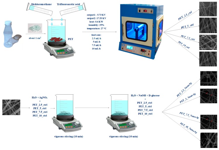
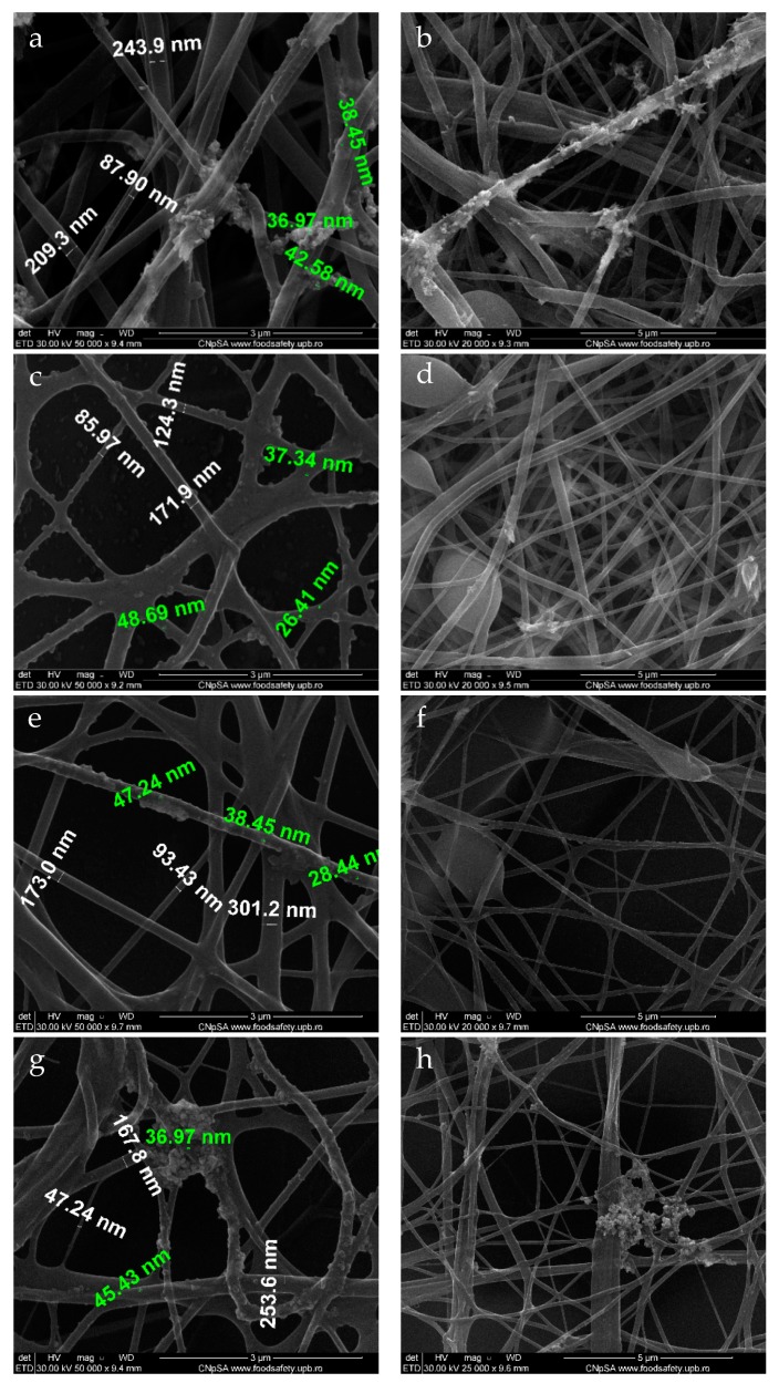
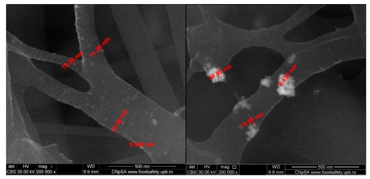
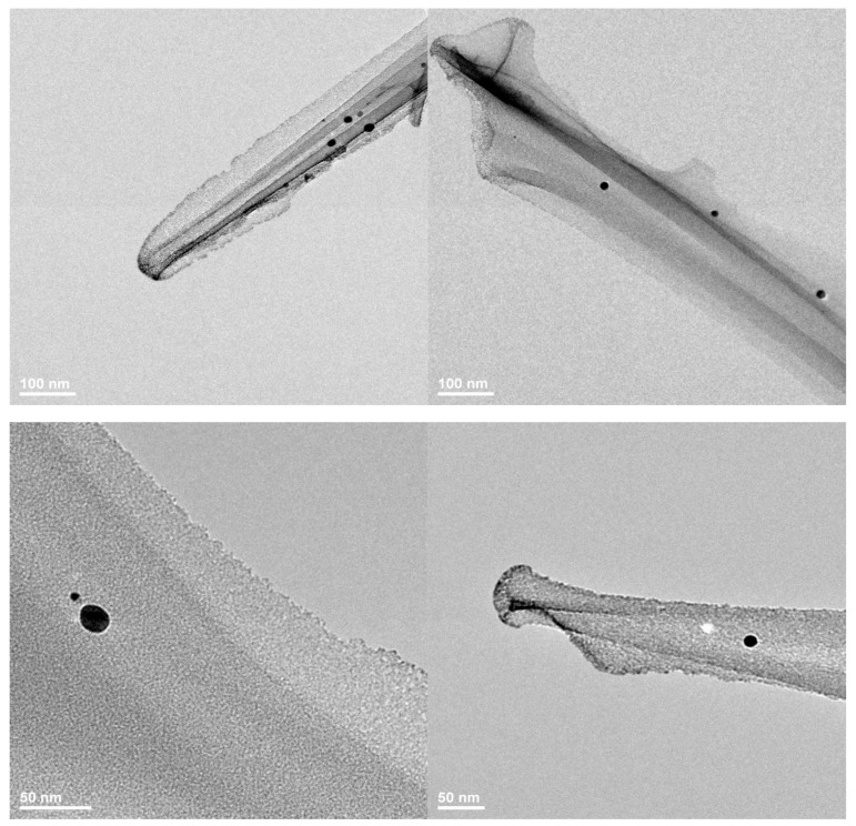
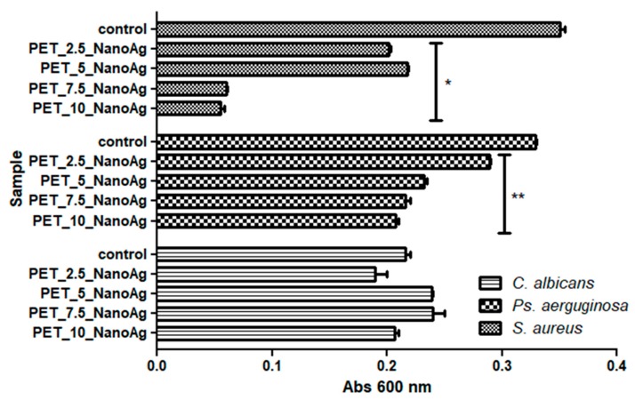
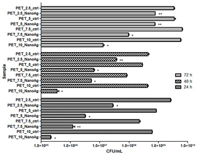
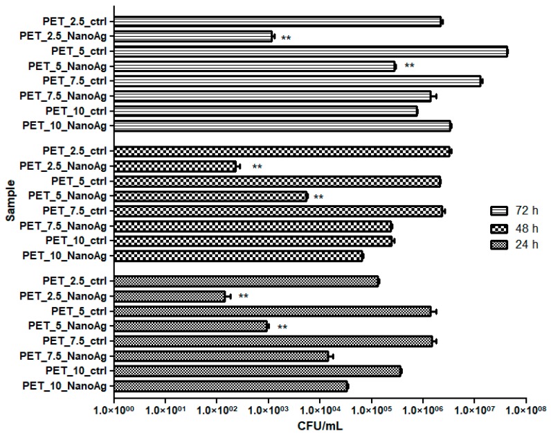
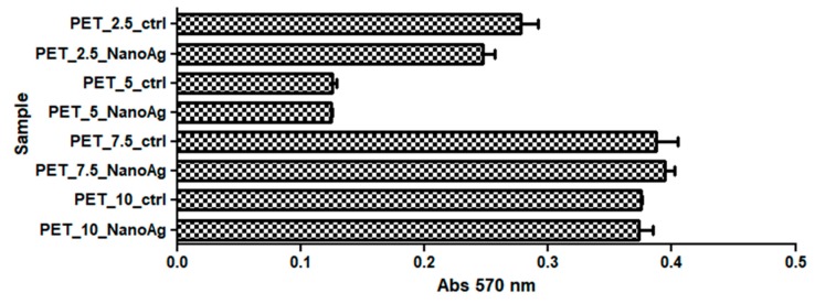
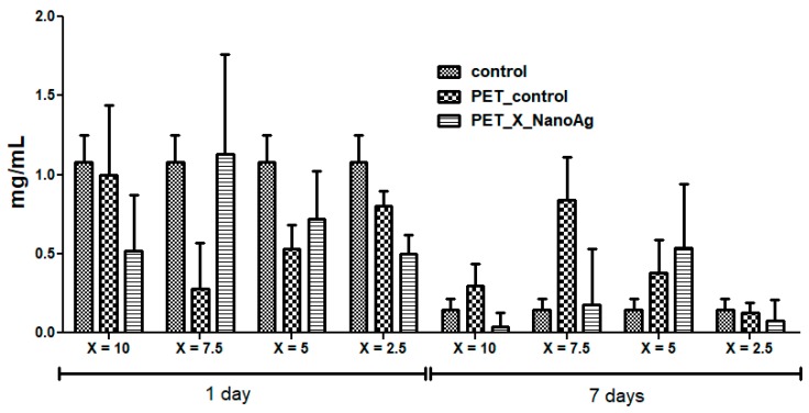
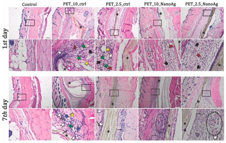
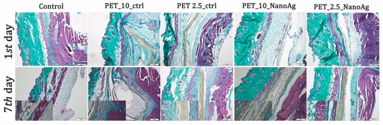
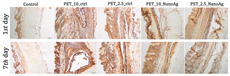
Polyethylene terephthalate (PET) is a major pollutant polymer, due to its wide use in food packaging and fiber production industries worldwide. Currently, there is great interest for recycling the huge amount of PET-based materials, derived especially from the food and textile industries. In this study, we applied the electrospinning technique to obtain nanostructured fibrillary membranes based on PET materials. Subsequently, the recycled PET networks were decorated with silver nanoparticles through the chemical reduction method for antimicrobial applications. After the characterization of the materials in terms of crystallinity, chemical bonding, and morphology, the effect against Gram-positive and Gram-negative bacteria, as well as fungal strains, was investigated. Furthermore, in vitro and in vivo biocompatibility tests were performed in order to open up potential biomedical applications, such as wound dressings or implant coatings. Silver-decorated fibers showed lower cytotoxicity and inflammatory effects and increased antibiofilm activity, thus highlighting the potential of these systems for antimicrobial purposes.

Keywords: PET; antimicrobial agents; biocompatibility; electrospinning; nanofibers; polyethylene terephthalate; silver nanoparticles.


Conflict of interest statement

The authors declare no conflicts of interest.

Figures

Scheme 1
Scheme 1
Synthesis process for control and silver-loaded polyethylene terephthalate (PET_X_ctrl and PET_X_NanoAg).
Figure 1
Figure 1
Transmission electron microscopy (TEM) images recorded for (a,b) silver nanoparticles (NanoAg) and selected area electron diffraction (SAED) pattern (c).
Figure 2
Figure 2
X-ray diffractogram recorded for NanoAg.
Figure 3
Figure 3
Fourier-transform infrared (FT-IR) spectra recorded for the silver-loaded polyethylene terephthalate (PET_X_NanoAg) samples.
Figure 4
Figure 4
SEM images recorded for PET_X_NanoAg at various flows: (a,b) PET_2.5_NanoAg; (c,d) PET_5_NanoAg; (e,f) PET_7.5_NanoAg; (g,h) PET_10_NanoAg. Green text—dimensions for nanoAg; white text—dimensions for PET fibers.
Figure 5
Figure 5
SEM images recorded in backscattering for PET_5_NanoAg.
Figure 6
Figure 6
TEM images recorded for PET_2.5_NanoAg.
Figure 7
Figure 7
Graphic representation of the recorded absorbance values of Staphylococcus aureus, Pseudomonas aeruginosa, and Candida albicans cultures, expressing the multiplication capacity of these cells after cultivation for 24 h in the presence of recycled PET_X_NanoAg materials. * p ≤ 0.001, ** p ≤ 0.05 after the comparison of control with NanoAg-containing PET fibers obtained by applying various flow rates).
Figure 8
Figure 8
Graphic representation of colony-forming units (CFU)/mL representing the number of S. aureus viable cells included in the monospecific biofilms developed on the surface of the materials, quantified after 24 h, 48 h, and 72 h at 37 °C. * p ≤ 0.001, ** p ≤ 0.05 comparing control PET and NanoAg PET obtained at the same flow rate.
Figure 9
Figure 9
Graphic representation of CFU/mL, representing the number of P. aeruginosa viable cells included in the monospecific biofilms developed on the surface of the materials, quantified after 24 h, 48 h, and 72 h at 37 °C. * p ≤ 0.001, ** p ≤ 0.05 comparing control PET and NanoAg PET obtained at the same flow rate.
Figure 10
Figure 10
Graphic representation of CFU/mL representing the number of C. albicans cells viable cells included in the monospecific biofilms developed on the surface of the materials, quantified after 24 h, 48 h, and 72 h at 37 °C. * p ≤ 0.001, ** p ≤ 0.05 comparing control PET and NanoAg PET obtained at the same flow rate.
Figure 11
Figure 11
Effects of PET_X_NanoAg on MTT (3-(4,5-dimethylthiazolyl-2)-2,5-diphenyltetrazolium bromide) specific activities in amniotic fluid stem cells (AFSC).
Figure 12
Figure 12
The effects of PET_X_NanoAg subcutaneous implantation in mice on the C-reactive protein (CRP) levels at 24 h and seven days post-surgery.
Figure 13
Figure 13
Representative histological images of PET_X_NanoAg mats-implanted sites in mice—days one and seven post-implantation. Neutrophils (black arrowhead); monocytes (green arrowhead); eosinophils (red arrowhead); macrophages (white arrowhead); plasma cells (purple arrowhead); giant cells (blue arrowhead); lymphocytes (yellow arrowhead); * implant (asterisk). Cells were stained with hematoxylin and eosin (H&E) stain. Scale bars = 200 and 20 μm.
Figure 14
Figure 14
Gomori’s trichrome stain of PET control and PET_X_NanoAg-implanted tissues.
Figure 15
Figure 15
Expression and specific distribution of tumor necrosis factor (TNF)-α at implantation site at 24 h and seven days after implantation; scale bar = 200 μm.

References

    1. Lu P., Ding B. Applications of electrospun fibers. Recent Pat. Nanotechnol. 2008;2:169–182. doi: 10.2174/187221008786369688. - DOI - PubMed
    1. Naghibzadeh M., Firoozi S., Nodoushan F.S., Adabi M., Khoradmehr A., Fesahat F., Esnaashari S.S., Khosravani M., Adabi M., Tavakol S., et al. Application of electrospun gelatin nanofibers in tissue engineering. Biointerface Res. Appl. Chem. 2018;8:3048–3052.
    1. Teleanu R.I., Gherasim O., Gherasim T.G., Grumezescu V., Grumezescu A.M., Teleanu D.M. Nanomaterial-based approaches for neural regeneration. Pharmaceutics. 2019;11:266. doi: 10.3390/pharmaceutics11060266. - DOI - PMC - PubMed
    1. Huang Z.-M., Zhang Y.Z., Kotaki M., Ramakrishna S. A review on polymer nanofibers by electrospinning and their applications in nanocomposites. Compos. Sci. Technol. 2003;63:2223–2253. doi: 10.1016/S0266-3538(03)00178-7. - DOI
    1. Zhang M., Zhao X., Zhang G., Wei G., Su Z. Electrospinning design of functional nanostructures for biosensor applications. J. Mater. Chem. B. 2017;5:1699–1711. doi: 10.1039/C6TB03121H. - DOI - PubMed

LinkOut - more resources