. 2019 Sep;39(9):1762-1775.
doi: 10.1161/ATVBAHA.119.312603. Epub 2019 Jul 18.

Caspase-1 Activation Is Related With HIV-Associated Atherosclerosis in an HIV Transgenic Mouse Model and HIV Patient Cohort

Affiliations

Caspase-1 Activation Is Related With HIV-Associated Atherosclerosis in an HIV Transgenic Mouse Model and HIV Patient Cohort

Alison C Kearns et al. Arterioscler Thromb Vasc Biol. 2019 Sep.

Abstract

Objective: Atherosclerotic cardiovascular disease (ASCVD) is an increasing cause of morbidity and mortality in people with HIV since the introduction of combination antiretroviral therapy. Despite recent advances in our understanding of HIV ASCVD, controversy still exists on whether this increased risk of ASCVD is due to chronic HIV infection or other risk factors. Mounting biomarker studies indicate a role of monocyte/macrophage activation in HIV ASCVD; however, little is known about the mechanisms through which HIV infection mediates monocyte/macrophage activation in such a way as to engender accelerated atherogenesis. Here, we experimentally investigated whether HIV expression is sufficient to accelerate atherosclerosis and evaluated the role of caspase-1 activation in monocytes/macrophages in HIV ASCVD. Approach and Results: We crossed a well-characterized HIV mouse model, Tg26 mice, which transgenically expresses HIV-1, with ApoE-/- mice to promote atherogenic conditions (Tg26+/-/ApoE-/-). Tg26+/-/ApoE-/- have accelerated atherosclerosis with increased caspase-1 pathway activation in inflammatory monocytes and atherosclerotic vasculature compared with ApoE-/-. Using a well-characterized cohort of people with HIV and tissue-banked aortic plaques, we documented that serum IL (interleukin)-18 was higher in people with HIV compared with non-HIV-infected controls, and in patients with plaques, IL-18 levels correlated with monocyte/macrophage activation markers and noncalcified inflammatory plaques. In autopsy-derived aortic plaques, caspase-1+ cells and CD (clusters of differentiation) 163+ macrophages correlated.

Conclusions: These data demonstrate that expression of HIV is sufficient to accelerate atherogenesis. Further, it highlights the importance of caspase-1 and monocyte/macrophage activation in HIV atherogenesis and the potential of Tg26+/-/ApoE-/- as a tool for mechanistic studies of HIV ASCVD.

Keywords: HIV infections; atherosclerosis; humans; macrophages; mice; monocytes.


Conflict of interest statement

Disclosures: ACK, FL, SD, JAR, EK, LTC, XP, JG, SM, AA, THB and XQ declare that no conflict of interest exists. JL had served as a consultant for Gilead Sciences and Viiv Healthcare, unrelated to this manuscript. MZ has participated in a scientific advisory board meeting for Roche diagnostic and received research funding from Gilead to her institution, not related to this manuscript. SG serves as a consultant for Theratechnologies.

Figures

Figure 1:
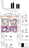
Figure 1:. Tg26+/−/ApoE−/− mice develop accelerated atherosclerosis on 8 week high fat diet.
(A) No difference in HIV transcript levels in multiple immune tissues from male and female Tg26+/−/ApoE−/− and Tg26+/− mice. To determine if HIV background effects HIV transcript levels qRT-PCR analysis of HIV mRNA expression represented as mean ± SEM of multiple tissues in 8 week old mice (n=8). Fold changes were normalized to heart HIV transcript levels. BM: bone marrow. P>0.05. Levels between all tissues were compared using one-way ANOVA. Specific immune tissues were compared by unpaired two-tailed t-test of Tg26+/−/ApoE−/− levels vs. Tg26+/− transcript levels. (B) Representative images of atherosclerosis of en face aorta with oil red O staining in male and female ApoE−/− and Tg26+/−/ApoE−/− mice on an atherogenic diet for 8 weeks. (C) Plaque of 8 week atherogenic diet en face analysis reported as a plaque area per total aortic area (Tg26+/−/ApoE−/− 13.01 ± 1.6% vs. ApoE−/− 8.959 ± 1.1%, P<0.05). (D) Representative images of aortic root plaque stained with oil red O staining in ApoE−/− and Tg26+/−/ApoE−/− mice on an atherogenic diet for 8 weeks. (E) Quantification of aortic root plaques as a percentage of the aortic sinus area (Tg26+/−/ApoE−/− 51.56 ± 1.8% vs. ApoE−/− 42.92 ± 3.3%, P<0.05). (F) Representative images of trichrome staining in ApoE−/− and Tg26+/−/ApoE−/− mice on an atherogenic diet for 8 weeks. (G) Significant increase in collagen content analyzed by trichrome staining reported as a percentage of the total plaque area in Tg26+/−/ApoE−/− 15.32 ± 3.0% vs. ApoE−/− 8.15 ± 1.73%, P<0.05. (H) BUN measured from serum of 8 week atherogenic mice reported in (mg/dL) no significant difference between Tg26+/−/ApoE−/− (26.33 ±1.04mg/dL; n=12) and ApoE−/− mice (29.82 ±1.39mg/dL; n=11). (I) No difference in serum cholesterol reported in mg/dL between Tg26+/−/ApoE−/− (1101 ±45.29 mg/dL) and ApoE−/− mice (1233 ±118.8 mg/dL) on 8 week atherogenic diet. (J) Triglycerides measured in serum from Tg26+/−/ApoE−/− (138.7 ±8.80 mg/dL) and ApoE−/− mice (139.3 ±18.29 mg/dL) on 8 week atherogenic diet. (K) Weight (g) of Tg26+/−/ApoE−/− (24.24 ±0.73g) and ApoE−/− mice (25.11 ±1.10g) after 8 weeks atherogenic diet. All graphs reported as mean ± SEM. *P < 0.05, vs ApoE-/−. Analyzed by two-tailed unpaired t-test.
Figure. 2:
Figure. 2:. Tg26+/−/ApoE−/− mice develop accelerated atherosclerosis on 8 month normal chow (A-F) and transgenic expression of HIV only in hematopoietic cells promotes the atherogenesis (G and H)
(A) Representative en face aorta analysis from 8 months of normal chow reported as a percentage of plaque per total aortic area. (B) Significant increase in en face aortic plaque in male Tg26+/−/ApoE−/− 7.12 ± 3.5% vs. male ApoE−/− 4.26 ±0.96% (1 tailed t-test P<0.05). (C) BUN measured from serum of 8 month normal chow mice reported in mg/dL. No significant difference between Tg26+/−/ApoE−/− (33.71±2.78mg/dL) and ApoE−/− mice (31.33 ±1.60mg/dL). (D) No difference in serum cholesterol reported in mg/dL between Tg26+/−/ApoE−/− (488.3 ±47.57mg/dL) and ApoE−/− mice (492.2±38.03mg/dL) on 8 month normal chow (E) Triglycerides measured in serum from Tg26+/−/ApoE−/− (191.7±27.51mg/dL) and ApoE−/− mice (198.7 ±24.40mg/dL) on 8 month normal chow showing no significant difference. (F) Weight (g) of Tg26+/−/ApoE−/− (27.46 ±0.95g) and ApoE−/− mice (29.07±0.86g) after 8 months’ normal chow showing no significant difference. (G) The plaque area in arch of the chimeric mice: the ApoE−/− mice reconstituted with the bone marrow of Tg26+/−/ApoE−/− mice is greater than that of ApoE−/− transplanted into ApoE−/− mice (14.95 ± 1.70 vs. 9.38 ± 1.27). (H) Real-time RT PCR analysis of HIV transcripts in Tg26+/−/ApoE−/− and chimeric mice. All graphs reported as mean ± SEM. Analyzed by two-tailed unpaired t-test. *P<0.05.
Figure 3.
Figure 3.. Expression of HIV increases caspase-1 activity in inflammatory monocytes in Tg26+/−/ApoE−/− mice.
PBMCs from male and female Tg26+/−/ApoE−/− and littermate ApoE−/− were collected and stained with anti-CD11b, -Ly6G, -Ly6C mouse Abs and FAM-FLICA-Caspase-1 Ab, and analyzed by flow cytometry. A-E. Representative flow cytometry depicting CD11b+ monocyte (MC) caspase-1 activity. (A) Single cells gated according to the forward scatter(FSC)-A and FSC-H. (B) White Blood Cells (WBCs) were then selected according to the FSC-A and side scatter (SSC)-A. (C) Neutrophils were gated out by selecting SSC-H low and CD11b+ cells and Ly6G low and CD11b+ cells. (D) Inflammatory monocytes were identified as CD11b+Ly6C+ high monocytes. (E) Representative histograms depicting caspase-1 activity as a percentage of total cells in CD11b+Ly6C+ monocytes from ApoE−/− and Tg26+/−/ApoE−/− mice respectively. (F) Two experiments with significant increased monocyte caspase-1 activity in Tg26+/−/ApoE−/− compared to ApoE−/− after 6 weeks on an atherogenic diet (38.40 + 10.05 vs. 20.70 +2.40%caspase-1+ cells, P<0.05 and 19.5 +0.98 vs. 16.18 +0.71% caspase-1+ cells, p<0.05 respectively). Analyzed by two-tailed unpaired t-test, *P<0.05.
Figure 4.
Figure 4.. HIV increases caspase-1 activity in the vasculature of Tg26+/−/ApoE−/− mice.
(A) Western blot of pro-caspase-1, active caspase-1 p20, and pro-IL-1β in atherosclerotic aortic arch from male and female mice on an atherogenic diet for 8 weeks. (B) Significant increase in the activation of caspase-1 in the vasculature as measured by the ratio of caspase-1 p20 to pro-caspase-1 in Tg26+/−/ApoE−/− mice (0.93 ±0.06) compared to in ApoE−/− mice (0.35 ±0.24). (C) A trend towards significantly higher pro-caspase-1 protein expression in the vasculature of Tg26+/−/ApoE−/− mice (1.21 ±0.30) compared to in ApoE−/− mice (0.46 ±0.20). (D) A trend towards significantly higher active caspase-1 p20 protein expression in Tg26+/−/ApoE−/− mice (1.17 ±0.36) compared to in ApoE−/− mice (0.26 ±0.23). Data depicted as mean ± SEM of values in the detectable range. Analyzed by two-tailed unpaired T-Test, *P<0.05, ** P<0.01.
Figure 5:
Figure 5:. Upstream and downstream caspase-1 pathway activation in Tg26+/−ApoE−/− mice.
(A) NLRP3 mRNA level in the spleen of male and female ApoE−/− and Tg26+/−ApoE−/− mice on atherogenic diet for 8 weeks. The NLRP3 mRNA transcript was first normalized by GAPDH and compared between two group. The expression level of NLRP3 in ApoE−/− group was set as 1. (Mean± s.e.m, *p<0.05 using two-tailed unpaired t-test. (B&C) Serum IL-1β and IL-18 levels measured in mice on atherogenic diet for 8 weeks using R&D systems ELISA. (B) Significant increase in serum IL-1β in Tg26+/−/ApoE−/− mice (20.74 ±4.19pg/mL) compared to in ApoE−/− mice (5.86 ±1.19pg/mL) measured by ELISA with orbital shaking to decrease the lower detection limit of the kit to 2.35 pg/mL. (C) A trend towards a significant increase in serum IL-18 in in Tg26+/−/ApoE−/− mice (584.4 ±74.91pg/mL) compared to ApoE−/− mice (421.7 ±46.07pg/mL). Data depicted as mean ± SEM of values in the detectable range. Analyzed by two-tailed unpaired T-Test, *P<0.05, ** P<0.01.
Figure 6:
Figure 6:. Circulating IL-18 levels increased in PWH and in patients with plaque.
Serum from PWH and non-HIV-infected controls was examined for IL-18 and IL-1β. (A) Log serum IL-18 was significantly elevated in the PWH compared to non-HIV-infected controls (5.59 ± 0.48 log pg/mL vs. 5.29 ± 0.51log pg/mL, P<0.0001). (B) Log serum IL-18 was significantly elevated in all subjects with plaque compared to those without plaque (5.58 ± 0.50 log pg/mL vs. 5.42 ± 0.49 log pg/mL, P=0.02). IL-1β levels were below the limit of detection (data not shown). Data reported as mean ± SD and were analyzed by two-tailed unpaired t-test.
Figure 7:
Figure 7:. The number of caspase-1+ cells and CD163+ macrophages correlate in tissue banked aortic plaques.
Immunohistochemical analyses of tissue banked aortas from HIV- and HIV+ specimens were performed for CD163 (M2- macrophages) and caspase-1. (A) Representative images of CD163+ macrophages and caspase-1+ cells in aortic plaques. (B) The numbers of caspase-1+ and CD163+ macrophages positively correlated in plaque (r=0.6, P= 0.02). Correlation was determined using Spearman non-parametric analysis.

References

    1. Freiberg MS, Chang CC, Kuller LH, et al. HIV infection and the risk of acute myocardial infarction. JAMA Intern Med. 2013;173:614–622. doi: 10.1001/jamainternmed.2013.3728. - DOI - PMC - PubMed
    1. Hsue PY, Hunt PW, Schnell A, Kalapus SC, Hoh R, Ganz P, Martin JN and Deeks SG. Role of viral replication, antiretroviral therapy, and immunodeficiency in HIV-associated atherosclerosis. AIDS (London, England). 2009;23:1059–1067. doi: 10.1097/QAD.0b013e32832b514b. - DOI - PMC - PubMed
    1. Kearns A, Gordon J, Burdo TH and Qin X. HIV-1-Associated Atherosclerosis: Unraveling the Missing Link. Journal of the American College of Cardiology. 2017;69:3084–3098. doi: 10.1016/j.jacc.2017.05.012. - DOI - PMC - PubMed
    1. Lo J, Abbara S, Shturman L, Soni A, Wei J, Rocha-Filho JA, Nasir K and Grinspoon SK. Increased prevalence of subclinical coronary atherosclerosis detected by coronary computed tomography angiography in HIV-infected men. AIDS (London, England). 2010;24:243–253. doi: 10.1097/QAD.0b013e328333ea9e. - DOI - PMC - PubMed
    1. Burdo TH, Lentz MR, Autissier P, Krishnan A, Halpern E, Letendre S, Rosenberg ES, Ellis RJ and Williams KC. Soluble CD163 made by monocyte/macrophages is a novel marker of HIV activity in early and chronic infection prior to and after anti-retroviral therapy. The Journal of infectious diseases. 2011;204:154–163. doi: 10.1093/infdis/jir214. - DOI - PMC - PubMed

Publication types