. 2019 Jul 17;19(1):166.
doi: 10.1186/s12866-019-1534-3.

Extracellular vesicles and soluble factors secreted by Escherichia coli Nissle 1917 and ECOR63 protect against enteropathogenic E. coli-induced intestinal epithelial barrier dysfunction

Affiliations

Extracellular vesicles and soluble factors secreted by Escherichia coli Nissle 1917 and ECOR63 protect against enteropathogenic E. coli-induced intestinal epithelial barrier dysfunction

Carina-Shianya Alvarez et al. BMC Microbiol. .

Abstract

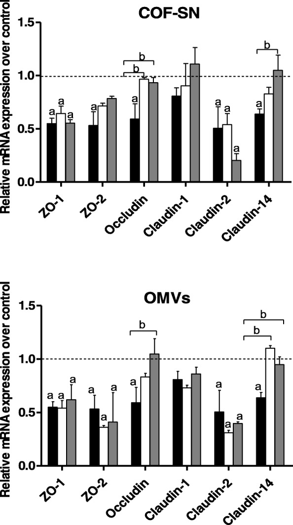
Background: Enteric pathogens have developed mechanisms to disrupt tight junctions and increase gut permeability. Many studies have analysed the ability of live probiotics to protect intestinal epithelial cells against tight junction damage caused by bacterial pathogens. Escherichia coli Nissle 1917 (EcN) is among the probiotics that positively modulates the intestinal epithelial barrier by regulating expression and distribution of tight junction proteins. We previously reported that regulation of ZO-1, claudin-14 and claudin-2 is mediated by EcN secreted factors, either free-released or associated with outer membrane vesicles (OMVs). Factors secreted by commensal ECOR63 elicited comparable effects in intact epithelial T-84 and Caco-2 cell monolayers.

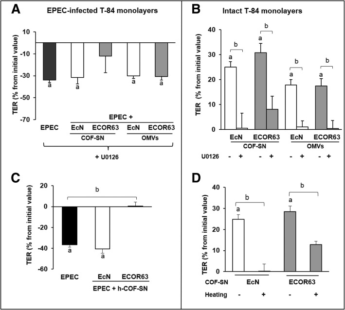
Results: Here we analyse the ability of OMVs and soluble secreted factors to protect epithelial barrier function in polarized T-84 and Caco-2 cells infected with enteropathogenic Escherichia coli (EPEC). Transepithelial electrical resistance, paracellular permeability, mRNA levels and subcellular distribution of tight junction proteins were monitored in the absence or presence of EcN and ECOR63 extracellular fractions. EPEC downregulated expression of ZO-1 ZO-2, occludin and claudin-14 and altered the subcellular localization of ZO-1, occludin and F-actin cytoskeleton. OMVs and soluble factors secreted by EcN and ECOR63 counteracted EPEC-altered transepithelial resistance and paracellular permeability, preserved occludin and claudin-14 mRNA levels, retained ZO-1 and occludin at tight junctions in the cell boundaries and ameliorated F-actin disorganization. Redistribution of ZO-1 was not accompanied by changes at mRNA level.

Conclusion: This study provides new insights on the role of microbiota secreted factors on the modulation of intestinal tight junctions, expanding their barrier-protective effects against pathogen-induced disruption.

Keywords: ECOR63; EPEC; Escherichia coli Nissle 1917; Intestinal epithelial barrier; Outer membrane vesicles; Tight junctions.


Conflict of interest statement

The authors declare that they have no competing interests.

Figures

Fig. 1
Fig. 1
OMVs and free-soluble factors (COF-SN) secreted by EcN and ECOR63 maintain barrier function in EPEC-infected cells. Confluent T-84 and Caco-2 cell monolayers grown in Transwell supports were infected with EPEC (MOI of 100) for 3 h in the absence or presence of EcN or ECOR63 OMVs (0.1 mg/ml) or COF-SN (0.5 mg/ml). Non-treated cells were processed in parallel as a control. Epithelial barrier function was analysed by measuring TER (a) and FD-4 flux (b) as markers of epithelial resistance and barrier permeability. a TER values were measured before and after 3-h infection. Data are presented as percentage of changes in TER (decrease) from the initial value. b After 3-h infection, cell monolayers were washed and treated apically with FD-4 (1 mg/ml). The fluorescence in the basolateral chamber was measured before and 1 h after the addition of FD-4. FD-4 flux values were calculated by subtracting the fluorescence intensity units (FI) measured at 0 h. Data were expressed as fold-change compared with non-infected control cells, whose values were normalized as 1. In all panels, data are from three independent biological experiments performed in triplicate. The TER baseline control values were 1290 ± 98 Ω.cm2 for T-84 monolayers and around 920 ± 80 Ω.cm2 for Caco-2 cells. a, Significance against untreated control cells (p ≤ 0.05); b, significance against control EPEC-infected cells (p ≤ 0.05)
Fig. 2
Fig. 2
Analysis of host and bacterial factors involved in the strengthening activity of EcN and ECOR63 secreted fractions. The strengthening activity of COF-SN and OMVs depends on the ERK1/2 signalling pathway both in EPEC-infected cells (a) and intact cell monolayers (b). a-b Before infection/stimulation with OMVs or COF-SN, T-84 cell monolayers were pre-treated for 15 min with the ERK1/2 inhibitor U0126 (25 μM). TER values were measured before and after 3-h treatment. c-d Effect of heat treatment on the strengthening activity of EcN and ECOR63 COF-SN. EPEC-infected (c) or intact (d) T-84 cell monolayers were incubated with heated COF-SN (h-COF-SN), and TER values were measured before and after 3-h treatment. In all panels, data are presented as percentage of changes in TER from the initial value from three independent biological experiments performed in triplicate. TER initial values were between 1100 and 1300 Ω.cm2. a, Significance against untreated control cells (p ≤ 0.02); b, significance against control EPEC-infected cells (a, c) or cells treated with EcN or ECOR63 control extracellular fractions (b, d) (p ≤ 0.05)
Fig. 3
Fig. 3
Effect of the secreted fractions (COF-SN and OMVs) from EcN and ECOR63 on the expression of TJ proteins in T-84 monolayers infected with EPEC. Intestinal epithelial cells were incubated for 3 h with EPEC at a MOI of 100 (black bars). Parallel infections were carried out in the presence of COF-SN (0.5 mg/ml) or OMVs (0.1 mg/ml) from EcN (white bars) or ECOR63 (gray bars). The relative mRNA levels of the indicated proteins were measured by RT-qPCR using β-actin as the reference gene. Data are presented as fold-change compared to untreated control cells (dotted line) from three independent biological experiments. a, Significance against untreated control cells (p ≤ 0.05); b, significance against EPEC-infected control cells (p ≤ 0.04)
Fig. 4
Fig. 4
Immunofluorescence staining of ZO-1 and occludin in Caco-2 cell monolayers incubated for 3 h with EPEC in the absence or presence of COF-SN or OMVs from the indicated bacterial strains. Analysis was performed by laser scanning confocal spectral microscope with 63x oil immersion objective lens, and images were captured with a Nikon color camera (8 bit). Images shown are representative from three independent biological experiments and are coded with Fire look-up table. Calibration bar is shown on the left. Scale bar, 20 μm
Fig. 5
Fig. 5
Fluorescence microscopy analysis of F-actin in Caco-2 cell monolayers incubated for 3 h with EPEC in the absence or presence of COF-SN or OMVs from the indicated bacterial strains. F-actin was stained with TRITC-labelled phalloidin (red) and analysis was performed as described for Fig. 4. Images shown are representative of three independent biological experiments. Scale bar: 10 μm
Fig. 6
Fig. 6
Schematic representation of mechanisms used by EPEC to disrupt the intestinal epithelial barrier and their prevention by EcN and ECOR63 secreted fractions (OMVs and soluble free-released factors)

References

    1. Turner JR. Intestinal mucosal barrier function in health and disease. Nat Rev Immunol. 2009;9:799–809. doi: 10.1038/nri2653. - DOI - PubMed
    1. Peterson LW, Artis D. Intestinal epithelial cells: regulators of barrier function and immune homeostasis. Nat Rev Immunol. 2014;14:141–153. doi: 10.1038/nri3608. - DOI - PubMed
    1. Chiba H, Osanai M, Murata M, Kojima T, Sawada N. Transmembrane proteins of tight junctions. Biochim Biophys Acta. 2008;1778:588–600. doi: 10.1016/j.bbamem.2007.08.017. - DOI - PubMed
    1. Lee SH. Intestinal permeability regulation by tight junctions: implication on inflammatory bowel diseases. Intest Res. 2015;13:11–18. doi: 10.5217/ir.2015.13.1.11. - DOI - PMC - PubMed
    1. González-Mariscal L, Tapia R, Chamorro D. Crosstalk of tight junction components with signaling pathways. Biochim Biophys Acta. 2008;1778:729–756. doi: 10.1016/j.bbamem.2007.08.018. - DOI - PubMed

Publication types

MeSH terms