. 2019 Jul 17;7(1):116.
doi: 10.1186/s40478-019-0768-5.

Retinal pathology in experimental optic neuritis is characterized by retrograde degeneration and gliosis

Affiliations

Retinal pathology in experimental optic neuritis is characterized by retrograde degeneration and gliosis

Praveena Manogaran et al. Acta Neuropathol Commun. .

Erratum in

Abstract

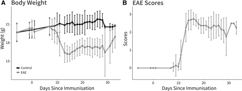
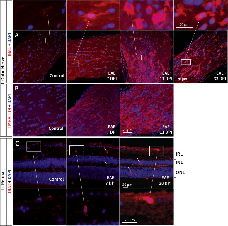
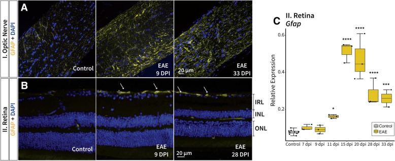
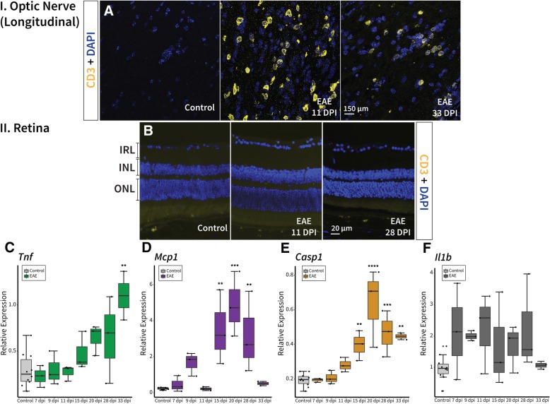
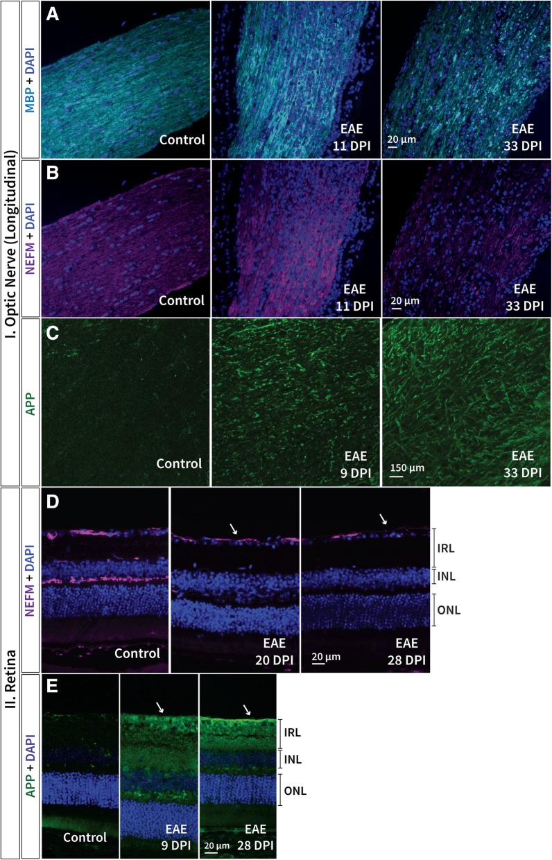
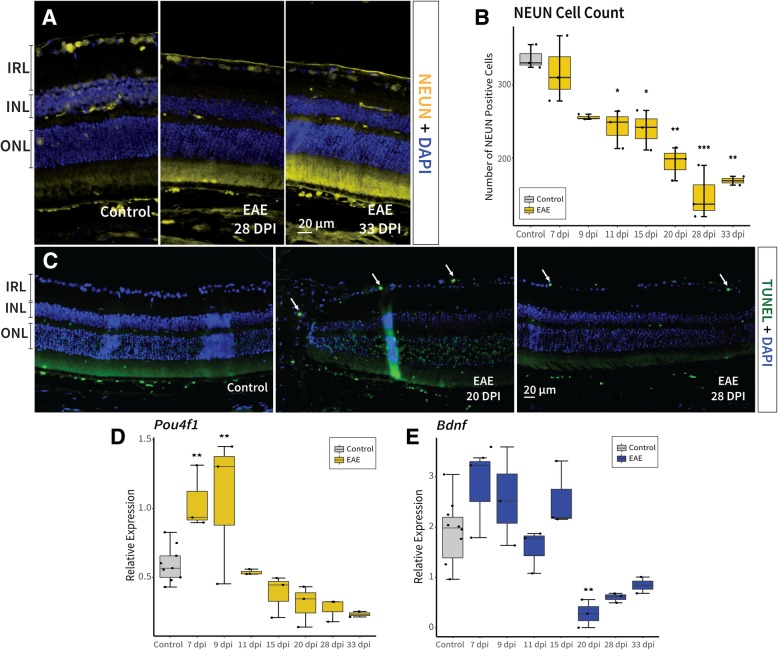
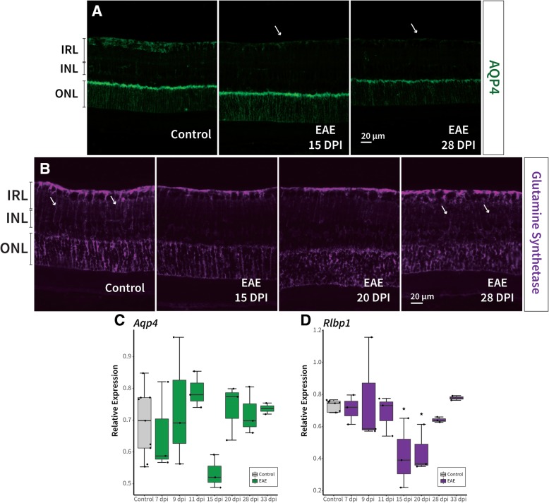
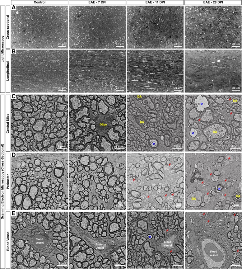
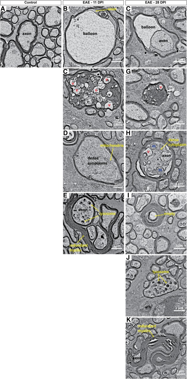
The exact mechanisms and temporal sequence of neurodegeneration in multiple sclerosis are still unresolved. The visual pathway including its unmyelinated retinal axons, can serve as a prototypic model of neurodegeneration in experimental optic neuritis. We conducted a longitudinal study combining retinal imaging through optical coherence tomography (OCT) with immunohistochemical analyses of retinal and optic nerve tissue at various time points in experimental autoimmune encephalomyelitis (EAE).Inner retinal layer (IRL) thickness was measured in 30 EAE and 14 healthy control C57BL/6 J mice using OCT. Distribution of marker proteins was assessed by immunofluorescence staining and retinal mRNA levels were assayed using real-time PCR. Histological morphology was evaluated on light and electron microscopy images.Signs of inflammatory edema 11 days post immunisation coincided with IRL thickening, while neuro-axonal degeneration throughout the disease course contributed to IRL thinning observed after 20 days post immunisation. Retinal pathology, including axonal transport impairment, was observed early, prior to cellular infiltration (i.e. T-cells) in the optic nerve 11 days post immunisation. Yet, the effects of early retinal damage on OCT-derived readouts were outweighed by the initial inflammatory edema. Early microglial activation and astrocytosis was detected in the retina prior to retinal ganglion cell loss and persisted until 33 days post immunisation. Müller cell reactivity (i.e. aquaporin-4 and glutamine synthetase decrease) presented after 11 days post immunisation in the IRL. Severe neuro-axonal degeneration was observed in the optic nerve and retina until 33 days post immunisation.Initial signs of retinal pathology subsequent to early glial activity, suggests a need for prophylactic treatment of optic neuritis. Following early inflammation, Müller cells possibly respond to retinal pathology with compensatory mechanisms. Although the majority of the IRL damage observed is likely due to retrograde degeneration following optic neuritis, initial pathology, possibly due to gliosis, may contribute further to IRL thinning. These results add morphological substrate to our OCT findings. The extent and rapid onset of axonal and neuronal damage in this model appears relevant for testing interventions scaled to human optic neuritis.

Keywords: Experimental autoimmune encephalomyelitis; Gliosis; Neuro-axonal degeneration; Optic neuritis; Optical coherence tomography; Retina.


Conflict of interest statement

PM has received a travel grant from Merck Sereno and Sanofi Genzyme. CAW has received a travel grant from Merck Sereno and Teva. CW-E has received travel grants from Merck Sereno and Sanofi Genzyme. SvS has received research grants from Novartis and Sanofi Genzyme, and consultancy and speaking fees from Biogen, Merck Serono, Novartis, Roche, Sanofi Genzyme, and Teva. ANS, CG, MS, and MR declare that they have no competing interests.

Figures

Fig. 1
Fig. 1
Clinical assessment of healthy and EAE induced C57BL/6 J mice. a Body weight over the observational period for both healthy control C57BL/6 J (black) and EAE induced (grey) mice, mean ± SD. b EAE clinical scores longitudinally in EAE induced mice, mean ± SD. EAE: experimental autoimmune encephalomyelitis.
Fig. 2
Fig. 2
Inner retinal layer thickness increased at onset and decreased at later time points in EAE. a Boxplots of the inner retinal layer (IRL) thickness over time in EAE (yellow) and healthy controls (blue). b Optical coherence tomography retinal B-scan of healthy C57BL/6 J mouse with segmented layers including the IRL, inner nuclear layer and outer plexiform layer. Inset includes a diagram of the IRL composed of retinal ganglion cells (RGC). The retinal nerve fiber layer consists of the unmyelinated axons of the RGCs, with their cell bodies and dendrites located within the ganglion cell layer and inner plexiform layer respectively. * p-values for comparison between EAE and control mice (**** p < 0.00001, *** p < 0.0001), § p-value for comparison between baseline and recovery in EAE mice (§§§§ p < 0.00001). EAE: experimental autoimmune encephalomyelitis, dpi: days post immunisation. Scale bars: 200 μm
Fig. 3
Fig. 3
Microgliosis presented early in EAE and persisted until the final observational time point. Increased microglial cell (IBA1) presence was observed as early as 7 days post immunisation (dpi) in both the a/b optic nerve and d retina (arrows), persisting until the final observational time point in EAE mice. b TMEM119 marked resident microglial cells observed at 7 dpi and 11 dpi in EAE mice, a while in the optic nerve, a significant increase in microglial cells at 11 dpi also coincided with cellular infiltration. The insets a/c display examples of microglial cells in controls (ramified: weak signal, long branched processes) and EAE mice (amoeboid: strong signal, round)
Fig. 4
Fig. 4
Astrocytosis appeared subsequent to microgliosis in EAE and also persisted until the final time point. Increased GFAP signal (associated with gliosis) presented at 9 dpi and also remained until 33 dpi in both the a optic nerve and the b retina (arrows). c mRNA expression of Gfap in EAE retinas significantly increased from 11 dpi to 33 dpi, tapering off at the final two observational time points. p-values for comparison between EAE and control mice (**** p < 0.00001, *** p < 0.0001, * p < 0.05). EAE: experimental autoimmune encephalomyelitis, GFAP: glial fibrillary acidic protein, IRL: inner retinal layer, INL: inner nuclear layer, ONL: outer nuclear layer
Fig. 5
Fig. 5
A strong inflammatory response was observed in the optic nerve and retina of EAE mice. a T-cells (CD3) initially appear 11 days post immunisation (dpi) along with the cellular infiltration (DAPI), subsiding at later time points but still remaining until 33 dpi in the optic nerve of EAE mice. b T-cells were not observed in the retina of EAE mice at any time point. Yet, c Tnf expression amplified from 20 to 33 dpi in EAE compared to healthy controls in the retina. Tnf is a proinflammatory cytokine involved in the innate immune response. Whereas, d mRNA expression of Mcp1 in the retina increased significantly between 15 dpi and 28 dpi in EAE mice. Mcp1 likely plays an amplifying role (rather than an initiating role) in EAE. e Caspase 1 (Casp1) increases significantly from 15 dpi to 33 dpi in EAE retinas. It has a crucial role in development of immune mediated inflammatory processes leading to central nervous system demyelination. However, f Il-1β, which is a cytokine activated by Caspase 1, was not different in retinal expression between EAE mice and healthy controls. p-values for comparison between EAE and control mice (**** p < 0.00001, *** p < 0.0001, ** p < 0.01). EAE: experimental autoimmune encephalomyelitis, TNF: tumor necrosis factor, MCP1: monocyte chemoattractant protein 1, IL-1β: interleukin-1β, IRL: inner retinal layer, INL: inner nuclear layer, ONL: outer nuclear layer. EAE: experimental autoimmune encephalomyelitis, IBA1: allograft inflammatory factor 1, TMEM119: transmembrane protein 119, IRL: inner retinal layer, INL: inner nuclear layer, ONL: outer nuclear layer
Fig. 6
Fig. 6
Demyelination and axonal pathology was perceived in both the retina and optic nerve in EAE. a Demyelination (MBP) in the optic nerve was first observed 11 days post immunisation (dpi) progressing until 33 dpi where profound signs of myelin disorganisation and possible myelin debris were observed. b Axonal degeneration (NEFM) was observed from 11 dpi until 33 dpi in the optic nerve of EAE mice. c APP, important for axonal transport, appears to increase as early as 9 dpi and also persisted until the final observational time point in EAE optic nerve sections. In the retina, d NEFM appears to decrease in the IRL (arrows) and in the outer plexiform layer over time in EAE mice; beginning later (20 dpi) than in the optic nerve. e APP in the retina appears to increase (arrows) as early as 9 dpi similar to presentation in the optic nerve for EAE mice. EAE: experimental autoimmune encephalomyelitis, MBP: myelin basic protein, NEFM: neurofilament-M, APP: Alzheimer precursor protein, IRL: inner retinal layer, INL: inner nuclear layer, ONL: outer nuclear layer
Fig. 7
Fig. 7
Neurodegeneration in the retina was observed throughout the observational period in EAE. a-b The number of NEUN positive cells decreased significantly from 11 days post immunisation (dpi) to 33 dpi in EAE mice compared to healthy controls. Furthermore, c apoptosis was observed using TUNEL staining from 20 to 33 dpi in the ganglion cell layer (arrows) of EAE mice. d Pou4f1, a transcription factor expressed in retinal ganglion cells first increased significantly at 7 and 9 dpi (possibly due to compensatory mechanisms within the cell) followed by a decrease from 20 to 33 dpi. e Bdnf retinal expression, which is an important neurotrophic factor for neuronal survival, also decreased at 20 and 28 dpi in EAE mice compared to healthy control, further exemplifying neurodegeneration. p-values for comparison between EAE and control mice (*** p < 0.0001, ** p < 0.01, * p < 0.05). EAE: experimental autoimmune encephalomyelitis, NEUN: neuronal nuclei, TUNEL: terminal deoxynucleotidyl transferase dUTP nick end labelling, IRL: inner retinal layer, POU4F1: POU domain class 4 transcription factor 1, BDNF: brain-derived neurotrophic factor, INL: inner nuclear layer, ONL: outer nuclear layer
Fig. 8
Fig. 8
Müller cell reactivity was detected following signs of optic neuritis. a A decrease in AQP4 signal (arrows) in the retinal nerve fibre layer was observed 15 and 28 days post immunisation (dpi) in the retina of EAE mice. b Glutamine synthetase, expressed in Müller cells, decreased in the Müller cell branched processes at 15 and 20 dpi but appeared to recover by 28 dpi in EAE mice. Arrows in the healthy control retina point to intact Müller cell processes, while in EAE, the arrows indicate the recovered processes that appear disorganized. c mRNA expression of Aqp4 decreased only at 15 dpi, while d Rlbp1 expression decreased significantly at 15 and 20 dpi in EAE mice retinas compared to healthy controls. p-values for comparison between EAE and control mice (* p < 0.05). EAE: experimental autoimmune encephalomyelitis, AQP4: aquaporin-4, RLBP1: retinaldehyde-binding protein 1, IRL: inner retinal layer, INL: inner nuclear layer, ONL: outer nuclear layer
Fig. 9
Fig. 9
Optic nerve pathology began near the perimeter and blood vessels and progressed into the center. In the optic nerve a-b, the fibres that surround the axon bundles appear to be disrupted by 11 days post immunisation (dpi) in EAE mice. a There was also a reduction in the number of axons present in EAE mice compared to healthy controls at 11 dpi and 28 dpi in the optic nerve. b In the longitudinal optic nerve sections, an overall disruption of axonal organisation was observed in EAE mice. c-e Scanning electron micrographs of cross-sectional optic nerves in healthy and EAE mice provide a closer look. c In central sections, signs of axonal degeneration were first observed 11 dpi, while at 28 dpi severe pathology was present throughout the entire optic nerve. d Near the perimeter of the optic nerve sections, strong axonal degeneration was observed at both 11 and 28 dpi in EAE mice compared to healthy controls. Severe loss of axons and large empty spaces were also observed at 28 dpi near the perimeter of the optic nerve. e Some demyelinated axons were observed along with axonal degeneration at 11 dpi near blood vessels, while at 28 dpi severe axonal loss was present in EAE mice compared to healthy controls. Red arrows: demyelinated axons, DA: degenerating axon, E: empty myelin sheath with no axon, Oligo: oligodendrocyte, EAE: experimental autoimmune encephalomyelitis. Electron micrograph image resolution: 8 nm/pixel
Fig. 10
Fig. 10
Examples of optic nerve pathology observed in EAE mice 11 and 28 days post immunisation. a Example of a healthy axonal fibre with normal myelination. b Swollen nerve fibres with a fragment of axon left and a large balloon produced from the splitting of the myelin sheaths was observed in EAE mice at 11 dpi. c Another large swollen nerve fibre with dark cytoplasm (likely a sign of severe degeneration) filled with numerous vacuoles at 11 dpi. d Many bloated myelinated axons were observed at 11 dpi with a dense axoplasm filled with primarily neurofilament. e A dystrophic axon where the axoplasm contains an accumulation of lysosomes. Redundant myelination can also be found around the axons at 11 dpi. f In EAE mice at 28 dpi, ballooning was also observed in some nerve fibres. g Nerve fibres with dark cytoplasm were identified at 28 dpi as well but appeared more shrunken than at 11 dpi. h At 28 dpi, large myelinated nerve fibres showed dense degeneration with numerous vacuoles and organelles within the cytoplasm, however they were not as large as fibres at 11 dpi. i Empty myelin sheaths with completely degenerated axons were visible at 28 dpi in EAE mice. j Dystrophic axons were also found at 28 dpi. k Redundant myelin was more common at 28 dpi, often forming around tiny fragments of axons or entirely on its own. v: vacuoles, m: mitochondria, EAE: experimental autoimmune encephalomyelitis. Electron micrograph image resolution: 8 nm/pixel

References

    1. Luessi F, Siffrin V, Zipp F (2012) Neurodegeneration in multiple sclerosis: novel treatment strategies. Expert Rev Neurother - PubMed
    1. Fairless R, Williams SK, Hoffmann DB, Stojic A, Hochmeister S, Schmitz F, Storch MK, Diem R. Preclinical retinal neurodegeneration in a model of multiple sclerosis. J Neurosci. 2012;32:5585–5597. doi: 10.1523/JNEUROSCI.5705-11.2012. - DOI - PMC - PubMed
    1. Hein K, Gadjanski I, Kretzschmar B, Lange K, Diem R, Sättler MB, Bähr M. An optical coherence tomography study on degeneration of retinal nerve fiber layer in rats with autoimmune optic neuritis. Invest Ophthalmol Vis Sci. 2012;53:157–163. doi: 10.1167/iovs.11-8092. - DOI - PubMed
    1. Green AJ, McQuaid S, Hauser SL, Allen IV, Lyness R. Ocular pathology in multiple sclerosis: retinal atrophy and inflammation irrespective of disease duration. Brain. 2010;133:1591–1601. doi: 10.1093/brain/awq080. - DOI - PMC - PubMed
    1. Jenkins TM, Toosy AT. Optic neuritis: the eye as a window to the brain. Curr Opin Neurol. 2017;30:61–66. doi: 10.1097/WCO.0000000000000414. - DOI - PubMed

Publication types

MeSH terms

LinkOut - more resources