. 2019 Aug 23;294(34):12610-12621.
doi: 10.1074/jbc.AC119.009977. Epub 2019 Jul 17.

Human ATG4 autophagy proteases counteract attachment of ubiquitin-like LC3/GABARAP proteins to other cellular proteins

Affiliations

Human ATG4 autophagy proteases counteract attachment of ubiquitin-like LC3/GABARAP proteins to other cellular proteins

Alexander Agrotis et al. J Biol Chem. .

Abstract

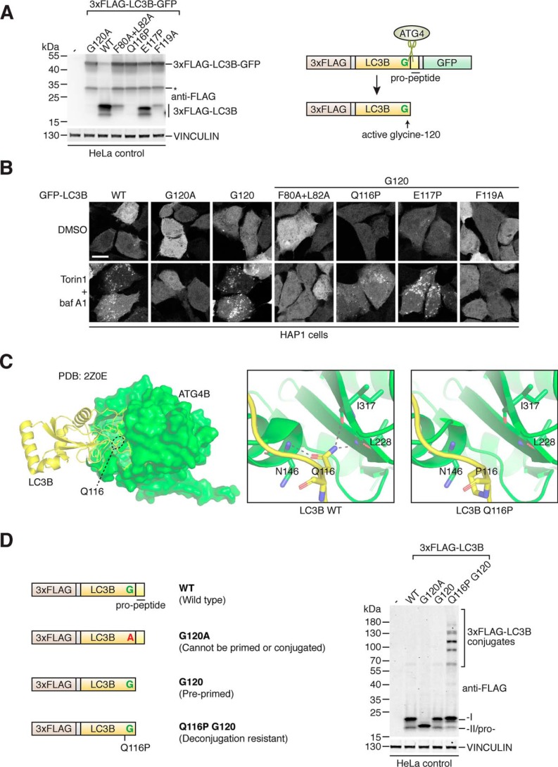
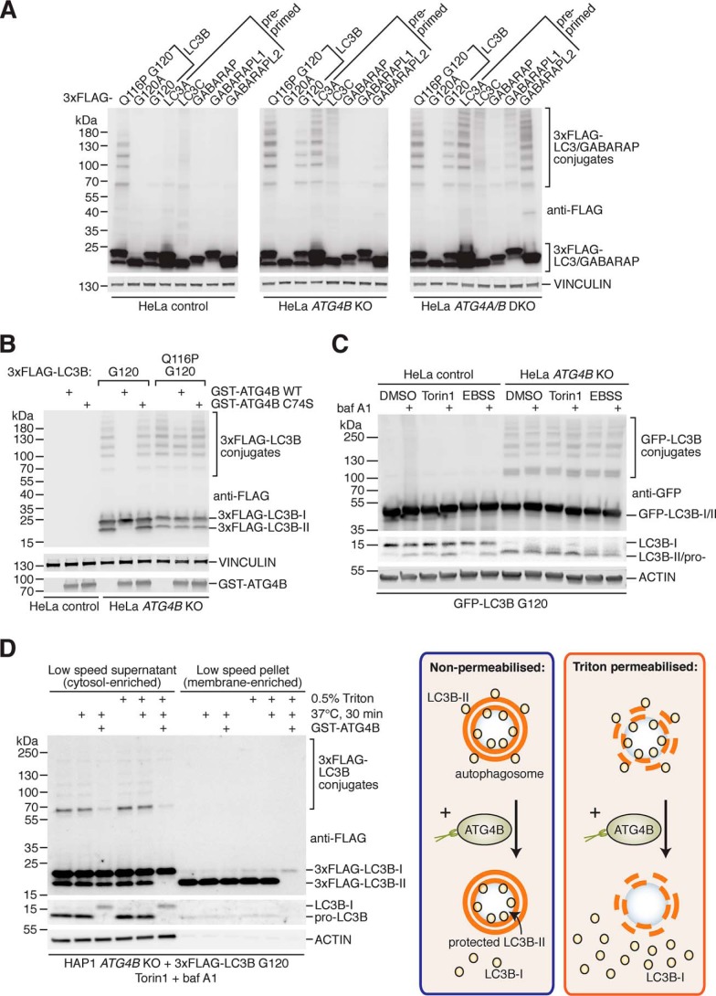
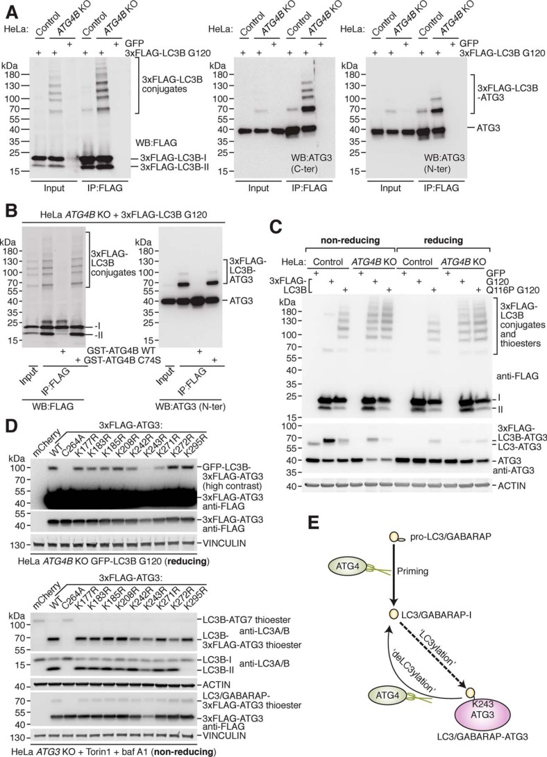
Microtubule-associated protein 1 light chain 3 α (LC3)/GABA type A receptor-associated protein (GABARAP) comprises a family of ubiquitin-like proteins involved in (macro)autophagy, an important intracellular degradation pathway that delivers cytoplasmic material to lysosomes via double-membrane vesicles called autophagosomes. The only currently known cellular molecules covalently modified by LC3/GABARAP are membrane phospholipids such as phosphatidylethanolamine in the autophagosome membrane. Autophagy-related 4 cysteine peptidase (ATG4) proteases process inactive pro-LC3/GABARAP before lipidation, and the same proteases can also deconjugate LC3/GABARAP from lipids. To determine whether LC3/GABARAP has other molecular targets, here we generated a pre-processed LC3B mutant (Q116P) that is resistant to ATG4-mediated deconjugation. Upon expression in human cells and when assessed by immunoblotting under reducing and denaturing conditions, deconjugation-resistant LC3B accumulated in multiple forms and at much higher molecular weights than free LC3B. We observed a similar accumulation when pre-processed versions of all mammalian LC3/GABARAP isoforms were expressed in ATG4-deficient cell lines, suggesting that LC3/GABARAP can attach also to other larger molecules. We identified ATG3, the E2-like enzyme involved in LC3/GABARAP lipidation, as one target of conjugation with multiple copies of LC3/GABARAP. We show that LC3B-ATG3 conjugates are distinct from the LC3B-ATG3 thioester intermediate formed before lipidation, and we biochemically demonstrate that ATG4B can cleave LC3B-ATG3 conjugates. Finally, we determined ATG3 residue Lys-243 as an LC3B modification site. Overall, we provide the first cellular evidence that mammalian LC3/GABARAP post-translationally modifies proteins akin to ubiquitination ("LC3ylation"), with ATG4 proteases acting like deubiquitinating enzymes to counteract this modification ("deLC3ylation").

Keywords: ATG4B; Atg8; GABARAPL2; LC3ylation; autophagy; cysteine protease; deconjugation; deubiquitylation (deubiquitination); post-translational modification; ubiquitin-conjugating enzyme (E2 enzyme).


Conflict of interest statement

The authors declare that they have no conflicts of interest with the contents of this article

Figures

Figure 1.
Figure 1.
Development of deconjugation-resistant LC3B mutant reveals novel LC3B conjugates in cells. A, LC3B priming assay in HeLa cells using transiently transfected 3xFLAG–LC3B–GFP construct and point mutants. Western blotting of lysates using anti-FLAG antibody is shown on the left-hand side (asterisk indicates nonspecific cleavage product) and schematic of construct processing by endogenous ATG4 in cells shown on right. B, confocal microscopy of GFP localization in HAP1 control cells transiently transfected with GFP–LC3B constructs and treated with DMSO or 250 nm Torin1 + 10 nm baf A1 for 3 h prior to fixation. Point mutations were introduced into the pre-primed G120 form of GFP–LC3B where indicated. Scale bar, 10 μm. C, location of LC3B Gln-116 residue within the binding pocket of ATG4B visualized using co-crystal structure PDB code 2Z0E (42), with LC3B displayed as a ribbon in yellow and ATG4B as surface model in green (left-hand side). Boxes show close up of H-bond interactions between LC3B Gln-116 and ATG4B residues Asn-146, Ile-317, and Leu-228 for WT (middle) and modeled Q116P mutation (right). D, 3xFLAG–LC3B constructs (schematics shown on left) were transiently expressed in HeLa control cells prior to lysis and Western blotting detection using anti-FLAG antibody (right).
Figure 2.
Figure 2.
Identification and characterization of LC3/GABARAP conjugates in ATG4-deficient cells. A, expression of pre-primed 3xFLAG-tagged LC3/GABARAP isoforms (LC3A G120, LC3C G126, GABARAP G116, GABARAPL1 G116, and GABARAPL2 G116) and LC3B mutants in control, ATG4B KO, and ATG4A/B DKO HeLa cells using transient transfection prior to Western blot analysis. Panels are vertically divided for presentation; all results are from the same membrane and displayed at the same exposure. B, digestion of 3xFLAG–LC3B conjugates from HeLa ATG4B KO cells. Lysates were prepared in IP buffer and subsequently incubated on ice (untreated samples) or at 37 °C for 1 h with purified recombinant GST–ATG4B WT or inactive (C74S) mutant at a final concentration of 0.02 mg/ml, prior to Western blotting. ATG4B antibody was used to detect GST–ATG4B. C, assessment of GFP–LC3B conjugates following autophagy modulation. HeLa control and ATG4B KO cells stably expressing GFP–LC3B G120 were treated with combinations of EBSS, DMSO, 250 nm Torin1, and 10 nm baf A1 for 3 h prior to lysis and Western blot analysis. Endogenous LC3B was detected using anti-LC3B antibody, and GFP–LC3B conjugates were detected using anti-GFP. D, cell fractionation and GST–ATG4B protease protection assessment of 3xFLAG–LC3B conjugates. HAP1 ATG4B KO cells transiently transfected with 3xFLAG–LC3B G120 were treated for 3 h with 250 nm Torin1 and 10 nm baf A1 prior to sample preparation, processing, and analysis by Western blotting (left). 3xFLAG–LC3B G120 was detected using anti-FLAG antibody, and endogenous LC3B was detected using anti-LC3B (consisting of pro-LC3B in untreated samples). On right is a schematic highlighting how the presence of detergent (Triton) disrupts protease protection as a positive control.
Figure 3.
Figure 3.
ATG3 protein is a target of LC3/GABARAP conjugation and ATG4-mediated deconjugation. A, enrichment of transiently expressed 3xFLAG–LC3B G120 from HeLa control and ATG4B KO cells by IP with anti-FLAG antibody prior to Western blotting with anti-FLAG antibody (left) and anti-ATG3 antibodies (middle, right panel). Cells were transfected with GFP as a negative control. B, 3xFLAG–LC3B G120 expressed in HeLa ATG4B KO cells was enriched by IP with anti-FLAG antibody prior to treatment with 0.02 mg/ml recombinant GST–ATG4B WT or C74S at 37 °C for 1 h and Western blotting using anti-FLAG and anti-ATG3 N-ter antibody. C, HeLa control and ATG4B KO cells were transfected with 3xFLAG–LC3B G120, 3xFLAG–LC3B Q116P G120, or GFP as a negative control prior to harvesting. Western blotting was performed to detect 3xFLAG–LC3B and ATG3 using the same lysates diluted with either nonreducing (β-mercaptoethanol excluded) or reducing (standard recipe) sample buffer. Thioester-linked species are detected under nonreducing conditions only. D, Western blotting of HeLa ATG4B KO GFP–LC3B G120 cells transiently transfected with 3xFLAG–ATG3 WT and point mutant constructs. Samples were lysed in buffer lacking NEM and processed under reducing conditions (upper panel). The same constructs were used to rescue HeLa ATG3 KO cells treated for 3 h with 250 nm Torin1 and 10 nm baf A1 and assessed by Western blotting under nonreducing conditions (lower panel). E, schematic of human ATG4 protease cellular function in protein deconjugation (deLC3ylation), which counteracts covalent attachment of LC3/GABARAP (LC3ylation) to ATG3 protein at residue Lys-243.

References

    1. Galluzzi L., Baehrecke E. H., Ballabio A., Boya P., Bravo-San Pedro J. M., Cecconi F., Choi A. M., Chu C. T., Codogno P., Colombo M. I., Cuervo A. M., Debnath J., Deretic V., Dikic I., Eskelinen E.-L., et al. (2017) Molecular definitions of autophagy and related processes. EMBO J. 36, 1811–1836 10.15252/embj.201796697 - DOI - PMC - PubMed
    1. Dikic I. (2017) Proteasomal and autophagic degradation systems. Annu. Rev. Biochem. 86, 193–224 10.1146/annurev-biochem-061516-044908 - DOI - PubMed
    1. Schulman B. A., and Harper J. W. (2009) Ubiquitin-like protein activation by E1 enzymes: the apex for downstream signalling pathways. Nat. Rev. Mol. Cell Biol. 10, 319–331 10.1038/nrm2673 - DOI - PMC - PubMed
    1. Stewart M. D., Ritterhoff T., Klevit R. E., and Brzovic P. S. (2016) E2 enzymes: more than just middle men. Cell Res. 26, 423–440 10.1038/cr.2016.35 - DOI - PMC - PubMed
    1. Akutsu M., Dikic I., and Bremm A. (2016) Ubiquitin chain diversity at a glance. J. Cell Sci. 129, 875–880 10.1242/jcs.183954 - DOI - PubMed

Publication types

Associated data