. 2019 Jul 30;116(31):15505-15513.
doi: 10.1073/pnas.1906253116. Epub 2019 Jul 17.

Structural characterization of an activin class ternary receptor complex reveals a third paradigm for receptor specificity

Affiliations

Structural characterization of an activin class ternary receptor complex reveals a third paradigm for receptor specificity

Erich J Goebel et al. Proc Natl Acad Sci U S A. .

Abstract

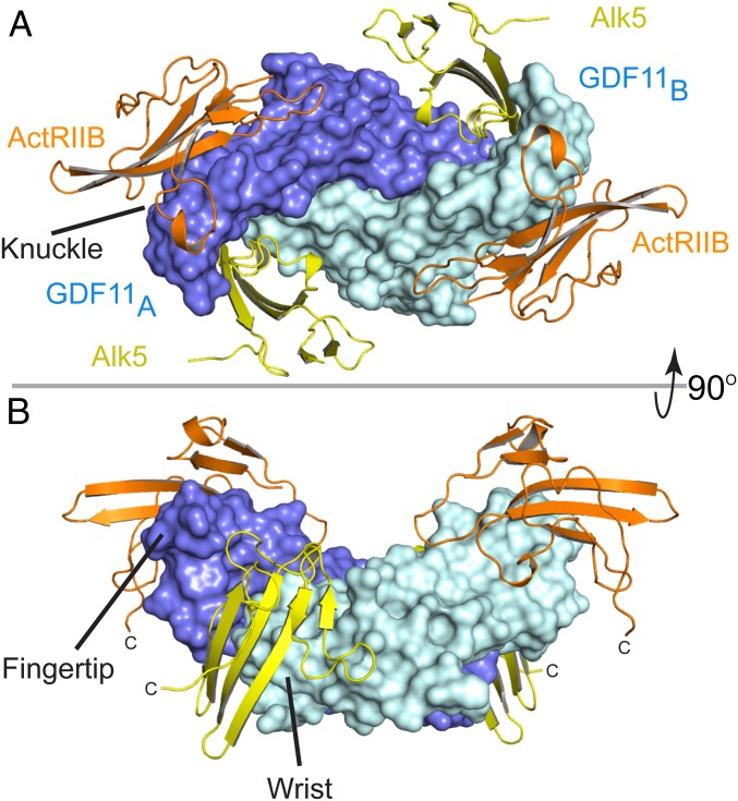
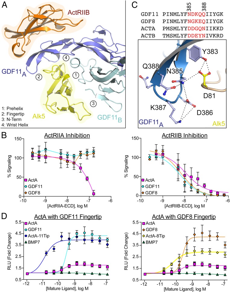
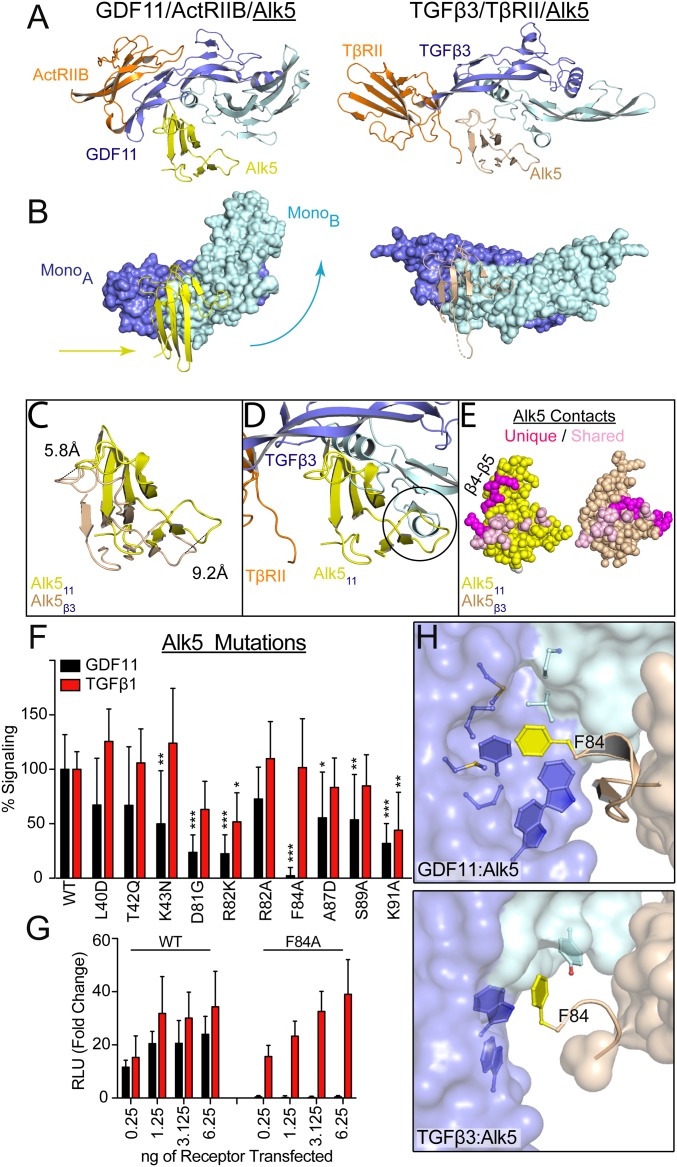
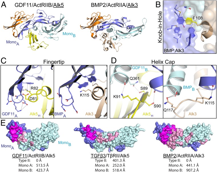
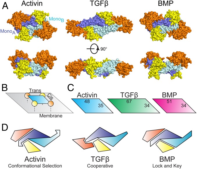
TGFβ family ligands, which include the TGFβs, BMPs, and activins, signal by forming a ternary complex with type I and type II receptors. For TGFβs and BMPs, structures of ternary complexes have revealed differences in receptor assembly. However, structural information for how activins assemble a ternary receptor complex is lacking. We report the structure of an activin class member, GDF11, in complex with the type II receptor ActRIIB and the type I receptor Alk5. The structure reveals that receptor positioning is similar to the BMP class, with no interreceptor contacts; however, the type I receptor interactions are shifted toward the ligand fingertips and away from the dimer interface. Mutational analysis shows that ligand type I specificity is derived from differences in the fingertips of the ligands that interact with an extended loop specific to Alk4 and Alk5. The study also reveals differences for how TGFβ and GDF11 bind to the same type I receptor, Alk5. For GDF11, additional contacts at the fingertip region substitute for the interreceptor interactions that are seen for TGFβ, indicating that Alk5 binding to GDF11 is more dependent on direct contacts. In support, we show that a single residue of Alk5 (Phe84), when mutated, abolishes GDF11 signaling, but has little impact on TGFβ signaling. The structure of GDF11/ActRIIB/Alk5 shows that, across the TGFβ family, different mechanisms regulate type I receptor binding and specificity, providing a molecular explanation for how the activin class accommodates low-affinity type I interactions without the requirement of cooperative receptor interactions.

Keywords: Alk5; GDF11; TGF-β superfamily; activin; ternary signaling complex.


Conflict of interest statement

Conflict of interest statement: T.B.T. is a consultant for Acceleron Pharma and Scientific Founder for Eclode. R.C. and R.K. are current employees of Acceleron Pharma with ownership interest in the company. A.N.E. and V.J.I. are current employees of Regeneron Pharmaceuticals with ownership interest in the company. The other authors report no competing interests.

Figures

Fig. 1.
Fig. 1.
Structure of GDF11/ActRIIB/Alk5 ternary complex. (A) GDF11/ActRIIB/Alk5 as viewed on the membrane surface. GDF11 has 2 monomers represented in slate (monomer A) and cyan (monomer B). ActRIIB-ECD is represented in orange, with Alk5-ECD in yellow. (B) Ninety-degree upward view of the ternary complex. ActRIIB binds at the convex knuckle region of GDF11 whereas Alk5 binds at the concave interface of GDF11 formed between the fingertip of monomer A and the wrist helix of monomer B GDF11.
Fig. 2.
Fig. 2.
Differences in receptor specificity for the activin class. (A) Ribbon representation of one half of the GDF11/ActRIIB/Alk5 complex with GDF11/Alk5 interfaces numbered and labeled accordingly. (B) Inhibition curves (CAGA-luciferase) for respective ligands following the titration of the ECD of ActRIIA (Left) or ActRIIB (Right). (C) Sequence alignment of activin class ligands with fingertip residues represented in red. Numbers correlate to residues in GDF11. Specific molecular interactions between the GDF11 fingertip and the Alk5 β4–β5 loop highlighting the formation of a hydrogen bonding network with dashed lines. (D) Split β-galactosidase dimerization assay (DiscoverX) specific for ActRIIB/Alk5 assembly. For B and D, each data point represents the mean ± SD of triplicate experiments measuring relative luminescence units (RLU) (B) or β-galactosidase activity (D). For B, 100% signaling represents uninhibited signaling of each respective ligand.
Fig. 3.
Fig. 3.
Differential binding of shared type I receptor Alk5 to GDF11 and TGFβ. (A) Ribbon showing 1 type I and 1 type II for GDF11/ActRIIB/Alk5 (PDB ID code 6MAC) and TGFβ3/TβRII/Alk5 (PDB ID code 3KFD), with orientation consistent with alignment of monomer A of the ligand (9). (B) Surface representation of GDF11 (Left) and TGFβ3 (Right) to highlight the relative positional differences of monomer B and Alk5. Arrows indicate the directional shift of GDF11 and Alk5 relative to TGFβ3. (C) Overlay of Alk5 bound to GDF11 (yellow) and TGFβ3 (sand), respectively. Alignment was performed using monomer A. Only the receptor is shown to highlight the shifts in both the β4–β5 loop and the N-terminal region. (D) Overlay of GDF11-bound Alk5 onto TGFβ3 complex using monomer A for superposition. Circle indicates a steric clash with the N terminus of the ligand that would occur if Alk5 were to bind TGFβ3 in a similar position as GDF11. (E) Surface interactions (within 5 Å) of Alk5 with both GDF11 and TFGβ3, with shared residues in pink and unique ligand-interacting residues in magenta. (F and G) Luciferase reporter assay in R1B L17 cells of GDF11 and TGFβ1 signaling following transient transfection of 1.25 ng of Alk5 variants and 2.5 ng (CAGA)12 promoter (F) or titration of Phe84-Ala (F84A) Alk5 transfection (G) (*P ≤ 0.05, **P ≤ 0.01, and ***P ≤ 0.001, 1-way ANOVA). (H) Differences in how F84 of Alk5 binds to GDF11 (Top) and TGFβ3 (Bottom).
Fig. 4.
Fig. 4.
Comparison of type I receptor engagement across the TGFβ family. (A) Ribbon showing 1 type I and 1 type II for GDF11/ActRIIB/Alk5 (PDB ID code 6MAC) and BMP2/ActRIIA/Alk3 (PDB ID code 2GOO), with orientation consistent with alignment of monomer A of the ligand. (B) Knob-in-hole motif observed between F108 of Alk3 and BMP2. (C and D) Comparison of the ligand fingertip-type I interface (C) and the ligand wrist helix cap (D) between GDF11/Alk5 (Left) and BMP2/Alk3 (Right). (E) Ligand surface representation of type I binding interface, with interacting residues on monomer A colored magenta and those on monomer B colored pink. Buried surface area calculations for each of the interfaces are shown as determined by using jsPISA.
Fig. 5.
Fig. 5.
Receptor assembly paradigms of the TGFβ superfamily. (A) Full signaling complex structures from across the TGFβ superfamily: activin (PDB ID code 6MAC), TGFβ (PDB ID code 3KFD), and BMP (PDB ID code 2GOO) (8, 9). Type II and type I receptors are represented in orange and yellow, respectively, with mature ligands in blue. (B) Cartoon representation of the receptor surface complex with cis- and trans-receptor pairs labeled. (C) Distances are calculated from the center of mass for each receptor for cis- and trans-receptor combinations. (D) Schematic representation depicting half the signaling complex for the 3 receptor binding mechanisms of TGFβ family ligands.

References

    1. Hinck A. P., Mueller T. D., Springer T. A., Structural biology and evolution of the TGF- b family. Cold Spring Harb. Perspect. Biol. 8, a022103 (2016). - PMC - PubMed
    1. Hinck A. P., Structural studies of the TGF-betas and their receptors–Insights into evolution of the TGF-beta superfamily. FEBS Lett. 586, 1860–1870 (2012). - PubMed
    1. Weiss A., Attisano L., The TGFbeta superfamily signaling pathway. Wiley Interdiscip. Rev. Dev. Biol. 2, 47–63 (2013). - PubMed
    1. Attisano L., et al. , Identification of human activin and TGFb type I receptors that form heteromeric kinase complexes with type II receptors. Cell 75, 671–680 (1993). - PubMed
    1. Wrana J. L., Attisano L., Wieser R., Ventura F., Massague J., Mechanism of activation of the TGF-β receptor. Nature 370, 341–347 (1994). - PubMed

MeSH terms

Substances

Associated data

LinkOut - more resources