. 2019 Sep 12;93(19):e00459-19.
doi: 10.1128/JVI.00459-19. Print 2019 Oct 1.

Recombinant Hepatitis E Viruses Harboring Tags in the ORF1 Protein

Affiliations

Recombinant Hepatitis E Viruses Harboring Tags in the ORF1 Protein

Dagmara Szkolnicka et al. J Virol. .

Abstract

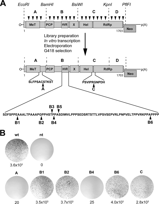
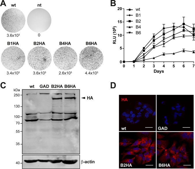
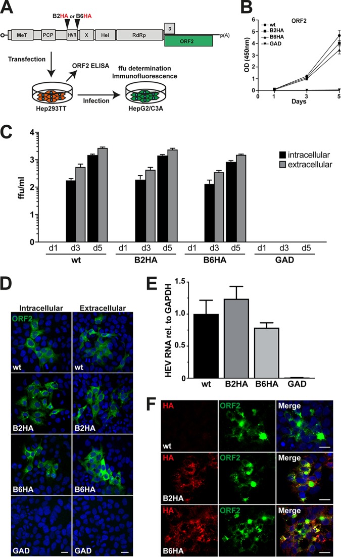
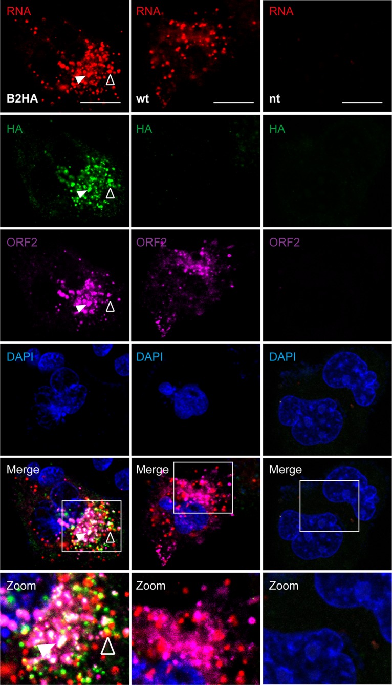
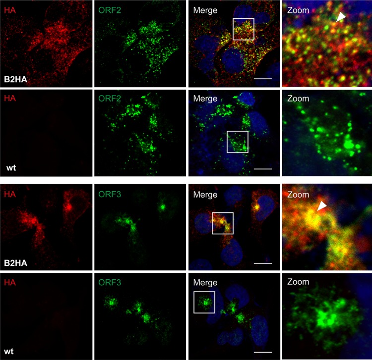
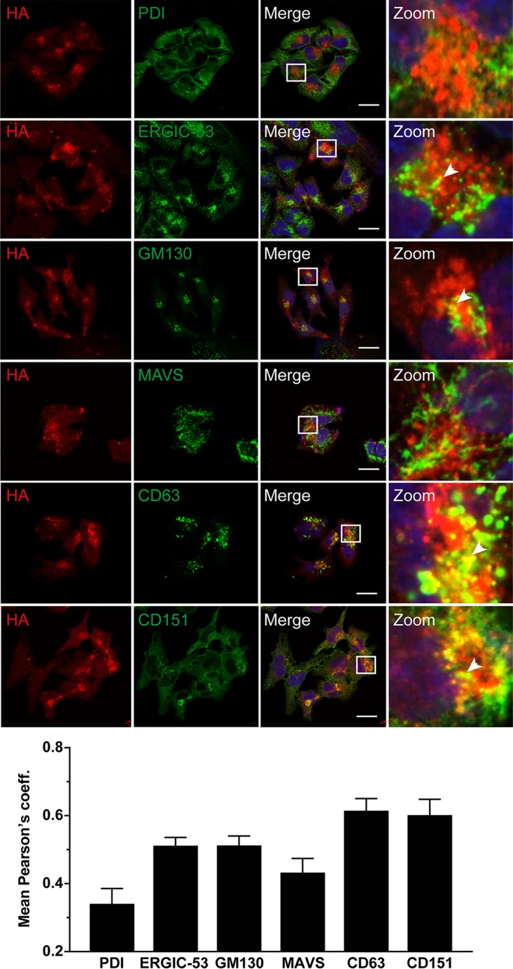
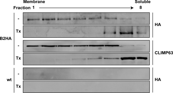
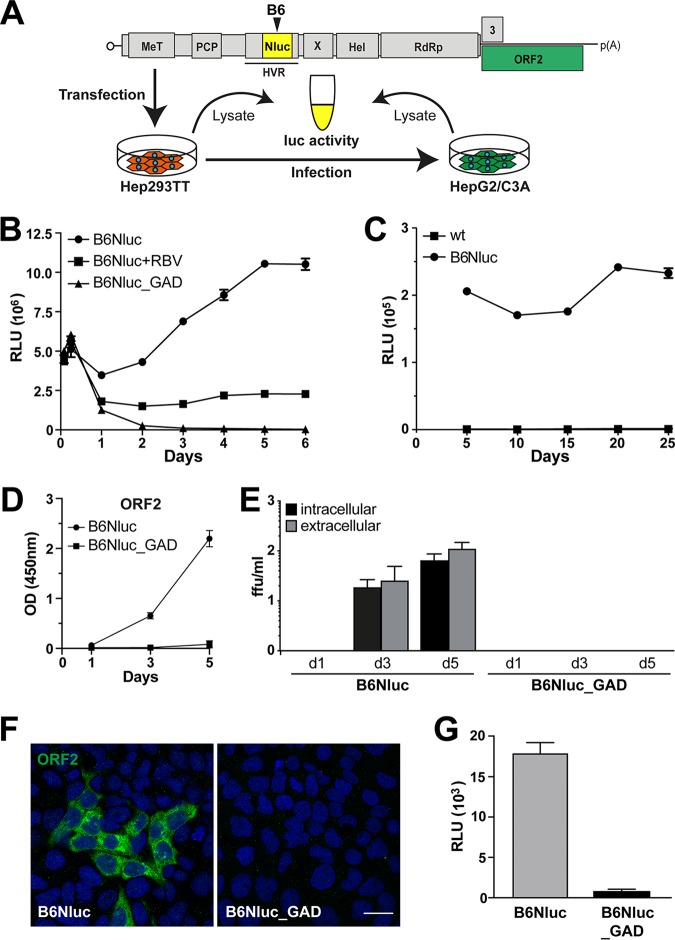
Hepatitis E virus (HEV) is one of the most common causes of acute hepatitis and jaundice in the world. Current understanding of the molecular virology and pathogenesis of hepatitis E is incomplete, due particularly to the limited availability of functional tools. Here, we report the development of tagged HEV genomes as a novel tool to investigate the viral life cycle. A selectable subgenomic HEV replicon was subjected to random 15-nucleotide sequence insertion using transposon-based technology. Viable insertions in the open reading frame 1 (ORF1) protein were selected in a hepatoblastoma cell line. Functional insertion sites were identified downstream of the methyltransferase domain, in the hypervariable region (HVR), and between the helicase and RNA-dependent RNA polymerase domains. HEV genomes harboring a hemagglutinin (HA) epitope tag or a small luciferase (NanoLuc) in the HVR were found to be fully functional and to allow the production of infectious virus. NanoLuc allowed quantitative monitoring of HEV infection and replication by luciferase assay. The use of HA-tagged replicons and full-length genomes allowed localization of putative sites of HEV RNA replication by the simultaneous detection of viral RNA by fluorescence in situ hybridization and of ORF1 protein by immunofluorescence. Candidate HEV replication complexes were found in cytoplasmic dot-like structures which partially overlapped ORF2 and ORF3 proteins as well as exosomal markers. Hence, tagged HEV genomes yield new insights into the viral life cycle and should allow further investigation of the structure and composition of the viral replication complex.IMPORTANCE Hepatitis E virus (HEV) infection is an important cause of acute hepatitis and may lead to chronic infection in immunocompromised patients. Knowledge of the viral life cycle is incomplete due to the limited availability of functional tools. In particular, low levels of expression of the ORF1 protein or limited sensitivity of currently available antibodies or both limit our understanding of the viral replicase. Here, we report the successful establishment of subgenomic HEV replicons and full-length genomes harboring an epitope tag or a functional reporter in the ORF1 protein. These novel tools should allow further characterization of the HEV replication complex and to improve our understanding of the viral life cycle.

Keywords: HEV; open reading frame 1 protein; positive-strand RNA virus; replicase; replication complex; replicon; transposon; viral life cycle.


Figures

FIG 1
FIG 1
Identification of functional insertion sites in HEV ORF1 protein. (A) Schematic outline of the transposon-based random insertion screen. The selectable subgenomic HEV replicon is represented with ORF1 domains designated methyltransferase (MeT), papain-like cysteine protease (PCP), hypervariable region (HVR), macrodomain (X), helicase (Hel), and RNA-dependent RNA polymerase (RdRp). Neo, neomycin phosphotransferase. MuA transposase-mediated random insertion was carried out on pUC-HEV83-2_Neo, followed by in vitro transcription of sublibraries A to D, electroporation into Hep293TT cells, and selection with G418. Sequencing identified transposon insertions at sites A, B1 to B6, and C, as denoted by the black arrowheads. Sequences are detailed in Table 1. (B) Replication efficiency of subgenomic HEV replicons harboring transposon insertions. Hep293TT cells were electroporated with in vitro-transcribed RNA from the parental pUC-HEV83-2_Neo replicon construct (wild type [wt]) or subgenomic replicons harboring transposon insertions. Nontransfected cells (nt) served as a control. After G418 selection, colonies were fixed and stained with crystal violet. Results from a representative experiment are shown, with the number of colonies per microgram of transfected RNA indicated below each plate.
FIG 2
FIG 2
Replication of HA-tagged subgenomic HEV replicons. (A) Replication efficiency of HA-tagged subgenomic HEV replicons. Hep293TT cells were fixed 12 days postelectroporation with in vitro-transcribed RNA from a subgenomic replicon construct (pUC-HEV83-2_Neo_B1-HA, pUC-HEV83-2_Neo_B2-HA, pUC-HEV83-2_Neo_B4-HA, or pUC-HEV83-2_Neo_B6-HA) and stained with crystal violet. The parental HEV83-2-27_Neo replicon (wild type [wt]) and nontransfected cells (nt) served as positive and negative controls, respectively. Results from a representative experiment are shown, with the number of colonies per microgram of transfected RNA indicated below each plate. (B) Culture supernatants from Hep293TT cells electroporated with in vitro-transcribed RNA from the parental pUC-HEV83-2_Gluc (wild type [wt]) or the pUC-HEV83-2_Gluc-derived replicon construct harboring 15-nucleotide transposon insertions in site B1, B2, B4, or B6 were analyzed for luciferase activity over 7 days. Gaussia luciferase activity was measured by adding 10 μl of supernatant from Hep293TT cells electroporated with luciferase HEV replicon constructs to 60 μl of substrate buffer (0.8 μM coelenterazine–PBS). Relative light units (RLU) were determined four times for each time point. (C and D) Immunoblotting (C) and immunofluorescence detection (D) of HA-tagged ORF1 protein in Hep293TT cells. Cells were electroporated with wild-type (wt), polymerase-defective (GAD), or HA-tagged (B2HA and B6HA) selectable subgenomic replicon constructs. (C) Protein lysates were prepared 5 days postelectroporation and separated by 8% SDS-PAGE, followed by sequential immunoblotting using rabbit MAb C29F4 against HA and mouse MAb AC-15 against β-actin. The arrowhead denotes HA-tagged ORF1 protein. Molecular weight markers are indicated on the left. (D) Cells were fixed 5 days postelectroporation and subjected to immunofluorescence with rabbit MAb C29F4 against HA as primary antibody and Alexa Fluor 594 anti-rabbit IgG as secondary antibody. Cell nuclei were counterstained with DAPI. The scale bar represents 20 μm.
FIG 3
FIG 3
Infectivity of HA-tagged full-length HEV genomes. (A) Schematic representation of the HEV genome (see Fig. 1 legend for abbreviations) and of the experimental workflow. Hep293TT cells were transfected with in vitro-transcribed RNA from full-length HEV 83-2-27 constructs. Levels of ORF2 protein in culture supernatants were measured by ELISA. Culture supernatants as well as cell lysates were used to infect naive HepG2/C3A cells, followed by determination of focus-forming units (ffu), quantitative RT-PCR, and immunofluorescence analyses. (B) HEV ORF2 release from electroporated Hep293TT cells was measured by ELISA. The y axis represents optical density (OD) values at 450 nm after blank subtraction. Error bars represent means ± standard deviations (SD) of triplicate measurements for each condition. (C) Culture supernatants and cell lysates were collected at day 1, 3, and 5 postelectroporation and used to infect naive HepG2/C3A cells, followed by determination of focus-forming units (ffu) as described in Materials and Methods. Infectivity was determined for parental (wild type [wt]), HA-tagged (B2HA or B6HA), and polymerase-defective (GAD) full-length genomes using MAb 1E6 against ORF2 protein. Means ± SD of results from 6 replicates for each condition are shown. (D) HepG2/C3A cells infected with either intracellular or extracellular virus samples were fixed at day 5 and subjected to immunofluorescence using MAb 1E6 against ORF2 protein followed by confocal analysis. Nuclei were counterstained with DAPI. The scale bars represent 20 μm. (E) HepG2/C3A cells inoculated with cell lysates obtained from Hep293TT cells transfected with parental (wild-type [wt]), B2HA, B6HA, or polymerase-defective (GAD) full-length genomes were analyzed 5 days postinfection by quantitative RT-PCR. Samples were normalized to GAPDH RNA, and results are expressed as relative expression levels compared to the parental genome. Error bars represent means ± SD of results from six replicates for each condition. (F) Immunofluorescence detection of HA epitope tag and ORF2 protein after infection. HepG2/C3A cells infected with wild-type (wt) or HA-tagged HEV (B2HA or B6HA) from Hep293TT cell lysates were subjected to immunofluorescence using rabbit MAb C29F4 against HA and mouse MAb 1E6 against ORF2 protein, followed by appropriate secondary antibodies (red and green, respectively). Cell nuclei were counterstained with DAPI (blue). The scale bar represents 20 μm.
FIG 4
FIG 4
Simultaneous detection of HEV RNA and HA-tagged ORF1 protein in cells replicating full-length genomes. Hep293TT cells transfected with full-length B2HA or wild-type (wt) HEV RNA were fixed 5 days posttransfection and subjected to fluorescence in situ hybridization (FISH) using the HEV-specific probe V-HEV-C2 (red), followed by immunofluorescence detection of HA-tagged ORF1 protein using rabbit MAb C29F4 against HA (green) and mouse MAb 1E6 against ORF2 protein (magenta). Cell nuclei were counterstained with DAPI (blue). Nontransfected cells (nt) served as negative control. White squares indicate fields shown in higher magnification at the bottom. The empty arrowhead denotes an example of HEV RNA and ORF1 protein colocalization while the filled arrowhead denotes colocalization of HEV RNA as well as ORF1 and ORF2 proteins. The scale bar represents 20 μm.
FIG 5
FIG 5
HA-tagged ORF1 protein partially colocalizes with ORF2 and ORF3 proteins. Hep293TT cells transfected with full-length B2HA or wild-type (wt) HEV RNA were fixed 5 days posttransfection and subjected to immunofluorescence using rabbit MAb C29F4 against HA (red) and either mouse MAb 1E6 against ORF2 or mouse recombinant MAb MRB198 against ORF3 protein (green). Cell nuclei were counterstained with DAPI (blue). White squares indicate fields shown in higher magnification on the right. Arrowheads denote examples of ORF1 protein colocalization with ORF2 or ORF3 protein. The scale bar represents 20 μm.
FIG 6
FIG 6
HA-tagged ORF1 protein partially colocalizes with exosomal markers. Hep293TT cells transfected with full-length B2HA or parental (wild type [wt]) HEV RNA were fixed 5 days posttransfection and subjected to immunofluorescence using rabbit MAb C29F4 against HA (red) and antibodies against markers for different cell compartments (green), including protein disulfide-isomerase (PDI) for the endoplasmic reticulum (ER), ERGIC-53 for the ER-Golgi intermediate compartment, GM130 for the Golgi apparatus, MAVS for mitochondria, and the tetraspanins CD63 and CD151 for exosomes (see Materials and Methods for abbreviations and the list of antibodies used). Cell nuclei were counterstained with DAPI (blue). White squares indicate fields shown in higher magnification on the right. Arrowheads denote examples of colocalization. The scale bar represents 20 μm. Means ± SEM of Pearson's correlation coefficients (coeff.) determined for each condition (n ≥ 10 cells each) are represented as histograms in the bottom panel.
FIG 7
FIG 7
HA-tagged ORF1 protein is membrane-associated. Hep293TT cells electroporated with full-length B2HA or parental (wild type [wt]) HEV RNA were lysed in a hypotonic buffer, followed by membrane flotation as described in Materials and Methods. Lysates were not treated (−) or treated with 1% Triton X-100 (Tx) prior to gradient centrifugation. Eight fractions were collected from the top and analyzed by immunoblotting using rabbit MAb C29F4 against the HA tag. Detection of CLIMP63 served as a control for an integral membrane protein.
FIG 8
FIG 8
HEV harboring a luciferase gene within ORF1 allows quantitative monitoring of viral replication. (A) Schematic representation of the HEV genome (see Fig. 1 legend for abbreviations) and of the experimental workflow. (B) Hep293TT cells were transfected with in vitro-transcribed RNA from full-length HEV 83-2-27 constructs with a NanoLuc insertion in ORF1 (B6Nluc). The antiviral activity of ribavirin (RBV) was evaluated at a concentration of 50 μM. A polymerase-defective mutant (B6Nluc_GAD) served as negative control. NanoLuc activity was measured at 2 and 6 h as well as at 1 to 6 days posttransfection. Mean relative light units (RLU) ± SD from triplicate experiments measured twice each are shown. (C) NanoLuc activity was measured in Hep293TT cells electroporated with parental (wild type [wt]) or B6Nluc full-length RNA over 25 days (5 passages). Each data point was obtained from 3 independent samples normalized to one million cells each. (D) HEV ORF2 protein release from Hep293TT cells electroporated with B6Nluc or B6Nluc_GAD full length RNA was measured by ELISA. The y axis represents the OD values at 450 nm after blank subtraction. Error bars represent means ± SD of results from triplicate measurements for each condition. (E) NanoLuc-tagged full-length HEV RNA produces infectious virus. Culture supernatants as well as cell lysates of Hep293TT cells electroporated with either B6Nluc or B6Nluc_GAD full-length RNA were collected at day 1 and day 3 as well as day 5 postelectroporation and used to infect naive HepG2/C3A cells, followed by determination of focus-forming units (ffu) to measure viral infectivity from intracellular or extracellular compartments as described in Materials and Methods. (F and G) Lysates from Hep293TT cells transfected with HEV83-2_B6Nluc or HEV83-2_B6Nluc_GAD RNA were used to infect naive HepG2/C3A cells. Infectivity was assessed by immunofluorescence 5 days postinfection using mouse MAb 1E6 against the ORF2 protein (F) or by luciferase assay (G). Cell nuclei were counterstained with DAPI (blue). The scale bar represents 20 μm. RLU levels were determined in triplicate 12-well plates measured twice each.

References

    1. Hoofnagle JH, Nelson KE, Purcell RH. 2012. Hepatitis E. N Engl J Med 367:1237–1244. doi:10.1056/NEJMra1204512. - DOI - PubMed
    1. Kamar N, Izopet J, Pavio N, Aggarwal R, Labrique A, Wedemeyer H, Dalton HR. 2017. Hepatitis E virus infection. Nat Rev Dis Primers 3:17086. doi:10.1038/nrdp.2017.86. - DOI - PubMed
    1. Rein DB, Stevens GA, Theaker J, Wittenborn JS, Wiersma ST. 2012. The global burden of hepatitis E virus genotypes 1 and 2 in 2005. Hepatology 55:988–997. doi:10.1002/hep.25505. - DOI - PubMed
    1. European Association for the Study of the Liver. 2018. Clinical practice guidelines on hepatitis E virus infection. J Hepatol 68:1256–1271. doi:10.1016/j.jhep.2018.03.005. - DOI - PubMed
    1. Dalton HR, Kamar N, van Eijk JJ, McLean BN, Cintas P, Bendall RP, Jacobs BC. 2016. Hepatitis E virus and neurological injury. Nat Rev Neurol 12:77–85. doi:10.1038/nrneurol.2015.234. - DOI - PubMed

Publication types

LinkOut - more resources