. 2019 Aug 5;39(8):BSR20191124.
doi: 10.1042/BSR20191124. Print 2019 Aug 30.

Eukaryotic initiation factor 3, subunit C silencing inhibits cell proliferation and promotes apoptosis in human ovarian cancer cells

Affiliations

Eukaryotic initiation factor 3, subunit C silencing inhibits cell proliferation and promotes apoptosis in human ovarian cancer cells

Fang Wen et al. Biosci Rep. .

Abstract

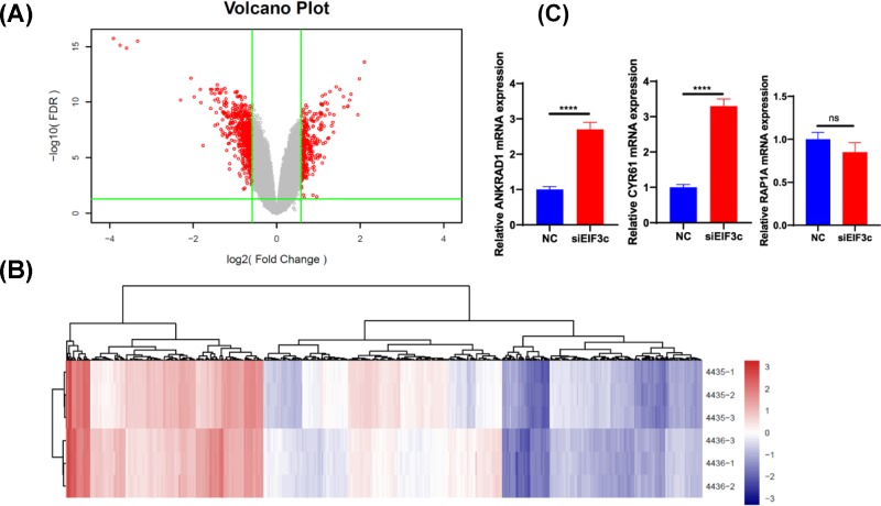
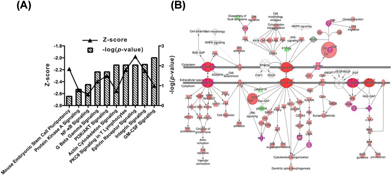
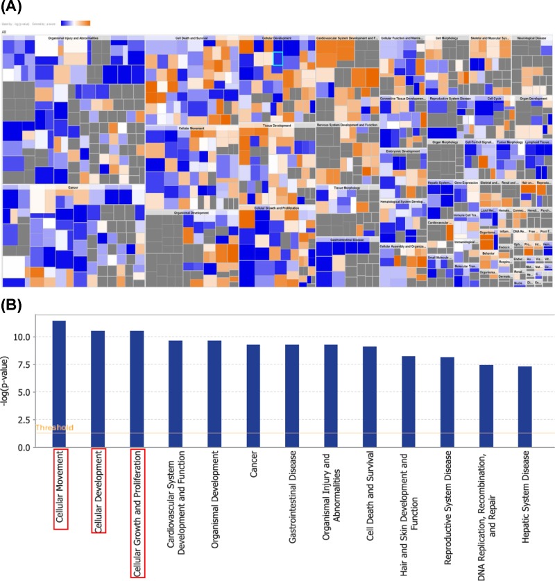
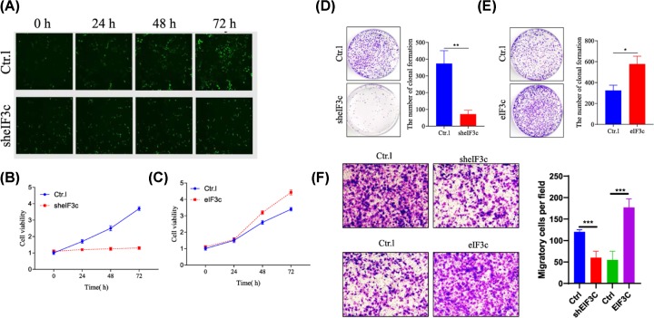
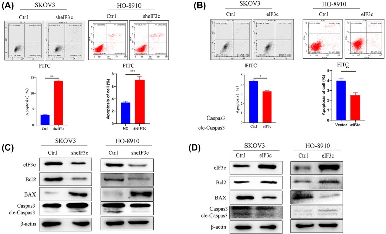
Ovarian cancer remains the leading cause of death among all gynaecological cancers, illustrating the urgent need to understand the molecular mechanisms involved in this disease. Eukaryotic initiation factor 3c (EIF3c) plays an important role in protein translation and cancer cell growth and proliferation, but its role in human ovarian cancer is unclear. Our results showed that EIF3c silencing significantly up-regulated 217 and down-regulated 340 genes. Ingenuity Pathway Analysis (IPA) indicated that the top differentially expressed genes are involved in 'Classical Pathways', 'Diseases and Functions' and 'Networks', especially those involved in signalling and cellular growth and proliferation. In addition, eIF3c silencing inhibited cellular proliferation, enhanced apoptosis and regulated the expression of apoptosis-associated proteins. In conclusion, these results indicate that by dysregulating translational initiation, eIF3c plays an important role in the proliferation and survival of human ovarian cancer cells. These results should provide experimental directions for further in-depth studies on important human ovarian cancer cell pathways.

Keywords: IPA; apoptosis; eIF3c; ovarian cancer cells,; proliferation.


Conflict of interest statement

The authors declare that there are no competing interests associated with the manuscript.

Figures

Figure 1
Figure 1. The clustering of differentially expressed genes in SKOV3 cells after elF3 silencing as visualized by volcano maps and heatmap analysis
(A) All the differential genes shown by volcano maps; the red part means significant difference. (B) The significantly different genes shown by volcano maps. (C) RT-PCR analysis the ANKRD1, RAP1A and CYR61 expression. The data showed that the expression level of ANKRD1 and CYR61 was significantly higher in the eIF3c-silenced group than the negative control group (P<0.05). There was no significant difference in RAP1A expression between the silenced group and the negative group (P>0.05). ***P<0.001, ****P<0.0001.
Figure 2
Figure 2. Differentially expressed ‘classical pathways’ after elF3 silencing as determined by iterative IPA
(A) Top nine ‘classical pathways’ suppressed by elF3 silencing are shown. Left-vertical axis indicates log of the calculated P-value. Right-vertical axis indicates the Z-score value. (B) The Z-score is negative, which indicates the pathways decreased after elF3 silencing. When the Ephrin Receptor signaling was inhibited, other relative pathways were also affected.
Figure 3
Figure 3. Diseases and functions analysis
(A) Colour-coded heatmap analysis of differentially expressed genes after elF3 silencing, clustered according to functions and diseases. Orange indicates up-regulated gene expression; grey indicates down-regulated gene expression and blue indicates unidentified genes. (B) The most statistically significant functions altered by elF3 silencing are shown.
Figure 4
Figure 4. SKOV3 cell growth after elF3 silencing
Cell growth was measured everyday for 5 days by GFP expression analysis, Cellgro cytometry and MTT staining. (A) Images of GFP expression in control and elF3 silenced SKOV3 cells over 5 days as observed by flourescence microscopy (100×). (B) The cell proliferation analyzed by MTT assay at knockdown elF3. (C) Overexpression of elf3, the cell proliferation analyzed by MTT assay. (D) The cell clonal formation analyzed at knockdown elF3. (E) Overexpression of elf3 in the cell clonal formation analysis. The proliferation of cells was significantly inhibited by eIF3C silencing. (F) The transwell assay showed that the invasive ability of cells decreased after silencing eIF3C. *P<0.05, **P<0.01, ***P<0.001, ****P<0.0001.
Figure 5
Figure 5. Silencing eif3c promoted SKOV3 and HO-8910 cells apoptosis
(A) Silencing of elF3 increased SKOV3 and HO-8910 apoptosis as evaluated by flow cytometery. (B) Overexpression of elF3 suppressed SKOV3 and HO-8910 apoptosis as evaluated by flow cytometery. (C) The expression of apoptosis protein (Bcl2, Bax, Caspase 3, cle-Caspase 3) at silencing of elF3. (D) The expression of apoptosis protein (Bcl2, Bax, Caspase 3, cle-Caspase 3) at overexpression of elF3. Taken together, down-regulation of eif3c promoted apoptosis in SKOV3 and HO-8910 cells. *P<0.05, **P<0.01, ***P<0.001, ****P<0.0001.

Similar articles

Cited by

References

    1. Torre L.A.et al. (2015) Global cancer statistics, 2012. CA Cancer J. Clin. 65, 87–10810.3322/caac.21262 - DOI - PubMed
    1. Temkin S.M. and Terplan M. (2015) Trends in relative survival for ovarian cancer from 1975-2011. Obstet. Gynecol. 126, 898.10.1097/AOG.0000000000001073 - DOI - PubMed
    1. Sholl L.M. (2015) Biomarkers in lung adenocarcinoma: a decade of progress. Arch. Pathol. Lab. Med. 139, 469–48010.5858/arpa.2014-0128-RA - DOI - PubMed
    1. Zhu T.et al. (2017) Aberration in translation initiation and associated diseases: role of the eukaryotic translation initiation factor 3A. Zhong Nan Da Xue Xue Bao Yi Xue Ban 42, 1204–1211 - PubMed
    1. Golob-Schwarzl N.et al. (2017) Separation of low and high grade colon and rectum carcinoma by eukaryotic translation initiation factors 1, 5 and 6. Oncotarget 8, 101224–10124310.18632/oncotarget.20642 - DOI - PMC - PubMed

Publication types

MeSH terms

Substances