Comparative Study
. 2019 Jul 17;9(1):10358.
doi: 10.1038/s41598-019-46775-8.

Sperm-borne miR-216b modulates cell proliferation during early embryo development via K-RAS

Affiliations
Comparative Study

Sperm-borne miR-216b modulates cell proliferation during early embryo development via K-RAS

Maíra Bianchi Rodrigues Alves et al. Sci Rep. .

Abstract

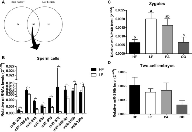
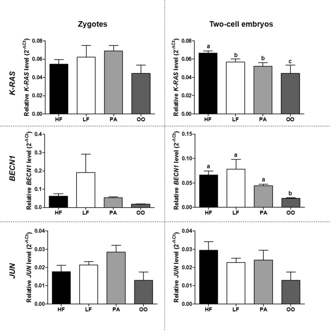
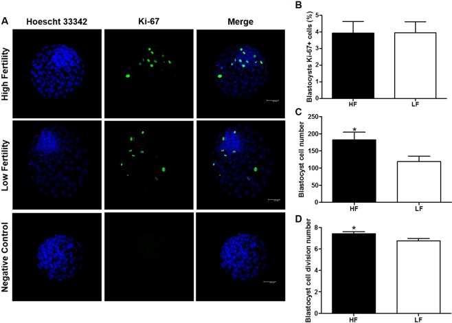
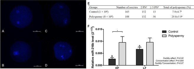
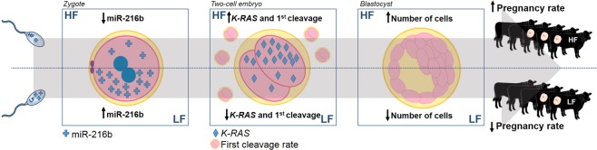
Semen fertilizing potential is dependent upon the morphological, functional and molecular attributes of sperm. Sperm microRNAs (miRNAs) were recently shown to hold promise regarding their association with different fertility phenotypes. However, their role in fertility regulation remains to be determined. We postulated that sperm miRNAs might regulate early embryonic development. From this perspective, sperm quality and 380 sperm miRNAs were investigated in frozen-thawed semen from high (HF; 54.3 ± 1.0% pregnancy rate) and low (LF; 41.5 ± 2.3%) fertility bulls. Out of nine miRNAs that showed different levels in sperm cells, miR-216b was present at lower levels in HF sperm cells and zygotes. Among miR-216b target genes (K-RAS, BECN1 and JUN), K-RAS, related to cell proliferation, revealed a higher level in HF two-cell embryos. First cleavage rate, blastocyst cell number and division number were also higher in HF. In addition, by using a model based on polyspermy embryos, we demonstrated an increase in miR-216b levels in zygotes associated with sperm cell entry. Our results shed light on a possible mechanism of paternal contribution involving sperm-borne miR-216b that modulates levels of miR-216b in zygotes and K-RAS in two-cell embryos. This modulation might regulate early development by interfering with the first cleavage and blastocyst quality.


Conflict of interest statement

M.F.S.F. is employed by Alta Genetics; M.B.R.A., R.P.A., T.H.C.D.B., S.A.F.R., C.B., F.V.M., J.C.S., F.P. and E.C.C.C. have nothing to declare.

Figures

Figure 1
Figure 1
MiRNA analysis in sperm cells and in in vitro-produced embryos. (A) Venn diagram of 298 detected miRNAs in sperm cells from high fertility (HF) or low fertility (LF) bulls from a profile of 380 miRNAs. (B) Relative levels of the differentially abundant miRNAs (P < 0.10) between sperm cells from high fertility and low fertility bulls. (C) MiR-216b relative level in zygotes from high fertility (HF), low fertility (LF), parthenogenetic (PA) and mature oocyte (OO) groups. (D) miR-216b relative level in two-cell embryos from high fertility (HF), low fertility (LF), parthenogenetic (PA) and mature oocyte (OO) groups. Asterisks (*) indicate difference between the groups with a significance level of P < 0.10. a,bDifferent letters indicate statistical difference (P < 0.05) between groups. All quantitative data are presented as means and SEM.
Figure 2
Figure 2
Analyses of miR-216b target genes in mature oocytes and in in vitro-produced embryos. K-RAS, BECN1 and JUN relative level in zygotes and two-cell embryos from high fertility (HF), low fertility (LF), parthenogenetic (PA) and mature oocyte (OO) groups. a,b,cDifferent letters indicate statistical difference (P < 0.05) between groups. All quantitative data are presented as means and SEM.
Figure 3
Figure 3
Blastocyst qualitative analysis. (A) Confocal representative photomicrographs of Ki-67 immunostaining in blastocysts from high fertility and low fertility groups and negative control. Scale bar: 50 μm. (B) Percentage of cells in high fertility (HF) and low fertility (LF) blastocysts that were marked with Ki-67 antibody. (C) Cell numbers in high fertility (HF) and low fertility (LF) blastocysts stained with Hoechst 33342. (D) Cell division number in high fertility (HF) and low fertility (LF) blastocysts. Asterisks (*) indicate difference between the groups with a significance level of P < 0.05. All quantitative data are presented as means and SEM.
Figure 4
Figure 4
Polyspermic embryo validation and analysis of miR-216b relative level. (AD) Representative fluorescence microscopy photomicrographs of pronuclei from zygotes of control and polyspermic groups. Scale bar: 100 μm. In (A,B), zygotes with two pronuclei. In (C,D), zygotes with more than three pronuclei. (E) Evaluation of pronucleus (PN) and polyspermy rate in bovine zygotes produced in vitro with control IVF (sperm concentration of 1 × 106 sperm/mL) or polyspermy IVF (sperm concentration of 8 × 106 sperm/mL). 12 PN: two pronuclei; 2 ≥ 3 PN: more than three pronuclei. (F) MiR-216b relative level in zygotes produced in vitro with control IVF (sperm concentration of 1 × 106 sperm/mL) or polyspermy IVF (sperm concentration of 8 × 106 sperm/mL) using semen samples from high (HF) and low (LF) fertility bulls. Capital letters indicate difference (P < 0.05) between groups (HF vs. LF) with the same treatment (Control). Asterisk (*) indicates difference (P < 0.05) between the treatments (control vs. polyspermy) in the same group. All quantitative data are presented as means and SEM.
Figure 5
Figure 5
Hypothetical schematic model. Schematic figure demonstrating that sperm cells from high fertility (HF) bulls might deliver a lower level of miR-216b to zygotes than sperm from low fertility (LF) bulls. Thus, zygotes from HF may display a lower miR-216b level than zygotes from LF and its target gene, K-RAS, may exhibit a higher level in two-cell embryos from HF than LF. Furthermore, this might result in a higher first cleavage rate in HF embryos. These changes are probably reflected in the higher HF blastocyst cell number, which might be beneficial to in vivo development and thereby could increase pregnancy rates in cattle.

References

    1. Vincent P, et al. Bovine semen quality control in artificial insemination centers. Anim. Reprod. 2012;9:153–165.
    1. Krawetz SA. Paternal contribution: new insights and future challenges. Nat. Rev. Genet. 2005;6:633–642. doi: 10.1038/nrg1654. - DOI - PubMed
    1. Zhang BR, Larsson B, Lundeheim N, Ha MGH. Prediction of bull fertility by combined in vitro assessments of frozen-thawed semen from young dairy bulls entering an AI-programme. Int. J. Androl. 1999;22:253–260. doi: 10.1046/j.1365-2605.1999.00178.x. - DOI - PubMed
    1. Oliveira BM, et al. Fertility and uterine hemodynamic in cows after artificial insemination with semen assessed by fluorescent probes. Theriogenology. 2014;82:767–772. doi: 10.1016/j.theriogenology.2014.06.007. - DOI - PubMed
    1. Oliveira LZ, et al. Assessment of field fertility and several in vitro sperm characteristics following the use of different Angus sires in a timed-AI program with suckled Nelore cows. Livest. Sci. 2012;146:38–46. doi: 10.1016/j.livsci.2012.02.018. - DOI

Publication types

Substances