. 2019 Jul 17;9(1):10387.
doi: 10.1038/s41598-019-46886-2.

RNA sequencing analysis revealed the induction of CCL3 expression in human intracranial aneurysms

Affiliations

RNA sequencing analysis revealed the induction of CCL3 expression in human intracranial aneurysms

Tomohiro Aoki et al. Sci Rep. .

Abstract

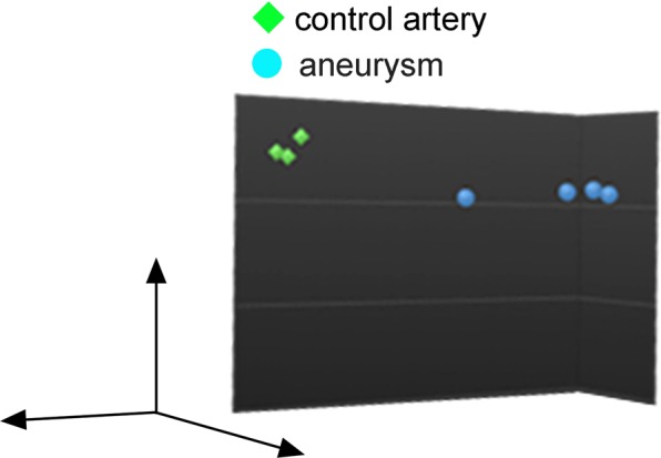
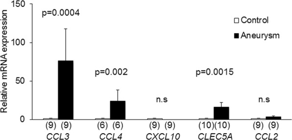
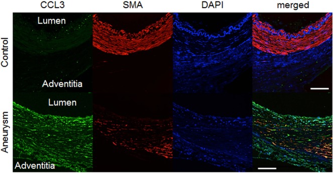
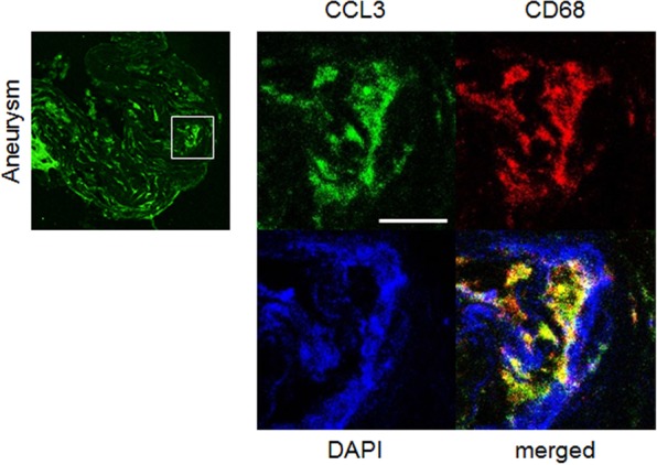
Intracranial aneurysm (IA) is a socially important disease as a major cause of subarachnoid hemorrhage. Recent experimental studies mainly using animal models have revealed a crucial role of macrophage-mediated chronic inflammatory responses in its pathogenesis. However, as findings from comprehensive analysis of unruptured human IAs are limited, factors regulating progression and rupture of IAs in humans remain unclear. Using surgically dissected human unruptured IA lesions and control arterial walls, gene expression profiles were obtained by RNA sequence analysis. RNA sequencing analysis was done with read count about 60~100 million which yielded 6~10 billion bases per sample. 79 over-expressed and 329 under-expressed genes in IA lesions were identified. Through Gene Ontology analysis, 'chemokine activity', 'defense response' and 'extracellular region' were picked up as over-represented terms which included CCL3 and CCL4 in common. Among these genes, quantitative RT-PCR analysis using another set of samples reproduced the above result. Finally, increase of CCL3 protein compared with that in control arterial walls was clarified in IA lesions. Findings of the present study again highlight importance of macrophage recruitment via CCL3 in the pathogenesis of IA progression.


Conflict of interest statement

This work was supported in part by Core Research for Evolutional Science and Technology Program (CREST) on ‘Mechanobiology’ from the Japan Agency for Medical Research and Development (AMED) (#JP18gm0810006, T.A.) and by the Coordination Fund from the Japanese Ministry for Education, Culture, Sports, Science and Technology (MEXT) and Astellas Pharma Inc. to Kyoto University (T.A. and S.N.). S.N. is supported by the Coordination Fund from MEXT and Astellas Pharma Inc. and T.A. was supported until 31th/March/2017. S.N. is a scientific advisor to Astellas Pharma. No potential conflicts of interest were disclosed by the other authors.

Figures

Figure 1
Figure 1
Principal component analysis of gene expressions in control arterial walls and intracranial aneurysms. Image from Principal component analysis of gene expressions in control arterial walls (n = 3) and intracranial aneurysms (n = 4) is shown, where genes expressed in primary culture of smooth muscle cells from human carotid artery were excluded.
Figure 2
Figure 2
Induction of CCL3, CCL4, CLEC5A mRNA expression in intracranial aneurysm lesions. Total RNA was purified from dissected control arterial walls and intracranial aneurysm lesions and mRNA expressions of CCL3, CCL4, CXCL10, CLEC5A and CCL2 were examined by quantitative RT-PCR analysis. The numbers of specimens analyzed are shown in parentheses. Statistical analysis was performed using a Mann-Whitney test. All bars indicate mean ± SEM. n.s; statistically not significant.
Figure 3
Figure 3
Induced expression of CCL3 protein in intracranial aneurysm lesions. Specimens from control arterial walls (Control) and intracranial aneurysm lesions (Aneurysm) were immunostained. Images of immunostaining for CCL3 (green), α-smooth muscle actin (SMA) as a marker of medial smooth muscle cell (red), nuclear staining by DAPI (blue) and merged ones are shown. Bar, 20 μm. Representative images from 2 (Control) and 4 (Aneurysm) independent specimens are shown in this figure and images from remaining samples are shown in Supplementary Fig. S2.
Figure 4
Figure 4
Induction of CCL3 in macrophages of intracranial aneurysm lesions. Images of immunostaining for CCL3 (green), CD68 as a marker for macrophages (red), nuclear staining by DAPI (blue) and merged ones are shown. Square in the left panel indicates the region magnified in right panels. Bar, 10 μm. Representative images from 4 independent experiments are shown.

References

    1. van Gijn J, Kerr RS, Rinkel GJ. Subarachnoid haemorrhage. Lancet. 2007;369:306–318. doi: 10.1016/S0140-6736(07)60153-6. - DOI - PubMed
    1. Morita A, et al. The natural course of unruptured cerebral aneurysms in a Japanese cohort. N Engl J Med. 2012;366:2474–2482. doi: 10.1056/NEJMoa1113260. - DOI - PubMed
    1. Aoki T, Koseki H, Miyata H, Abekura Y, Shimizu K. [Intracranial Aneurysm as an Inflammation-related Disease] No shinkei geka. Neurological surgery. 2018;46:275–294. doi: 10.11477/mf.1436203718. - DOI - PubMed
    1. Aoki T, Yamamoto R, Narumiya S. Targeting macrophages to treat intracranial aneurysm. Oncotarget. 2017;8:104704–104705. doi: 10.18632/oncotarget.21368. - DOI - PMC - PubMed
    1. Turjman AS, Turjman F, Edelman ER. Role of fluid dynamics and inflammation in intracranial aneurysm formation. Circulation. 2014;129:373–382. doi: 10.1161/CIRCULATIONAHA.113.001444. - DOI - PMC - PubMed

Publication types