Review
. 2019 Jun 27:13:532.
doi: 10.3389/fnins.2019.00532. eCollection 2019.

Motor Neuron Susceptibility in ALS/FTD

Affiliations
Review

Motor Neuron Susceptibility in ALS/FTD

Audrey M G Ragagnin et al. Front Neurosci. .

Abstract

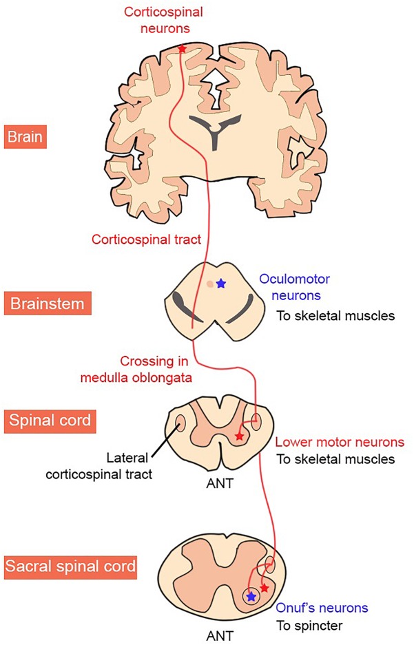
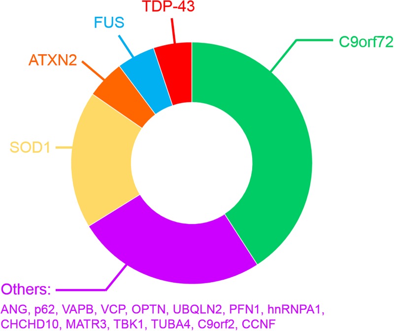
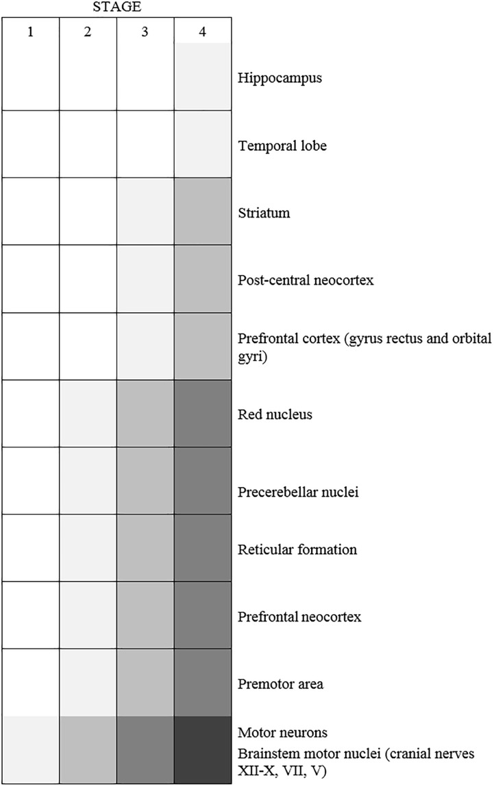
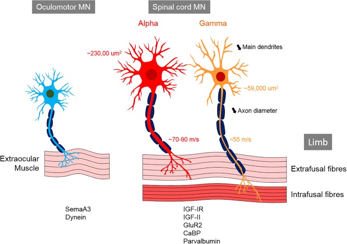
Amyotrophic lateral sclerosis (ALS) is a neurodegenerative disease characterized by the death of both upper and lower motor neurons (MNs) in the brain, brainstem and spinal cord. The neurodegenerative mechanisms leading to MN loss in ALS are not fully understood. Importantly, the reasons why MNs are specifically targeted in this disorder are unclear, when the proteins associated genetically or pathologically with ALS are expressed ubiquitously. Furthermore, MNs themselves are not affected equally; specific MNs subpopulations are more susceptible than others in both animal models and human patients. Corticospinal MNs and lower somatic MNs, which innervate voluntary muscles, degenerate more readily than specific subgroups of lower MNs, which remain resistant to degeneration, reflecting the clinical manifestations of ALS. In this review, we discuss the possible factors intrinsic to MNs that render them uniquely susceptible to neurodegeneration in ALS. We also speculate why some MN subpopulations are more vulnerable than others, focusing on both their molecular and physiological properties. Finally, we review the anatomical network and neuronal microenvironment as determinants of MN subtype vulnerability and hence the progression of ALS.

Keywords: amyotrophic lateral sclerosis; fast and slow motor units; frontotemporal dementia; neurodegeneration; selective vulnerability.


Figures

FIGURE 1
FIGURE 1
Organization of the human corticospinal tract. MN groups vulnerable and resistant to degeneration in ALS are shown in red and blue, respectively.
FIGURE 2
FIGURE 2
Frequency of mutated genes in FALS patients.
FIGURE 3
FIGURE 3
Schematic diagram representing the typical spread of neurodegeneration following an initial onset in motor neurons in ALS patients (n = 76 patients) (Brettschneider et al., 2013). Shading represents TDP-43 pathology.
FIGURE 4
FIGURE 4
Reported differences between the vulnerable (ventral spinal cord MNs) and resistant (oculomotor) motor neurons in ALS. The surface area and axonal conduction velocities referred to here were obtained from studies in cats (Westbury, 1982). The α-MNs innervate highly contracting extrafusal fibers, whereas γ-MNs innervate intrafusal fibers that contract much less; oculomotor neurons innervate the extraocular muscles in the orbit. α-MNs are larger than γ-MNs and oculomotor neurons and possess more dendritic trees. α-MNs are further subdivided based on their size and function. The proteins listed at the bottom of the figure are those enriched in each MN population.
FIGURE 5
FIGURE 5
Diagram showing a hypothetic cascade of cellular events leading to neurodegeneration and neuronal death in motor neurons in ALS/FTD. This schematic diagram summarizes the key features occurring in vulnerable MNs. Resistant MNs are protected by the expression of a genes controlling cellular mechanisms that are defective in ALS/FTD (RNA dysfunction, ER stress, mitochondrial defects, protein transport dysfunction, dysregulation of neuronal excitability and excitotoxicity). These processes can be exacerbated by age, environmental and genetic mutations.

References

    1. Acevedo-Arozena A., Kalmar B., Essa S., Ricketts T., Joyce P., Kent R., et al. (2011). A comprehensive assessment of the SOD1G93A low-copy transgenic mouse, which models human amyotrophic lateral sclerosis. Dis. Model Mech. 4 686–700. 10.1242/dmm.00723 - DOI - PMC - PubMed
    1. Adal M. N., Barker D. (1965). Intramuscular branching of fusimotor fibres. J. Physiol. 177 288–299. 10.1113/jphysiol.1965.sp007592 - DOI - PMC - PubMed
    1. Aizawa H., Sawada J., Hideyama T., Yamashita T., Katayama T., Hasebe N., et al. (2010). TDP-43 pathology in sporadic ALS occurs in motor neurons lacking the RNA editing enzyme ADAR2. Acta Neuropathol. 120 75–84. 10.1007/s00401-010-0678-x - DOI - PubMed
    1. Al Sultan A., Waller R., Heath P., Kirby J. (2016). The genetics of amyotrophic lateral sclerosis: current insights. Degen. Neurol. Neuromusc. Dis. 2016 49–64. - PMC - PubMed
    1. Alexander G. M., Erwin K. L., Byers N., Deitch J. S., Augelli B. J., Blankenhorn E. P., et al. (2004). Effect of transgene copy number on survival in the G93A SOD1 transgenic mouse model of ALS. Brain Res. Mol. Brain Res. 130 7–15. 10.1016/j.molbrainres.2004.07.002 - DOI - PubMed