

Thyroid Hormone Receptor Alpha Is Required for Thyroid Hormone-Dependent Neural Cell Proliferation During Tadpole Metamorphosis
- PMID: 31316462
- PMCID: PMC6610206
- DOI: 10.3389/fendo.2019.00396
Thyroid Hormone Receptor Alpha Is Required for Thyroid Hormone-Dependent Neural Cell Proliferation During Tadpole Metamorphosis
Abstract
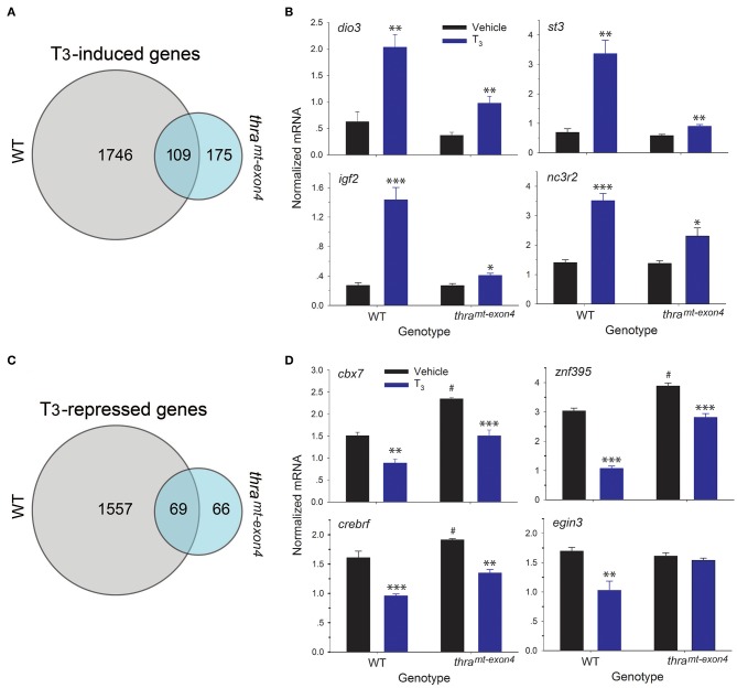
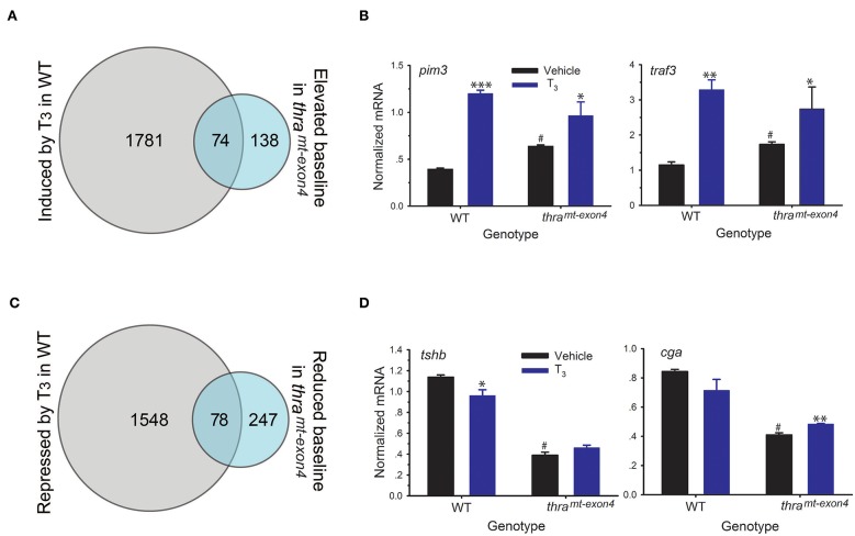
Thyroid hormone (T3) plays several key roles in development of the nervous system in vertebrates, controlling diverse processes such as neurogenesis, cell migration, apoptosis, differentiation, and maturation. In anuran amphibians, the hormone exerts its actions on the tadpole brain during metamorphosis, a developmental period dependent on T3. Thyroid hormone regulates gene transcription by binding to two nuclear receptors, TRα and TRβ. Our previous findings using pharmacological and other approaches supported that TRα plays a pivotal role in mediating T3 actions on neural cell proliferation in Xenopus tadpole brain. Here we used Xenopus tropicalis (X. tropicalis) tadpoles with an inactivating mutation in the gene that encodes TRα to investigate roles for TRα in mitosis and gene regulation in tadpole brain. Gross morphological analysis showed that mutant tadpoles had proportionally smaller brains, corrected for body size, compared with wildtype, both during prometamorphosis and at the completion of metamorphosis. This was reflected in a large reduction in phosphorylated histone 3 (pH3; a mitosis marker) immunoreactive (ir) nuclei in prometamorphic tadpole brain, when T3-dependent cell proliferation is maximal. Treatment of wild type premetamorphic tadpoles with T3 for 48 h induced gross morphological changes in the brain, and strongly increased pH3-ir, but had no effect in mutant tadpoles. Thyroid hormone induction of the direct TR target genes thrb, klf9, and thibz was dysregulated in mutant tadpoles. Analysis of gene expression by RNA sequencing in the brain of premetamorphic tadpoles treated with or without T3 for 16 h showed that the TRα accounts for 95% of the gene regulation responses to T3.
Keywords: Xenopus; development; knockout (KO); metamorphosis; neurogenesis; thyroid hormone (T3).
Figures


Similar articles
-
Analysis of Thyroid Hormone Receptor α-Knockout Tadpoles Reveals That the Activation of Cell Cycle Program Is Involved in Thyroid Hormone-Induced Larval Epithelial Cell Death and Adult Intestinal Stem Cell Development During Xenopus tropicalis Metamorphosis.Thyroid. 2021 Jan;31(1):128-142. doi: 10.1089/thy.2020.0022. Epub 2020 Jul 1. Thyroid. 2021. PMID: 32515287 Free PMC article.
-
A unique role of thyroid hormone receptor β in regulating notochord resorption during Xenopus metamorphosis.Gen Comp Endocrinol. 2019 Jun 1;277:66-72. doi: 10.1016/j.ygcen.2019.03.006. Epub 2019 Mar 6. Gen Comp Endocrinol. 2019. PMID: 30851299 Free PMC article.
-
Organ-Specific Requirements for Thyroid Hormone Receptor Ensure Temporal Coordination of Tissue-Specific Transformations and Completion of Xenopus Metamorphosis.Thyroid. 2020 Feb;30(2):300-313. doi: 10.1089/thy.2019.0366. Epub 2020 Jan 23. Thyroid. 2020. PMID: 31854240 Free PMC article.
-
Essential and subtype-dependent function of thyroid hormone receptors during Xenopus metamorphosis.Vitam Horm. 2023;123:503-523. doi: 10.1016/bs.vh.2023.02.004. Epub 2023 Mar 25. Vitam Horm. 2023. PMID: 37717996 Free PMC article. Review.
-
Autoinduction of nuclear receptor genes and its significance.J Steroid Biochem Mol Biol. 1993 Aug;46(2):105-19. doi: 10.1016/0960-0760(93)90286-6. J Steroid Biochem Mol Biol. 1993. PMID: 8664159 Review.
Cited by
-
Cre-loxP-mediated genetic lineage tracing: Unraveling cell fate and origin in the developing heart.Front Cardiovasc Med. 2023 Feb 27;10:1085629. doi: 10.3389/fcvm.2023.1085629. eCollection 2023. Front Cardiovasc Med. 2023. PMID: 36923960 Free PMC article. Review.
-
Temporal and spatial transcriptomic dynamics across brain development in Xenopus laevis tadpoles.G3 (Bethesda). 2022 Jan 4;12(1):jkab387. doi: 10.1093/g3journal/jkab387. G3 (Bethesda). 2022. PMID: 34751375 Free PMC article.
-
DNA methylation dynamics underlie metamorphic gene regulation programs in Xenopus tadpole brain.Dev Biol. 2020 Jun 15;462(2):180-196. doi: 10.1016/j.ydbio.2020.03.013. Epub 2020 Mar 31. Dev Biol. 2020. PMID: 32240642 Free PMC article.
-
Transcriptional responses to injury of regenerative lung alveolar epithelium.iScience. 2022 Aug 3;25(8):104843. doi: 10.1016/j.isci.2022.104843. eCollection 2022 Aug 19. iScience. 2022. PMID: 35996586 Free PMC article.
-
Metamorphic gene regulation programs in Xenopus tropicalis tadpole brain.PLoS One. 2023 Jun 29;18(6):e0287858. doi: 10.1371/journal.pone.0287858. eCollection 2023. PLoS One. 2023. PMID: 37384728 Free PMC article.
References
-
- Flamant F, Gauthier K, Richard S. Genetic investigation of thyroid hormone receptor function in the developing and adult brain. In: Douglas Forrest ST. editor. Current Topics in Developmental Biology: Nuclear Receptors in Development and Disease, Vol. 125. Cambridge, MA: Academic Press; Elsevier; (2017). p. 303–35. - PubMed
-
- Legrand J. Hormones thyroidiennes et maturation du systeme nerveux. J Physiol. (1982) 78:603–52. - PubMed
-
- Shi YB. Amphibian Metamorphosis: From Morphology to Molecular Biology. New York, NY: Wiley-Liss; (2000).
LinkOut - more resources
Full Text Sources
Molecular Biology Databases
Research Materials