. 2019 Aug;80(4):424-430.
doi: 10.1055/s-0038-1676036. Epub 2018 Nov 19.

Spontaneous Otogenic Pneumocephalus: Case Series and Update on Management

Affiliations

Spontaneous Otogenic Pneumocephalus: Case Series and Update on Management

Michael Eggerstedt et al. J Neurol Surg B Skull Base. 2019 Aug.

Abstract

Objectives This study is aimed to report the largest independent case series of spontaneous otogenic pneumocephalus (SOP) and review its pathophysiology, clinical presentation, and treatment. Design Four patients underwent a middle cranial fossa approach for repair of the tegmen tympani and tegmen mastoideum. A comprehensive review of the literature regarding this disease entity was performed. Setting U.S. tertiary academic medical center. Participants: Patients presenting to the lead author's clinic or to the emergency department with radiographic evidence of SOP. Symptoms included headache, otalgia, and neurologic deficits. Main Outcome Measures Patients were assessed for length of stay, postoperative length of stay, and neurologic outcome. Three of four patients returned to their neurologic baseline following repair. Results Four patients were successfully managed via a middle cranial fossa approach to repairing the tegmen mastoideum. Conclusion The middle cranial fossa approach is an effective strategy to repair defects of the tegmen mastoideum. SOP remains a clinically rare disease, with little published information on its diagnosis and treatment.

Keywords: middle cranial fossa; otogenic; pneumocephalus; tegmen.


Conflict of interest statement

Conflict of Interest None.

Figures

Fig. 1
Fig. 1
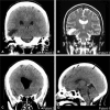
Selected images from computed tomography (CT) imaging of the brain upon presentation from patients 1 and 2. (A) Axial CT brain from patient 1, showing loculated focal moderate size pneumocephalus in the left temporal region with mild mass-effect on the adjacent brain parenchyma. (B) Coronal CT brain from patient 1. Additional imaging of the temporal bones revealed subtle defect in the left tegmen mastoideum adjacent to the pneumocephalus. (C) Axial CT brain from patient 2, showing moderate dilatation of the ventricular system with a large amount of intraventricular pneumocephalus and scattered foci subarachnoid pneumocephalus. (D) Coronal CT brain from patient 2 showing large air pocket in the right middle cranial fossa displacing the right temporal lobe and right temporal horn.
Fig. 2
Fig. 2
Selected images from CT and magnetic resonance imaging (MRI) imaging on presentation from patient 3 and 4 (sisters). (A) Coronal CT brain at the level of the external auditory canal from patient 3 showing cystic area (black arrow), immediately superior to the tegmen, with mild surrounding edema in the right temporal lobe. (B) Coronal T2 weighted MRI brain from patient 3 showing the same well-defined CSF-filled cystic lesion (black arrow) in the right temporal lobe representing a porencephalic cyst. Additional imaging in from this patient revealed that this cyst communicated with the right temporal horn of the lateral ventricle. (C) Coronal CT brain from patient 3 showing pneumocephalus in the right frontal horn of the lateral ventricle. (D) Sagittal CT brain from patient 4 showing small foci of pneumocephalus (black arrows) noted along left anterior temporal and left frontoparietal convexities. Additional imaging in this patient revealed a prominent osseous defect is noted along the left anterior/medial tegmen tympani, moderate fluid collection in the left tympanic cavity, left mastoid antrum, and air cells. CSF, cerebrospinal fluid.

References

    1. Abbati S G, Torino R R. Spontaneous intraparenchymal otogenic pneumocephalus: a case report and review of literature. Surg Neurol Int. 2012;3:32. - PMC - PubMed
    1. Stevens S M, Lambert P R, Rizk H, McIlwain W R, Nguyen S A, Meyer T A. Novel radiographic measurement algorithm demonstrating a link between obesity and lateral skull base attenuation. Otolaryngol Head Neck Surg. 2015;152(01):172–179. - PubMed
    1. Goldmann R W. Pneumocephalus as a consequence of barotrauma. JAMA. 1986;255(22):3154–3156. - PubMed
    1. Markham J W. The clinical features of pneumocephalus based upon a survey of 284 cases with report of 11 additional cases. Acta Neurochir (Wien) 1967;16(01):1–78. - PubMed
    1. Dandy W E. Pneumocephalus (intracranial pneumatocele or aerocele) Arch Surg. 1926;12:949–982.