. 2019 Apr 19;6(12):1900218.
doi: 10.1002/advs.201900218. eCollection 2019 Jun 19.

Expanding the Functional Scope of the Fmoc-Diphenylalanine Hydrogelator by Introducing a Rigidifying and Chemically Active Urea Backbone Modification

Affiliations

Expanding the Functional Scope of the Fmoc-Diphenylalanine Hydrogelator by Introducing a Rigidifying and Chemically Active Urea Backbone Modification

Vasantha Basavalingappa et al. Adv Sci (Weinh). .

Abstract

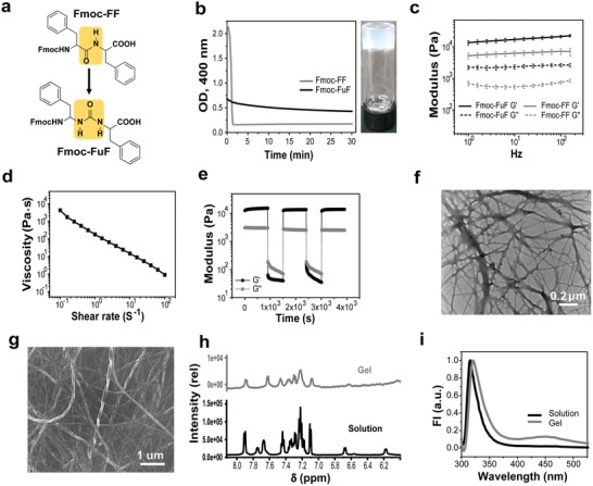
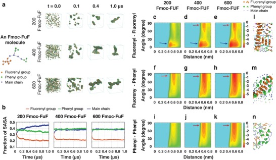
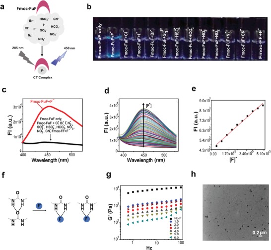
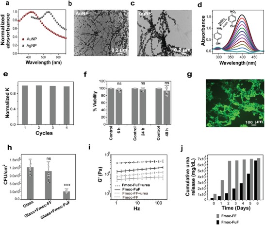
Peptidomimetic low-molecular-weight hydrogelators, a class of peptide-like molecules with various backbone amide modifications, typically give rise to hydrogels of diverse properties and increased stability compared to peptide hydrogelators. Here, a new peptidomimetic low-molecular-weight hydrogelator is designed based on the well-studied N-fluorenylmethoxycarbonyl diphenylalanine (Fmoc-FF) peptide by replacing the amide bond with a frequently employed amide bond surrogate, the urea moiety, aiming to increase hydrogen bonding capabilities. This designed ureidopeptide, termed Fmoc-Phe-NHCONH-Phe-OH (Fmoc-FuF), forms hydrogels with improved mechanical properties, as compared to those formed by the unmodified Fmoc-FF. A combination of experimental and computational structural methods shows that hydrogen bonding and aromatic interactions facilitate Fmoc-FuF gel formation. The Fmoc-FuF hydrogel possesses properties favorable for biomedical applications, including shear thinning, self-healing, and in vitro cellular biocompatibility. Additionally, the Fmoc-FuF, but not Fmoc-FF, hydrogel presents a range of functionalities useful for other applications, including antifouling, slow release of urea encapsulated in the gel at a high concentration, selective mechanical response to fluoride anions, and reduction of metal ions into catalytic nanoparticles. This study demonstrates how a simple backbone modification can enhance the mechanical properties and functional scope of a peptide hydrogel.

Keywords: anion sensing; antifouling materials; metal nanoparticles; peptide self‐assembly; peptidomimetics; urea slow release; ureidopeptides.


Conflict of interest statement

The authors declare no conflict of interest.

Figures

Figure 1
Figure 1
Characterization of the Fmoc‐FuF hydrogel. a) Chemical structure of Fmoc‐FF and Fmoc‐FuF hydrogelators, highlighting (yellow) the unmodified (Fmoc‐FF) or modified (Fmoc‐FuF) backbone. b) Left: OD kinetics at 400 nm. Right: Photograph of the semitransparent Fmoc‐FuF hydrogel. c) Frequency sweep measurement for Fmoc‐FuF and Fmoc‐FF hydrogels. Data represent mean ± standard deviation (SD, n = 3 hydrogels per condition). d) Flow sweep measurement showing shear‐thinning behavior of Fmoc‐FuF hydrogel. e) Five‐step loop time sweep measurement showing the thixotropic nature of Fmoc‐FuF hydrogel. f) TEM and g) HRSEM images of Fmoc‐FuF fibers. h) Partial 1H NMR spectrum, showing the NH and aromatic regions, in the solution and gel states. i) Fluorescence emission spectrum of Fmoc‐FuF in solution and gel states (λex = 285 nm).
Figure 2
Figure 2
Self‐assembly mechanism of Fmoc‐FuF revealed by microsecond‐long CG‐MD simulations of 200, 400, and 600 molecules. a) Snapshots of aggregates at four timepoints. The CG representation of an Fmoc‐FuF molecule is shown for clarity. b) Time evolution of the SASA fraction of fluorenyl, phenyl, and main chain groups. c–k) The FEL of worm‐like and branched gel‐like assemblies as a function of the centroid distance and the angle between the two aromatic rings in three different ring pairs. The basins are marked by arrows. l–n) Stacking patterns of fluorenyl–fluorenyl, fluorenyl–phenyl, and phenyl–phenyl ring pairs.
Figure 3
Figure 3
Selective anion binding by Fmoc‐FuF. a) Schematic representation of selective binding of F by Fmoc‐FuF, forming a charge‐transfer complex. b) Photograph of Fmoc‐FuF (5 × 10⁻⁶ m) DMSO solutions after the addition of 500 equiv. of various aqueous anion solutions under UV illumination (λex = 365 nm). c) Fluorescence emission spectra of Fmoc‐FuF (5 × 10⁻⁶ m) DMSO solutions upon the addition of 450 equiv. of various aqueous anion solutions (λex = 285 nm). d) Fluorescence intensity (FI) of Fmoc‐FuF (5 × 10⁻⁶ m) in DMSO solutions upon titration with 0–1000 equiv. of aqueous F solution (λex = 285 nm). e) Plot of log[FI] versus F concentration. Red line is a linear fit, R 2 = 0.99. f) Schematic illustration of breakage of Fmoc‐FuF hydogel by F. Dashed lines represent hydrogen bonds. g) Frequency sweep measurement of Fmoc‐FuF hydrogels (0.5 wt%) containing increasing F concentration (0–6.0 equiv. of F). h) TEM image of Fmoc‐FuF gel (0.5 wt%) containing 6.0 equiv. of F.
Figure 4
Figure 4
Diverse functionalities of the Fmoc‐FuF hydrogel. a) UV–visible spectra showing SPR bands of AuNPs or AgNPs formed in situ in the hydrogel (hydrogels were diluted threefold prior to measurement). b,c) TEM images of b) AuNP‐ and c) AgNP‐decorated fibers. d) Time‐dependent UV–vis spectra and e) corresponding normalized rate constant for repeated reaction cycles of the reduction of 4‐NP to 4‐AP, catalyzed by AuNP‐containing Fmoc‐FuF xerogel. f) Cell viability as determined by XTT assay, performed at different timepoints on 3T3 fibroblast cells cultured in naïve medium (control) or medium preincubated with Fmoc‐FuF hydrogel. Data represent mean ± SD from three independent experiments. Each timepoint was compared to control using Student's t‐test, P > 0.05 for all comparisons, indicating no statistically significant differences (ns). g) Representative Live/Dead staining of 3T3 fibroblast cells after 48 h of incubation on an Fmoc‐FuF hydrogel scaffold, showing a high abundance of living cells (green) and the negligible presence of dead cells (red). h) Bacterial adhesion to bare glass and glass surfaces coated with either Fmoc‐FF or Fmoc‐FuF xerogels, as per CFUs' count. Difference between Fmoc‐FuF coating and bare glass is significant (P < 0.001), difference between Fmoc‐FF coating and bare glass is not significant (ns, P > 0.05), as per Student's t‐test. Data represent mean ± SD from three independent experiments. i) Comparison of G′ values of Fmoc‐FuF and Fmoc‐FF hydrogels with or without encapsulated urea. Data represent mean ± SD (n = 3 hydrogels per condition). j) Comparison of cumulative urea release from urea‐containing Fmoc‐FuF (black) or Fmoc‐FF (gray) hydrogels, measured over a period of 6 days.

References

    1. Hauser C. A. E., Zhang S., Chem. Soc. Rev. 2010, 39, 2780. - PubMed
    1. Rubert Pérez C. M., Stephanopoulos N., Sur S., Lee S. S., Newcomb C., Stupp S. I., Ann. Biomed. Eng. 2015, 43, 501. - PMC - PubMed
    1. Adams D. J., Macromol. Biosci. 2011, 11, 160. - PubMed
    1. De Leon‐Rodriguez L. M., Hemar Y., Mo G., Mitra A. K., Cornish J., Brimble M. A., Acta Biomater. 2017, 47, 40. - PubMed
    1. Collier J. H., Segura T., Biomaterials 2011, 32, 4198. - PMC - PubMed

LinkOut - more resources