Machine learning for semi-automated classification of glioblastoma, brain metastasis and central nervous system lymphoma using magnetic resonance advanced imaging
- PMID: 31317002
- PMCID: PMC6603356
- DOI: 10.21037/atm.2018.08.05
Machine learning for semi-automated classification of glioblastoma, brain metastasis and central nervous system lymphoma using magnetic resonance advanced imaging
Abstract
Background: Differentiating glioblastoma, brain metastasis, and central nervous system lymphoma (CNSL) on conventional magnetic resonance imaging (MRI) can present a diagnostic dilemma due to the potential for overlapping imaging features. We investigate whether machine learning evaluation of multimodal MRI can reliably differentiate these entities.
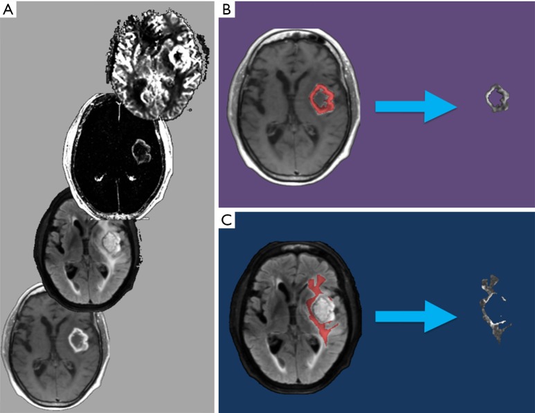
Methods: Preoperative brain MRI including diffusion weighted imaging (DWI), dynamic contrast enhanced (DCE), and dynamic susceptibility contrast (DSC) perfusion in patients with glioblastoma, lymphoma, or metastasis were retrospectively reviewed. Perfusion maps (rCBV, rCBF), permeability maps (K-trans, Kep, Vp, Ve), ADC, T1C+ and T2/FLAIR images were coregistered and two separate volumes of interest (VOIs) were obtained from the enhancing tumor and non-enhancing T2 hyperintense (NET2) regions. The tumor volumes obtained from these VOIs were utilized for supervised training of support vector classifier (SVC) and multilayer perceptron (MLP) models. Validation of the trained models was performed on unlabeled cases using the leave-one-subject-out method. Head-to-head and multiclass models were created. Accuracies of the multiclass models were compared against two human interpreters reviewing conventional and diffusion-weighted MR images.
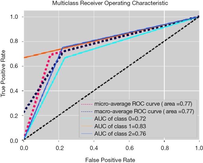
Results: Twenty-six patients enrolled with histopathologically-proven glioblastoma (n=9), metastasis (n=9), and CNS lymphoma (n=8) were included. The trained multiclass ML models discriminated the three pathologic classes with a maximum accuracy of 69.2% accuracy (18 out of 26; kappa 0.540, P=0.01) using an MLP trained with the VpNET2 tumor volumes. Human readers achieved 65.4% (17 out of 26) and 80.8% (21 out of 26) accuracies, respectively. Using the MLP VpNET2 model as a computer-aided diagnosis (CADx) for cases in which the human reviewers disagreed with each other on the diagnosis resulted in correct diagnoses in 5 (19.2%) additional cases.
Conclusions: Our trained multiclass MLP using VpNET2 can differentiate glioblastoma, brain metastasis, and CNS lymphoma with modest diagnostic accuracy and provides approximately 19% increase in diagnostic yield when added to routine human interpretation.
Keywords: Brain tumor; classification; machine learning; magnetic resonance imaging (MRI); neuroradiology.
Conflict of interest statement
Conflicts of Interest: The authors have no conflicts of interest to declare.
Figures