. 2019 Jun;7(11):240.
doi: 10.21037/atm.2019.04.79.

Polydopamine coating promotes early osteogenesis in 3D printing porous Ti6Al4V scaffolds

Affiliations

Polydopamine coating promotes early osteogenesis in 3D printing porous Ti6Al4V scaffolds

Lan Li et al. Ann Transl Med. 2019 Jun.

Abstract

Background: Titanium implants are widely used in orthopedic and dental for more than 30 years. Its stable physicochemical properties and mechanical strength are indeed appropriate for implantation. However, the Bioinertia oxidized layer and higher elastic modulus often lead to the early implantation failure.

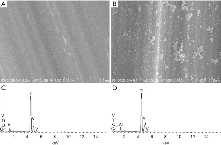
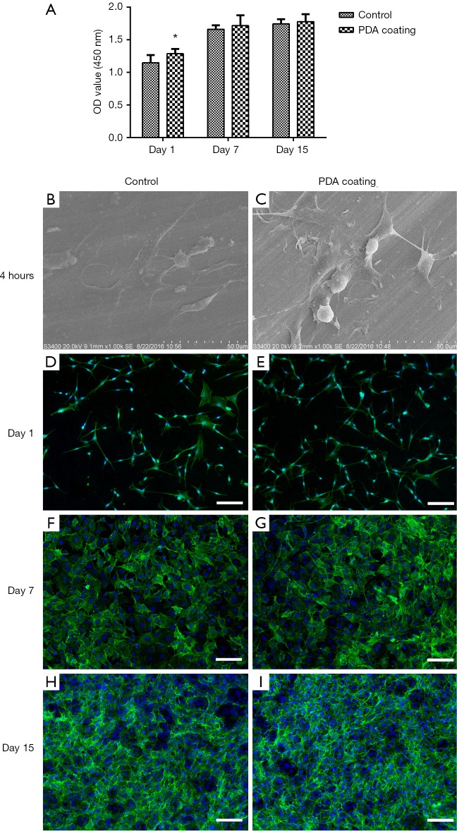
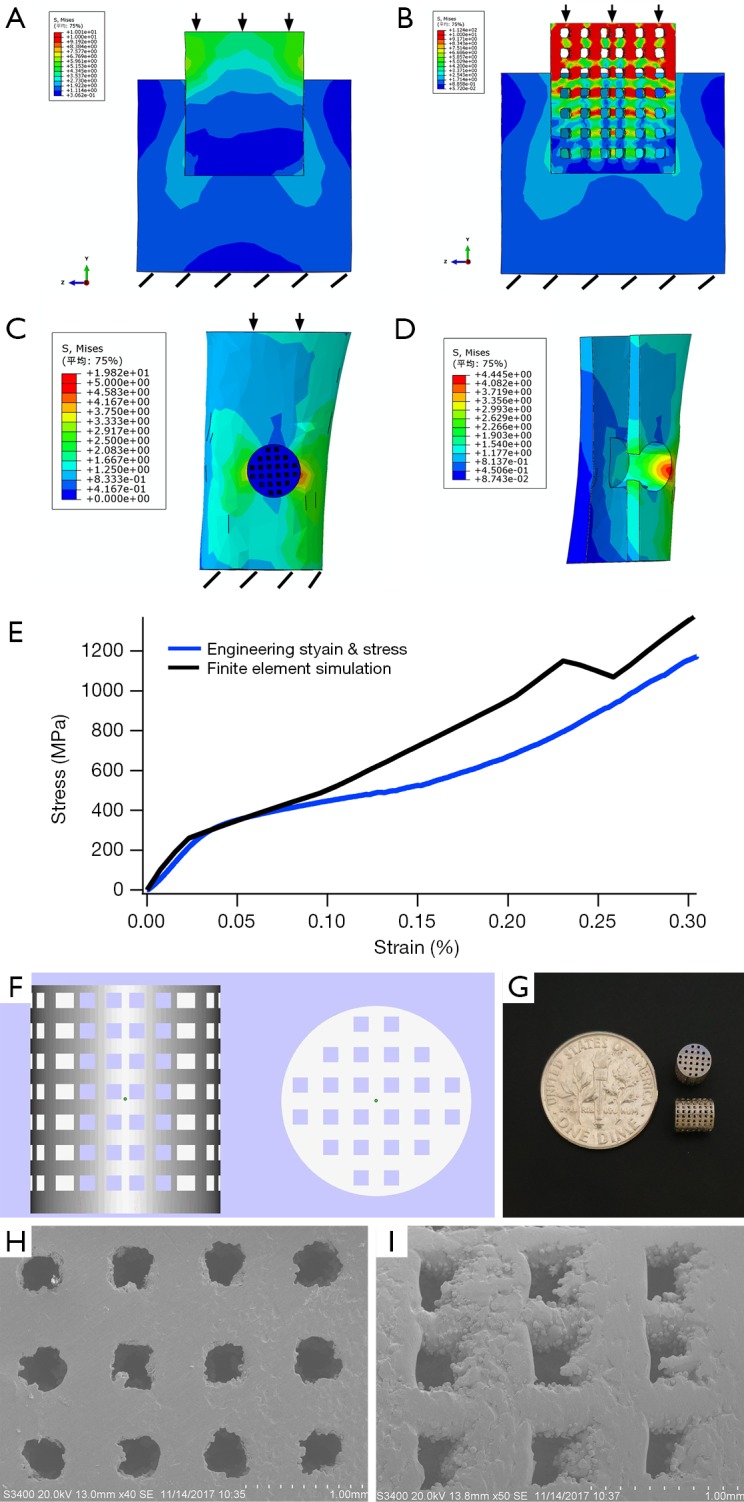
Methods: In this study, we proposed a simple design of porous structure to minimize the disparity between scaffold and natural bone tissue, and introduced a one-step reaction to form a polydopamine (PDA) layer on the surface of titanium for the purpose of improving osteogenesis as well. The porous scaffolds with pore size of 400 µm and porosity of 44.66% were made by additive manufacturing. The cell behavior was tested by seeding MC3T3-E1 cells on Ti6Al4V films for 15 days. The biomechanical properties were then analyzed by finite element (FE) method and the in vivo osteogenesis effect was accordingly evaluated by implanting the scaffolds for 5 weeks in rabbits.

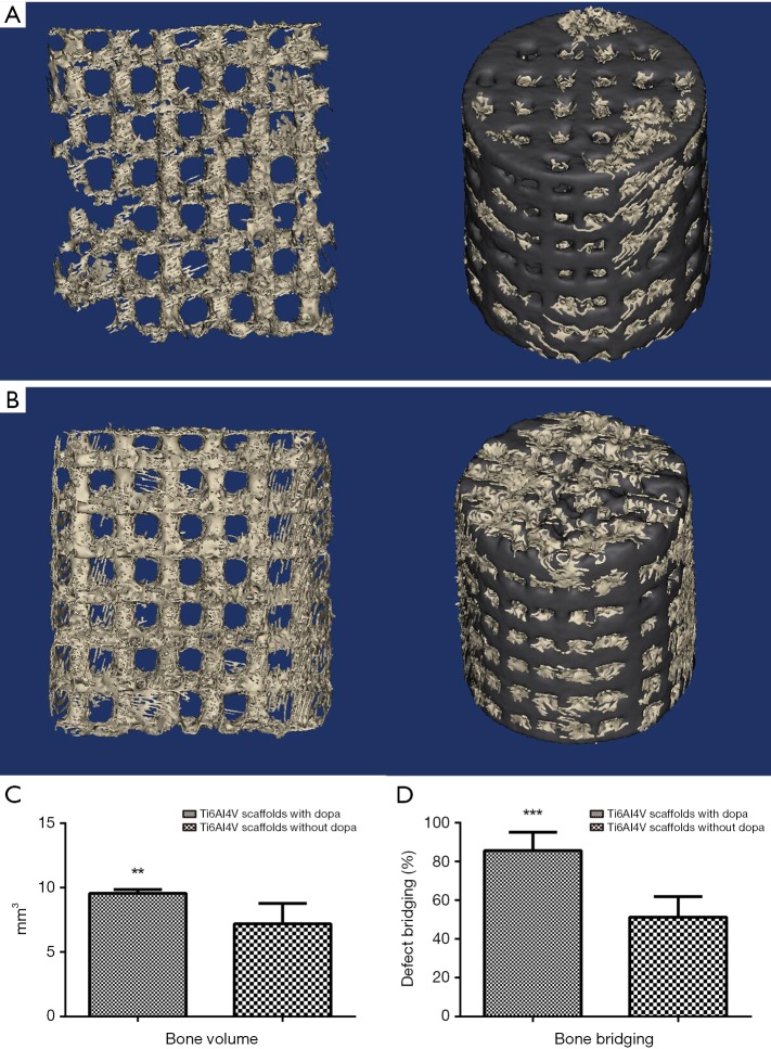
Results: According to the achieved results, it was revealed that the immersion for 40 min with dopamine could significantly improve the cell adhesion. The proposed method for design of porous structure can avoid the stress shielding effect and bone growth inside the PDA coating scaffolds, which were observed at the early stage of bone healing process.

Conclusions: It can be concluded that the proposed PDA coating method is effective in promoting early osteogenesis, as well as being easy to operate, and can be helpful in the future clinical application of titanium implants.

Keywords: 3D printing; Ti6Al4V scaffold; finite element simulation (FE simulation); polydopamine (PDA); surface modification.


Conflict of interest statement

Conflicts of Interest: The authors have no conflicts of interest to declare.

Figures

Figure 1
Figure 1
The results of SEM and EDS. (A) The SEM image of Ti6Al4V film without PDA coating; (B) the SEM image of Ti6Al4V film with PDA coating; (C) the EDS spectra of blank Ti6Al4V film; (D) the EDS spectra of Ti6Al4V film with PDA coating. PDA, polydopamine; SEM, scanning electron microscope; EDS, energy dispersive spectrometer.
Figure 2
Figure 2
The results of cell behavior. (A) The CCK-8 results after 1, 7, and 15 d; (B,C) SEM micrographs showing MC3T3-E1 cell morphology cultured on unmodified Ti films and PDA coating films after 4 h of adhesion; (D,E) immunofluorescence staining showing cell morphology cultured on unmodified Ti films and PDA coating films after 1 d of adhesion; (F,G) 7 d of adhesion, and (H,I) 15 d of adhesion. Scale bar: 100 µm. PDA, polydopamine; SEM, scanning electron microscope.
Figure 3
Figure 3
The results of FE simulation and 3D printing entities. (A) The stress transmission on solid scaffold; (B) the stress transmission on porous scaffold; (C) the general view of stress distribution after implantation with porous scaffold; (D) the internal view of stress distribution around the scaffold; (E) the arrow represented the direction of the force, and the oblique line represented the immobilization place; (F) the compression modulus of the porous scaffold, the black line represented the FE simulation result, and the blue line showed the compression test result; (G) the lateral and top views of the porous structure; (H) the general view of the 3D printing scaffolds, The SEM micrographs of the top view and (I) the lateral view of the scaffold. FE, finite element; SEM, scanning electron microscope.
Figure 4
Figure 4
The results of new-born bone tissue evaluation. The 3D reconstruction of control group (A) and PDA coating group (B), the black part represented the scaffold and the silver part represented the newly formed bone tissue. The bone volume/total volume in two groups (C), the bone bridging in two groups (D). Data were shown as mean ± SD, with **, P<0.01; ***, P<0.001. PDA, polydopamine.
Figure 5
Figure 5
The results of histological analysis. The position of selected sections (A), the Goldner trichrome staining (B) and the green fluorescence (C) of PDA coating group in section 1, the Goldner trichrome staining (D) and the green fluorescence (E) of PDA coating group in section 2, the Goldner trichrome staining (F) and the green fluorescence (G) of control group in section 1, Goldner trichrome staining (H) and the green fluorescence (I) of control group in section 2. In Goldner trichrome staining, the color of blue-green represents the regenerating bony tissue. The green fluorescence represented the newly formed bone tissue in the fluorescence images. PDA, polydopamine.

References

    1. Meyrueis JP, Cazenave A. Consolidation des fractures. EMC - Rhumatologie-Orthopedie 2004;1:138-62.
    1. Chien CY, Liu TY, Kuo WH, et al. Dopamine-assisted immobilization of hydroxyapatite nanoparticles and RGD peptides to improve the osteoconductivity of titanium. J Biomed Mater Res A 2013;101:740-7. 10.1002/jbm.a.34376 - DOI - PubMed
    1. Khan SN, Tomin E, Lane JM. Clinical applications of bone graft substitutes. Orthop Clin North Am 2000;31:389-98. 10.1016/S0030-5898(05)70158-9 - DOI - PubMed
    1. Ryan GE, Pandit AS, Apatsidis DP. Porous titanium scaffolds fabricated using a rapid prototyping and powder metallurgy technique. Biomaterials 2008;29:3625-35. 10.1016/j.biomaterials.2008.05.032 - DOI - PubMed
    1. Sidambe AT. Biocompatibility of Advanced Manufactured Titanium Implants-A Review. Materials 2014;7:8168. 10.3390/ma7128168 - DOI - PMC - PubMed