. 2019 Jun 16:2019:3808432.
doi: 10.1155/2019/3808432. eCollection 2019.

Eya2 Is Overexpressed in Human Prostate Cancer and Regulates Docetaxel Sensitivity and Mitochondrial Membrane Potential through AKT/Bcl-2 Signaling

Affiliations

Eya2 Is Overexpressed in Human Prostate Cancer and Regulates Docetaxel Sensitivity and Mitochondrial Membrane Potential through AKT/Bcl-2 Signaling

Zhongyuan Liu et al. Biomed Res Int. .

Abstract

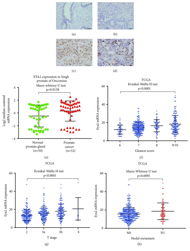
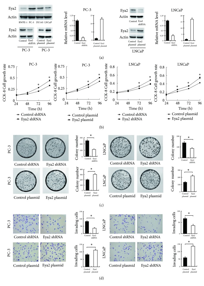
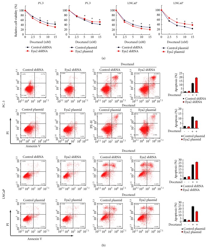
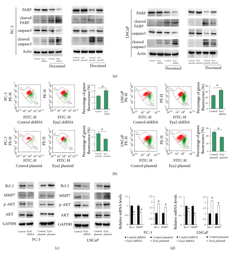
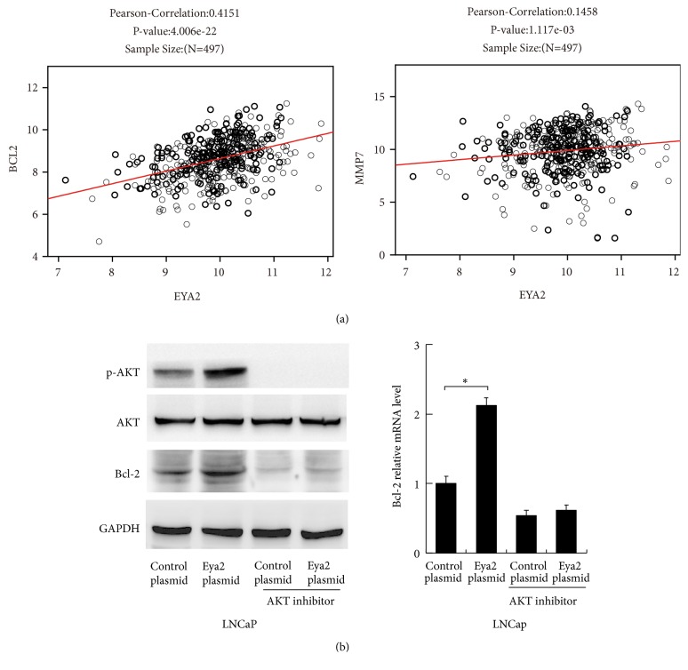
The aberrant expression of Eya2 has been observed in a wide range of cancer types. However, the clinical significance and biological effects of EYA2 in human prostate cancer remain unknown. In this study, we showed that increased levels of Eya2 protein correlated with advanced TNM stage, T stage, and a higher Gleason score. Data from the Cancer Genome Atlas (TCGA) prostate cohort consistently revealed that Eya2 mRNA was positively correlated with a higher Gleason score, higher T stage, and positive nodal metastasis in prostate cancer. Furthermore, data from the Oncomine database showed increased levels of EYA2 mRNA expression in prostate cancer tissues compared with normal tissues. Eya2 protein expression was also higher in prostate cancer cell lines compared with a normal RWPE-1 cell line. We selected LNCaP and PC-3 cell lines for plasmid overexpression and shRNA knockdown. CCK-8, colony formation, and Matrigel invasion assays demonstrated that the overexpression of Eya2 promoted proliferation, colony number, and invasion while Eya2 shRNA inhibited proliferation rate, colony formation, and invasion ability. CCK-8 and Annexin V assays showed that Eya2 reduced sensitivity to docetaxel and docetaxel-induced apoptosis while Eya2 shRNA showed the opposite effects. The overexpression of Eya2 also downregulated the cleavage of caspase3 and PARP while Eya2 depletion upregulated caspase3 and PARP cleavage. Notably, JC-1 staining demonstrated that Eya2 upregulated mitochondrial membrane potential. We further revealed that the overexpression of Eya2 upregulated Bcl-2, matrix metalloproteinase 7 (MMP7), and AKT phosphorylation. Accordingly, data from the TCGA prostate cohort indicated that EYA2 mRNA was positively correlated with the expression of Bcl-2 and MMP7. The inhibition of AKT attenuated EYA2-induced Bcl-2 upregulation. In conclusion, our data demonstrated that Eya2 was upregulated in prostate cancers. EYA2 promotes cell proliferation and invasion as well as cancer progression by regulating docetaxel sensitivity and mitochondrial membrane potential, possibly via the AKT/Bcl-2 axis.


Figures

Figure 1
Figure 1
Eya2 expression in prostate cancers. (a) Negative or weak immunostaining of Eya2 in normal prostate tissue. (b) Negative Eya2 staining in prostate cancer. (c) Moderate nuclear Eya2 staining in a prostate cancer. (d) Strong nuclear Eya2 immunostaining in a case of prostate cancer. (e) Analysis of the Singh Prostate dataset of Oncomine. Eya2 mRNA was significantly elevated in prostate cancers compared with normal prostate tissues. (f) Eya2 mRNA in prostate cancers (TCGA). Eya2 mRNA positively correlated with Gleason score. (g) Eya2 mRNA was positively correlated with T stage (TCGA dataset). (h) Eya2 mRNA expression was significantly higher in prostate cancers with nodal metastasis (TCGA dataset). Statistical methods and p values are indicated in the figure.
Figure 2
Figure 2
Eya2 regulates the proliferation and invasion of prostate cancer. (a) Western blot analysis of Eya2 protein in prostate cancer cell lines (LNCaP, PC-3, and DU145) and RWPE-1 (normal prostate cell line): there were higher levels of Eya2 protein in cancer cell lines than the normal cell line. Eya2 transfection significantly increased the levels of Eya2 protein and mRNA levels in PC-3 and LNCaP cells. The application of Eya2 shRNA significantly reduced the levels of Eya2 protein and mRNA levels in PC-3 and LNCaP cells. (b) Eya2 transfection led to an increase in proliferation rate in both PC-3 and LNCaP cell lines, while Eya2 shRNA knockdown reduced the proliferation rate. (c) Colony formation demonstrated that the overexpression of Eya2 upregulated colony number in both PC-3 and LNCaP cell lines, while Eya2 shRNA knockdown downregulated colony number. (d) Matrigel invasion assays demonstrated that the overexpression of Eya2 in both PC-3 and LNCaP cell lines increased the number of invading cells, while Eya2 shRNA knockdown decreased invading cell numbers. p<0.05.
Figure 3
Figure 3
Eya2 regulates docetaxel sensitivity and apoptosis. (a) PC-3 and LNCaP cells experiencing Eya2 transfection/shRNA knockdown were treated with different concentration of docetaxel (2.5, 5, 10, and 15 nM). CCK-8 assays were used to determine cell viability. The overexpression of Eya2 led to an increase in cell viability in both cell lines while the depletion of Eya2 led to downregulated cell viability. (b) PC-3 and LNCaP cells experiencing Eya2 transfection/shRNA knockdown were treated with 5nM docetaxel for 24 h. Annexin V/PI staining showed that Eya2 overexpression inhibited the docetaxel-induced rate of apoptosis. Eya2 shRNA increased the rate of docetaxel-induced apoptosis. ∗ p < 0.05.
Figure 4
Figure 4
Eya2 regulates mitochondrial membrane potential and related proteins. (a) The overexpression of Eya2 inhibited the expression of cleaved-caspase3, cleaved-PARP and upregulated caspase3, PARP in docetaxel treated cells. The knockdown of Eya2 using shRNA led to an upregulation in the levels of cleaved-caspase3, cleaved-PARP and downregulated caspase3, PARP in docetaxel treated cells. (b) JC-1 staining was used to determine Δψm. JC-1 is normally visualized as green when Δψm is reduced. Our JC-1 staining results showed that the overexpression of Eya2 upregulated the Δψm (as shown by a reduction in the proportion of green staining) while Eya2 shRNA reduced Δψm (as shown by an increased proportion of green staining) after CDDP treatment. (c) Protein levels of Bcl-2, MMP7, p-AKT, and AKT in prostate cancer cells transfected with the Eya2 plasmid and shRNA; the overexpression of Eya2 increased the expression of Bcl-2, MMP7, and p-AKT proteins while the application of shRNA caused levels of these proteins to decrease. (d) Realtime PCR showed that the overexpression of Eya2 o increased the mRNA levels of Bcl-2 and MMP7 in prostate cancer cells while the application of shRNA caused a reduction in the expression of these proteins. p<0.05.
Figure 5
Figure 5
Eya2 upregulates Bcl-2 via the AKT signaling pathway. (a) We identified positive associations between Eya2 mRNA and Bcl-2/MMP7 mRNA in 497 cases of prostate cancer (Pearson's correlation analysis, RNA-seq data was obtained from the TCGA database). (b) Protein levels of Bcl-2, p-AKT, and AKT in LNCaP cells treated with Eya2 and the AKT inhibitor, perifosine. Realtime PCR analysis of Bcl-2 mRNA in LNCaP cells treated with Eya2 plasmid and the AKT inhibitor, perifosine. Eya2 overexpression failed to upregulate levels of Bcl-2 mRNA and protein in cells treated with the AKT inhibitor.

References

    1. Siegel R. L., Miller K. D., Jemal A. Cancer statistics, 2017. CA: A Cancer Journal for Clinicians. 2017;67(1):7–30. doi: 10.3322/caac.21387. - DOI - PubMed
    1. Moore T. H. M., King A. J. L., Evans M., Sharp D., Persad R., Huntley A. L. Supportive care for men with prostate cancer: why are the trials not working? a systematic review and recommendations for future trials. Cancer Medicine. 2015;4(8):1240–1251. doi: 10.1002/cam4.446. - DOI - PMC - PubMed
    1. Hudson S. V., O'Malley D. M., Miller S. M. Achieving optimal delivery of follow-up care for prostate cancer survivors: improving patient outcomes. Patient Related Outcome Measures. 2015;6:75–90. doi: 10.2147/PROM.S49588. - DOI - PMC - PubMed
    1. Goodwin J., Kothari V., Drake J., et al. DNA-PKcs-mediated transcriptional regulation drives prostate cancer progression and metastasis. Cancer Cell. 2015;28(1):97–113. doi: 10.1016/j.ccell.2015.06.004. - DOI - PMC - PubMed
    1. Domingo-Domenech J., Vidal S. J., Rodriguez-Bravo V., et al. Suppression of acquired docetaxel resistance in prostate cancer through depletion of notch- and hedgehog-dependent tumor-initiating cells. Cancer Cell. 2012;22(3):373–388. doi: 10.1016/j.ccr.2012.07.016. - DOI - PMC - PubMed

MeSH terms