Review
. 2019 Oct;76(20):4043-4070.
doi: 10.1007/s00018-019-03199-x. Epub 2019 Jul 17.

Stem cells in tissues, organoids, and cancers

Affiliations
Review

Stem cells in tissues, organoids, and cancers

Xusheng Wang. Cell Mol Life Sci. 2019 Oct.

Abstract

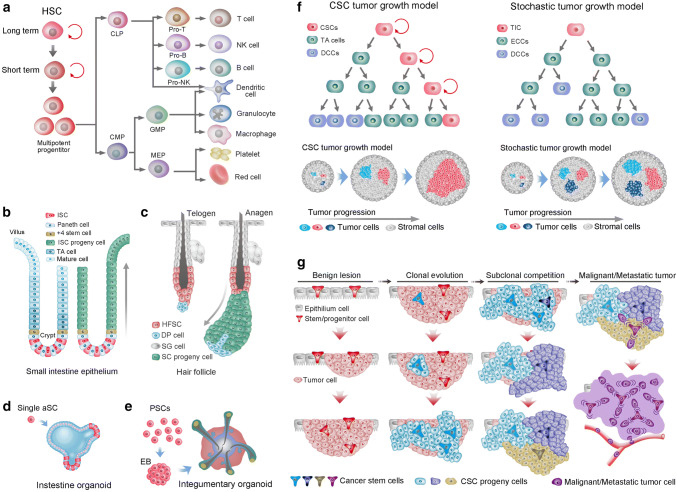
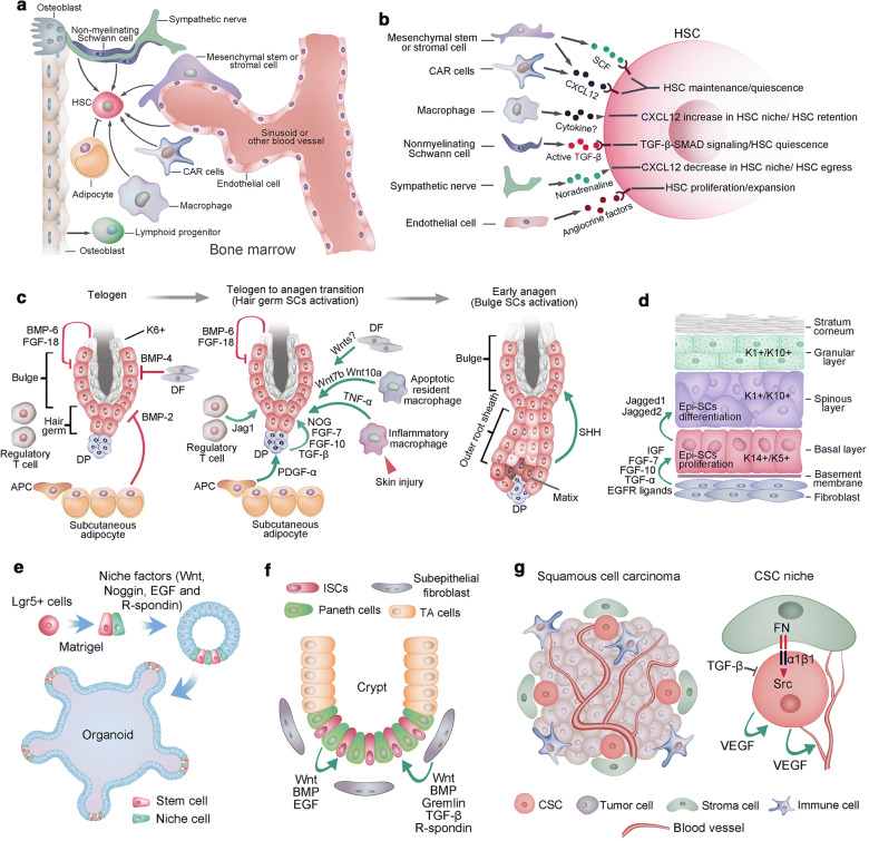
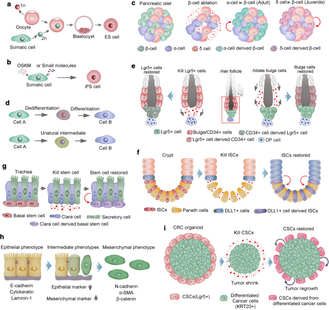
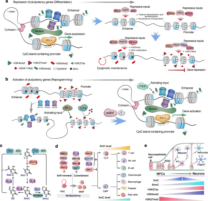
Stem cells give rise to all cells and build the tissue structures in our body, and heterogeneity and plasticity are the hallmarks of stem cells. Epigenetic modification, which is associated with niche signals, determines stem cell differentiation and somatic cell reprogramming. Stem cells play a critical role in the development of tumors and are capable of generating 3D organoids. Understanding the properties of stem cells will improve our capacity to maintain tissue homeostasis. Dissecting epigenetic regulation could be helpful for achieving efficient cell reprograming and for developing new drugs for cancer treatment. Stem cell-derived organoids open up new avenues for modeling human diseases and for regenerative medicine. Nevertheless, in addition to the achievements in stem cell research, many challenges still need to be overcome for stem cells to have versatile application in clinics.

Keywords: Cancer stem cell; Organoid; Stem cell; Stem cell niche.


Figures

Fig. 1
Fig. 1
Stem cells in tissues, organoids, and cancers. a Differentiation of hematopoietic stem cells (HSCs). HSCs are composed of long-term and short-term self-renewing stem cells and multipotent progenitors. The multipotent progenitors give rise to common lymphoid progenitors (CLPs) and common myeloid progenitors (CMPs). Subsequently, CMPs and CLPs develop into myeloid and lymphoid lineages of blood cells, respectively. Both CMPs and CLPs can generate all dendritic cells in mice. GMPs, Granulocyte macrophage precursors. b Intestinal stem cells (ISCs) at the base of the crypt generate rapidly proliferating TA cells in the lower half of the crypt. TA cells subsequently differentiate into the mature lineages of the surface epithelium (left). Lineage tracing showed that ISCs could repopulate the epithelium in 5–7 days (right). c The hair follicle stem cells (HFSCs) reside in the bulge region and maintain quiescence during the telogen phase (left). HFSCs generate all cycling portions of hair follicles in the anagen phase (right). d Single Lgr5 stem cell from small-intestinal crypts build crypt villus organoids in 3D culture. e A homogeneous population of mouse pluripotent stem cells generates skin organoids in vitro, which stratify with epidermal and dermal layers, and generates de novo hair follicles in a process that recapitulates embryonic hair folliculogenesis. f Two models of tumor growth. In the hierarchical model of tumor growth, only CSCs exhibit self-renewal capacity, whereas TA cells confer limited proliferative potential and subsequently differentiate into nonproliferative cancer cells (left upper). In the clonal assay, CSCs present dominant clonal expansion, whereas TA cells exhibit limited clonal expansion capacity (left lower). In the stochastic model of tumor growth, all cancer cells are equipotent and undergo either self-renewal or differentiation into nonproliferative cancer cells stochastically (right upper). In the clonal assay, all equipotent cancer cells showed similar clonal expansion capacity (right lower). g The hypothesis of clonal evolution in tumor progression. First, oncogenic stimulation insults a stem cell (alternatively, a progenitor or even a differentiated cell) of healthy epithelium, resulting in the generation of benign lesions with genetic homogeneity (benign lesion). Further evolution of the cells in the benign lesion generates a more invasive and malignant clone in the primary tumor (clonal evolution). Subsequently, subclone competition within the malignant subclones leads to further transformation, and genetically heterogeneous subclones coexist within the tumor (subclonal competition). Then, a final mutational insult leads to the tumor being thoroughly turned over by the malignant and metastatic cells that all behave as cancer stem cells
Fig. 2
Fig. 2
Stem cell niche. a. Various cell types in bone marrow play roles in regulating HSC maintenance, including mesenchymal stem/stromal cells, endothelial cells, CAR cells, macrophages, sympathetic neurons and nonmyelinating Schwann cells. However, adipocytes exhibit a negative effect on HSC maintenance. b. HSC niche cells contribute to HSC maintenance via the release of different factors. c. The quiescence of hair follicle SCs at the bulge and hair germ was maintained by a set of factors, including BMP6 and FGF-18 from K6+ bulge cells, BMP4 produced by dermal fibroblasts (DFs), and BMP2 expressed by subcutaneous adipocytes. At the onset of anagen, the activation factors prevailed, including noggin (NOG), FGF-7, FGF-10 and TGF-β2 produced by dermal papillae (DP) and PDGF-α derived from adipocyte precursor cells (APCs). Wnt7b and Wnt10a from apoptotic resident macrophages and Jag1 from regulatory T cells also contribute to the activation of hair follicle SCs. After skin injury, TNF-a from inflammatory macrophages could induce the activation of hair follicle SCs. At the beginning of a new hair cycle, SCs in the bulge remain quiescent until SHH is expressed by the TAC matrix. d. The epidermis is a stratified structure that is composed of the basal cell layer and the underneath basement membrane, spinous layer, granular layer and stratum corneum layer. Self-renewing and proliferating epidermal stem cells are located within the basal layer. Secreted factors, such as dermal fibroblasts, promote the self-renewal of cells in the basal layer. These factors include IGF, FGF-7, FGF-10, EGF ligands and TGF-α. Epidermal stem cells generate columnar units that undergo terminal differentiation via jagged activated Notch signaling. e. The essential components for intestine Lgr5+ stem cells to generate self-renewing epithelial organoids in vitro, including laminin-rich Matrigel, a cocktail of niche factors including Wnt, Noggin, R-spondin 1 and EGF that recapitulate the ISC niche in vivo. f. Paneth cells and subepithelial fibroblasts at the crypt bottom constitute the niche for intestinal stem cells. g. The CSC niche of squamous cell carcinoma. CSCs of squamous cell carcinoma are frequently found at the tumor-stroma interface (left). Extracellular matrix ligands, such as fibronectin (FN), could activate αβ1 integrins, resulting in hyperactivated focal adhesion kinase (FAK) and its associated tyrosine kinase Src, leading to the proliferation of CSCs. However, TGF-β signaling maintains the quiescence of CSCs. Moreover, VEGF secreted by CSCs could enhance CSC proliferation and promote the formation of new blood vessels
Fig. 3
Fig. 3
Stem cell plasticity. a Somatic cell nuclear transfer (SCNT) for nuclear reprogramming. The nucleus of a somatic cell (diploid, 2n) is transplanted into an enucleated oocyte (haploid, 1n). In the oocyte, the somatic cell nucleus is reprogrammed; thus, the cells derived from it are pluripotent stem cells. b Transcription factor transduction (Oct4, Sox2, Klf4 and cMyc, OSKM) or small molecule-induced pluripotent stem (iPS) cells. c Two transdifferentiation models. The first model presumes that a cell must first dedifferentiate into a precursor stage before it converts to a lineage. In the second model, cells transdifferentiate to generate new cells directly, in some cases mediated via an unnatural intermediate phase and in which genetic programming of two cell types is simultaneous. d Pancreatic islets have β-, α- and δ-cells. In adult mouse islets, α-cells transdifferentiate directly into insulin-producing cells after ablation of β-cells. However, in juvenile islets, δ-cells generate β-cells following ablation of β-cells. e During homeostasis, the hair follicle stem cell compartment is maintained by distinct stem cells, and ablated bulge cells (CD34+) can be replenished by cells in both the upper pilosebaceous unit and the hair germ (Lgr5+). Correspondingly, CD34+ bulge stem cells could compensate for the loss of Lgr5+ stem cells in hair germ. f Crypt stem cells give rise to all cell lineages in the mammalian intestinal epithelium during homeostasis. Radiation injury ablates ISCs, which stimulate dedifferentiation of DLL1+ cells to generate new ISCs. g Basal stem cells in the trachea give rise to differentiated secretory cells and Clara during homeostasis. Ablation of basal stem cells induces the dedifferentiation of Clara cells and generates new basal stem cells; h EMT is a transition of polarized epithelial cells into mobile mesenchymal cells. Several commonly used markers of epithelial and mesenchymal cells are listed. i Lgr5+ CSCs proliferate and differentiate into KRT20+ CRCs at steady state, depletion of CSCs results in the reduction of tumor size, some KRT20+ cells convert to Lgr5+ CSCs, and tumor regrowth occurs
Fig. 4
Fig. 4
Epigenetic regulation of stem cells. a Active regulatory elements are typically enriched for 5hmC, H3K27ac, H3K4me, and bound Mediator complex. During gene repression, the activating histone modifications are eliminated, and repressive marks, such as H3K27me3 and nucleosomal compaction, are established. In pluripotent stem cells, multiple enhancers combined with master pluripotency transcription factors, such as OCT4, SOX2 and KLF4, establish a super-enhancer, which supports the activation of pluripotency genes. The absence of master pluripotency factors could induce the disassembly of the enhancer–promoter complex in the assembly of repressive inputs. The nucleosome remodeling and deacetylase (NuRD) complex induces nucleosome formation at the binding region of pluripotency factors and alters the histone modifications that correspond to transcriptional activity. Similar disassembly of activating modifications occurs in the promoter by PRC2-associated histone demethylases KDM2B and KDM5A. Furthermore, H3K9me2 or H3K9me3 is deposited in the enhancer by G9A in complex with GLP and SETDB1, and DNA methylation by DNMT3A. Equally, PRC2 deposits H3K27me3 in promoters, which initiate chromatin compaction by recruiting the canonical PRC1 complex and monoubiquitylate H2AK119 (H2AK119ub). K27ac, Lys27 acetylation; Pol II, RNA polymerase II. PRC2, Polycomb repressive complex 2. GLP, G9A-like protein. DNMT3A, DNA methyltransferase 3A. CCTC-binding factor (CTCF). DNMT, DNA methyltransferase; HDAC, histone deacetylase; HAT, histone acetyltransferase; MED, mediator complex; MBD, methyl-DNA-binding domain protein; POL II, RNA polymerase II; TET, ten eleven translocation dioxygenase; TrxG, trithorax group complex. TF, transcription factor. b In differentiated cells, H3K27me3 and H2AK119ub are enriched in CpG island-containing promoters of some pluripotency genes. At the initiation of reprogramming, OCT4, SOX2 and KLF4 (known as OSK) bind to partial motif sequences of some select enhancers to engage pioneer factor-like activity. These OSK binding sites are modified with H3K4me1 and H3K4me2. OSK binding also promotes H3K4 methylation at the promoter via the MLL component WDR5 and is associated with local erasure of H3K27me3 by the histone demethylase UTX. Subsequently, OSK cooperates with unknown factors to establish a canonical enhancer architecture, including H3K4me2 and H3K27ac, and stable topological cognation to the promoter via the cohesin and Mediator complexes. Embryonic stem cell-specific BAF (esBAF) further stabilizes the enhancer-promoter complex, and the activating inputs predominate at this stage and direct the expression of the target genes. LSD1, Lys-specific demethylase 1; MBD3, methyl CpG-binding domain protein 3; MLL, mixed lineage leukemia. Pol II, RNA polymerase II, WDR5, WD repeat-containing protein 5. c The mechanisms of DNA methylation and demethylation. Cytosine is converted to 5mC by DNMTs. TET1 is responsible for the conversion of 5mC to 5hmC. Three TET family proteins could subsequently oxidize 5hmC to 5fC (5-formylcytosine) and then to 5caC (5-carboxylcytosine). Moreover, 5hmC can be converted to 5hmU (5-hydroxymethyluracil) by deaminase activation-induced cytidine deaminase (AID) and apolipoprotein B mRNA-editing enzyme catalytic polypeptides (APOBECs). 5fC, 5caC and 5hmU can be excised by thymine DNA glycosylase and replaced by an unmodified cytosine via the base-excision repair (BER) pathway. d Key epigenetic regulators that are involved in HSC self-renewal and lineage commitment during differentiation, and DNA methylation levels changes during hematopoietic lineage commitment. CLP, common lymphoid progenitor; CMP, common myeloid progenitor; DNMT, DNA methyltransferase; DNA me, DNA methylation; GLP, G9A-like protein. GMP, granulocyte–monocyte progenitor; MPP, multipotent progenitor; PRC, polycomb repressive complex; TET, Ten-eleven translocation; TrxG, Trithorax group. e In the embryonic neurogenesis of mice, neuroepithelial cells develop into radial glial cells (RGCs) around embryonic day 14. RGCs can either produce neurons directly or give rise to intermediate progenitor cells (IPCs), which in turn generate neurons. During later embryonic development, RGCs also give rise to oligodendrocytes (OligoD) and astrocytes
Fig. 5
Fig. 5
Stem cell application and perspectives. a Two distinct procedures to collect HSCs for transplantation. HSCs are isolated from donor blood cells, in which the HSCs are mobilized with G-CSF, GM-SCF or plerixafor, and enriched with HSCs marker of CD34+/CD38−. Alternatively, protocols have established to produce HSCs ether from endothelial cells, or from human pluripotent stem cells (PSCs), these two protocols treated initiate cells with overlapping cocktails of transcription factors. The primary HSCs need to receive as-yet-unknown extracellular signals for further maturation. b MSC immunosuppressive capacity and immunogenicity are affected by levels of systemic or local inflammatory cytokines. High immunosuppressive potential of MSCs is achieved via suppression of T cell activation and inhibition of antigen-presenting cell (APC) maturation. Whereas, MSCs that do not tip the balance toward immunosuppression are prone to immunogenicity and result in immune detection and destruction, as debris from apoptotic MSCs are processed by APCs in the context of danger signals. The rate of immune detection of allogeneic MSCs is determined by the balance between relative expression of immunogenic and immunosuppressive factors in MSCs. IFN-γ, interferon gamma; MHC, major histocompatibility complex; PGE2, prostaglandin E2; sHLA-G5, soluble human leukocyte antigen-g5; TCR, T cell receptor; TGF-β, transforming growth factor beta; TNF-α, tumor necrosis factor alpha; TSG-6; TNF-stimulated gene 6 protein. c Organoids generated from patient-derived healthy and tumor tissues can be genetically characterized and used for drug screening, and can be cryopreserved and stored in living organoid biobanks. Organoids developed from healthy tissue of the same patient can be used to screen drugs that are less toxic to healthy cells while selectively kill tumor cells. Moreover, hepatocyte organoid cultures may be used to test for hepatotoxicity. In this schematic example, drug C could specifically kills tumor organoids and does not show hepatotoxicity, and thus it seems most suitable for treating the patient. d CSC model of cancer relapse. Intrinsic and extrinsic mechanism contribute to the CSCs resistance to the medical therapy, in addition, non-CSCs may convert to CSCs and replenish the CSCs pool, ether CSCs drug resistance or replenish result in cancer relapse. The CSC model suggests that inhibiting CSC self-renewal, inducing CSC specific cell death, inducing CSC differentiation or targeting CSC niche would lead to the depletion of the CSCs pool and subsequent tumor regression. Nevertheless, if the CSC is reversed from no-CSCs, further specific and no-specific therapies will be needed the for the final regression of tumor. e The principle of interspecies blastocyst complementation for the generation of human–animal chimaeras. Human PSC-derived organ could help to solve the severe shortage of organ donors. Additionally, Human–animal chimaeras could be useful for modeling human diseases and for testing the efficacy and safety of a candidate drug in vivo. f The principle of tissue complementation chimera. In this example, human-pig integumentary chimera was achieved via transplanting human skin progenitors to the skin incision of newborn pig. The engraftment of human progenitors will develop to mature human skin tissue with appendage organs, such as hair follicle

References

    1. Becker AJ, Mc CE, Till JE. Cytological demonstration of the clonal nature of spleen colonies derived from transplanted mouse marrow cells. Nature. 1963;197:452–454. - PubMed
    1. Kretzschmar K, Watt FM. Lineage tracing. Cell. 2012;148(1–2):33–45. - PubMed
    1. Adams GB, Scadden DT. The hematopoietic stem cell in its place. Nat Immunol. 2006;7(4):333–337. - PubMed
    1. de Rooij DG. Proliferation and differentiation of spermatogonial stem cells. Reproduction. 2001;121(3):347–354. - PubMed
    1. Tumbar T, Guasch G, Greco V, Blanpain C, Lowry WE, Rendl M, Fuchs E. Defining the epithelial stem cell niche in skin. Science. 2004;303(5656):359–363. - PMC - PubMed

MeSH terms