. 2021 Aug;28(4):1271-1280.
doi: 10.1007/s12350-019-01818-5. Epub 2019 Jul 17.

Assessment of myocardial viability with [15O]water PET: A validation study in experimental myocardial infarction

Affiliations

Assessment of myocardial viability with [15O]water PET: A validation study in experimental myocardial infarction

Maria Grönman et al. J Nucl Cardiol. 2021 Aug.

Abstract

Background: Assessment of myocardial viability is often needed in patients with chest pain and reduced ejection fraction. We evaluated the performance of reduced resting MBF, perfusable tissue fraction (PTF), and perfusable tissue index (PTI) in the assessment of myocardial viability in a pig model of myocardial infarction (MI).

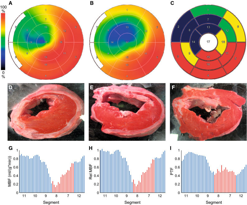
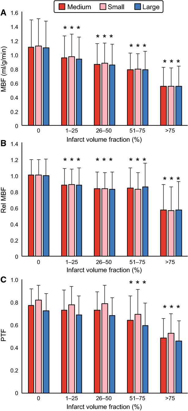
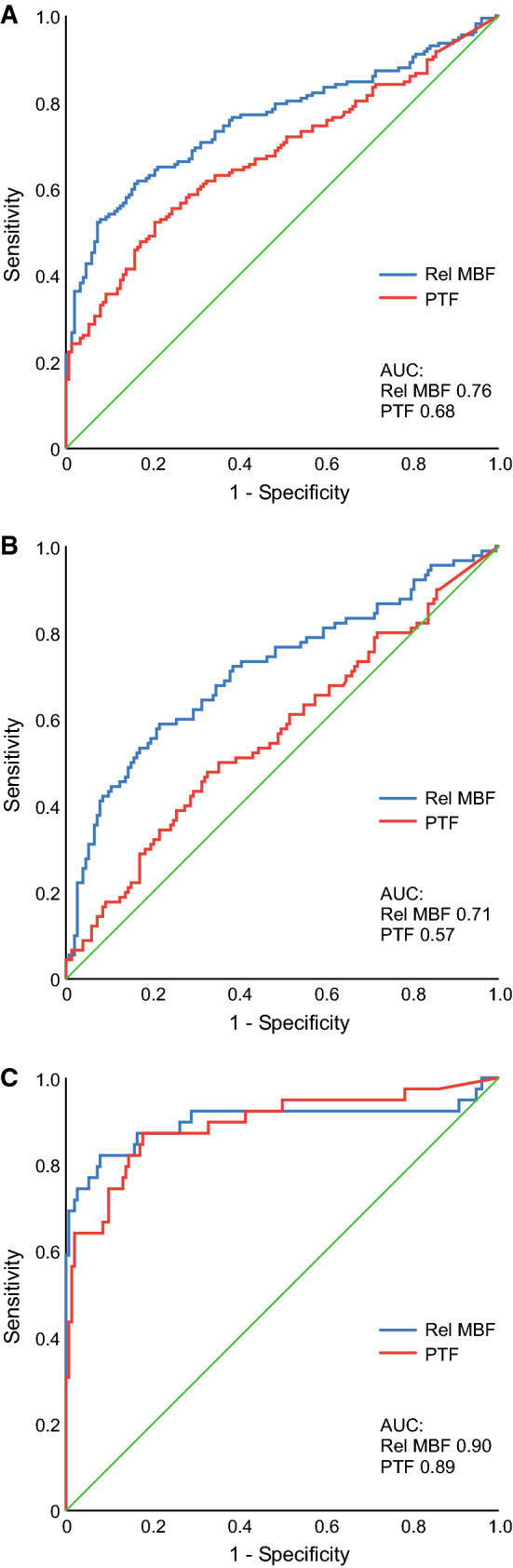
Methods and results: Pigs underwent resting [15O]water PET perfusion study 12 weeks after surgical (n = 16) or 2 weeks after catheter-based (n = 4) occlusion of the proximal left anterior descending coronary artery. MBF, PTF, and PTI were compared with volume fraction of MI in matched segments as assessed by triphenyl tetrazolium chloride staining of LV slices. MBF and PTF were lower in infarcted than non-infarcted segments. Segmental analysis of MBF showed similar area under the curve (AUC) of 0.85, 0.86, and 0.90 with relative MBF, PTF, and PTI for the detection of viable myocardium defined as infarct volume fraction of < 75%. Cut-off values of relative MBF of ≥ 67% and PTF of ≥ 66% resulted in accuracies of 90% and 81%, respectively.

Conclusions: Our results indicate that resting MBF, PTF, and PTI based on [15O]water PET perfusion imaging are useful for the assessment of myocardial viability.

Keywords: PET; Radiowater; myocardial infarction; myocardial perfusion imaging; viability.


Figures

Figure 1
Figure 1
Representative polar maps of resting myocardial blood flow (MBF) by [15O]water PET (A) and perfusable tissue fraction (PTF) by [15O]water PET (B). Polar maps were normalized to their own maximum. (C) shows grading of segmental volume fraction of myocardial infarction (red = 0%, yellow = 1%-24%, green = 25%-49%, and blue = > 75%) based on TTC staining of myocardial slices at the apical (D), papillary muscle (E), and basal (F) levels. Viable myocardium is stained red and non-viable infarcted white by TTC. MBF and PTF are reduced in segments with myocardial infarction. (G), (H), and (I) show examples of circumferential profiles of MBF, relative MBF, and PTF, respectively. Sectors with transmural infarction are marked with red color
Figure 2
Figure 2
Segmental values of absolute myocardial blood flow (MBF, A), relative MBF (B), and perfusable tissue fraction (PTF, C) by [15O]water PET measured using three different ROI thickness: medium = ROI width defined automatically by the software program, small = ROI width manually reduced to completely fit inside the myocardium, large = ROI width manually increased from the medium ROI. *P < 0.05 vs. segments with infarct volume fraction 0
Figure 3
Figure 3
Receiver operating characteristics (ROC) curve analysis of segmental relative myocardial blood flow (Rel MBF) and perfusable tissue fraction (PTF) by [15O]water PET in the detection of any myocardial infarction in the corresponding segment (A), subendocardial infarction (infarct volume fraction 1%-49%, B), or non-viable tissue (infarct volume fraction ≥ 75%, C)
Figure 4
Figure 4
Receiver operating characteristics (ROC) curve analysis of relative myocardial blood flow (Rel MBF) and perfusable tissue fraction (PTF) by [15O]water in the detection of myocardial viability with infarct volume fraction of < 50% (A) and with infarct volume fraction of < 75% (B)
Figure 5
Figure 5
Receiver operating characteristics (ROC) curve analysis of relative myocardial blood flow (Rel MBF), perfusable tissue fraction (PTF), and perfusable tissue index (PTI) by [15O]water in the detection of myocardial viability with infarct volume fraction of < 50% (A) and with infarct volume fraction of < 75% (B)

References

    1. Elsasser A, Schlepper M, Klovekorn W-P, Cai W-J, Zimmermann R, Muller K-D, et al. Hibernating myocardium: An incomplete adaptation to ischemia. Circulation. 1997;96:2920–2931. doi: 10.1161/01.CIR.96.9.2920. - DOI - PubMed
    1. Bax JJ, Delgado V. Myocardial viability as integral part of the diagnostic and therapeutic approach to ischemic heart failure. J Nucl Cardiol. 2015;22:229–245. doi: 10.1007/s12350-015-0096-5. - DOI - PMC - PubMed
    1. Schinkel AFL, Bax JJ, Poldermans D, Elhendy A, Ferrari R, Rahimtoola S. Hibernating myocardium: Diagnosis and patient outcomes. Curr Probl Cardiol. 2007;32:375–410. doi: 10.1016/j.cpcardiol.2007.04.001. - DOI - PubMed
    1. Iida H, Kanno I, Takahashi A, Miura S, Murakami M, Takahashi K, et al. Measurement of absolute myocardial blood flow with H215O and dynamic positron-emission tomography. Strategy for quantification in relation to the partial-volume effect. Circulation. 1988;78:104–115. doi: 10.1161/01.CIR.78.1.104. - DOI - PubMed
    1. Harms HJ, Nesterov SV, Han C, Danad I, Leonora R, Raijmakers P, et al. Comparison of clinical non-commercial tools for automated quantification of myocardial blood flow using oxygen-15-labelled water PET/CT. Eur Heart J. 2014;15:431–441. - PubMed

Substances