. 2019 Aug 1;5(8):1195-1204.
doi: 10.1001/jamaoncol.2019.1549.

Comparison of Biomarker Modalities for Predicting Response to PD-1/PD-L1 Checkpoint Blockade: A Systematic Review and Meta-analysis

Affiliations

Comparison of Biomarker Modalities for Predicting Response to PD-1/PD-L1 Checkpoint Blockade: A Systematic Review and Meta-analysis

Steve Lu et al. JAMA Oncol. .

Abstract

Importance: PD-L1 (programmed cell death ligand 1) immunohistochemistry (IHC), tumor mutational burden (TMB), gene expression profiling (GEP), and multiplex immunohistochemistry/immunofluorescence (mIHC/IF) assays have been used to assess pretreatment tumor tissue to predict response to anti-PD-1/PD-L1 therapies. However, the relative diagnostic performance of these modalities has yet to be established.

Objective: To compare studies that assessed the diagnostic accuracy of PD-L1 IHC, TMB, GEP, and mIHC/IF in predicting response to anti-PD-1/PD-L1 therapy.

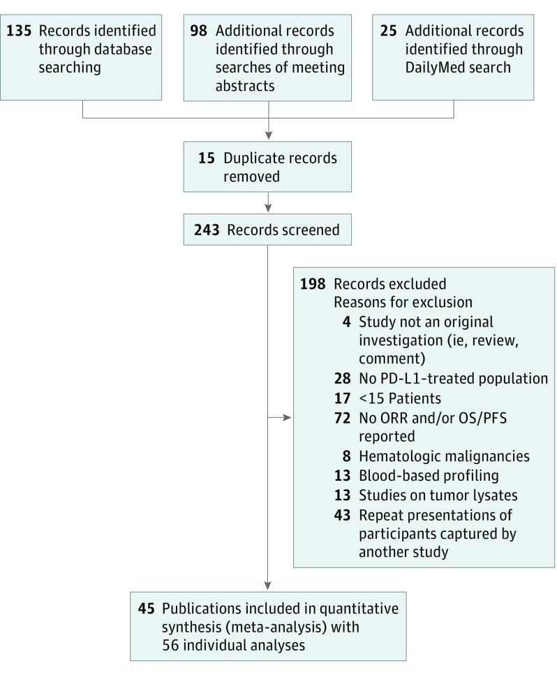
Evidence review: A search of PubMed (from inception to June 2018) and 2013 to 2018 annual meeting abstracts from the American Association for Cancer Research, American Society of Clinical Oncology, European Society for Medical Oncology, and Society for Immunotherapy of Cancer was conducted to identify studies that examined the use of PD-L1 IHC, TMB, GEP, and mIHC/IF assays to determine objective response to anti-PD-1/PD-L1 therapy. For PD-L1 IHC, only clinical trials that resulted in US Food and Drug Administration approval of indications for anti-PD-1/PD-L1 were included. Studies combining more than 1 modality were also included. Preferred Reporting Items for Systematic Reviews and Meta-analysis guidelines were followed. Two reviewers independently extracted the clinical outcomes and test results for each individual study.

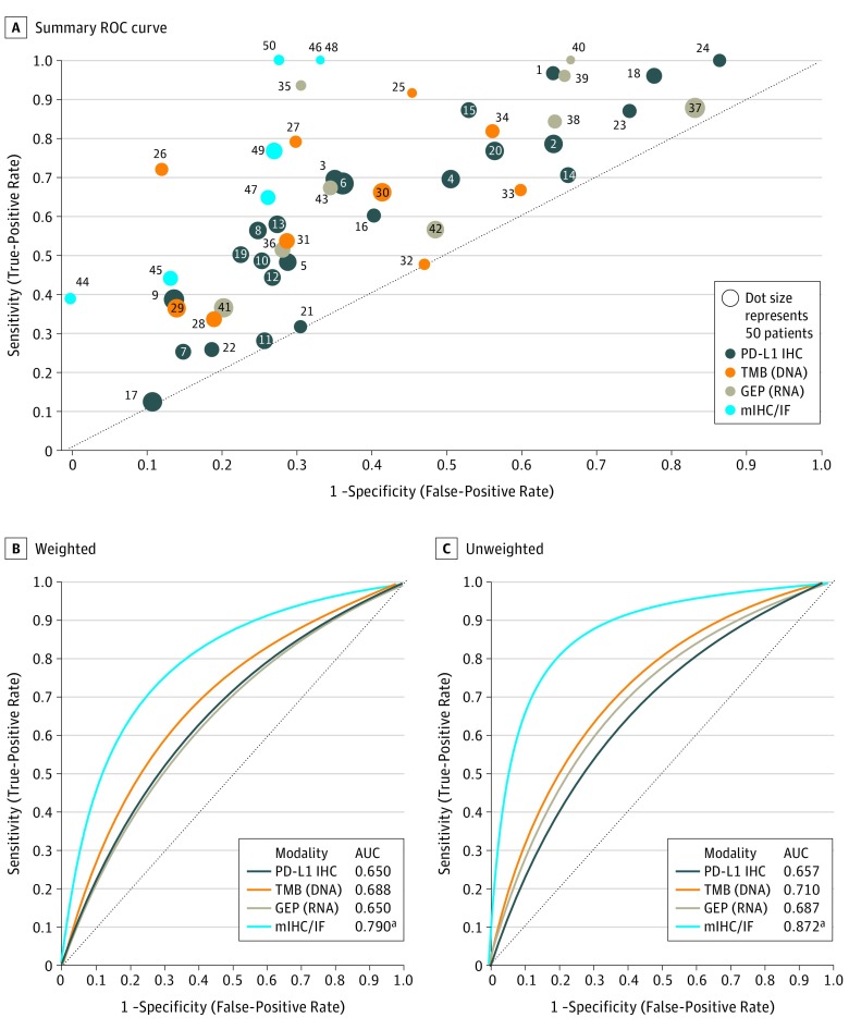
Main outcomes and measures: Summary receiver operating characteristic (sROC) curves; their associated area under the curve (AUC); and pooled sensitivity, specificity, positive and negative predictive values (PPV, NPV), and positive and negative likelihood ratios (LR+ and LR-) for each assay modality.

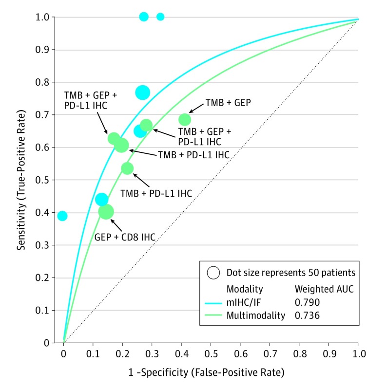
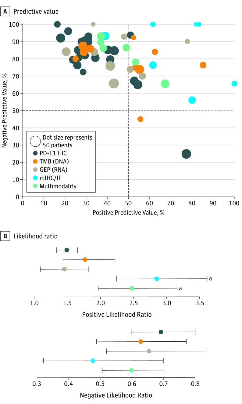
Results: Tumor specimens representing over 10 different solid tumor types in 8135 patients were assayed, and the results were correlated with anti-PD-1/PD-L1 response. When each modality was evaluated with sROC curves, mIHC/IF had a significantly higher AUC (0.79) compared with PD-L1 IHC (AUC, 0.65, P < .001), GEP (AUC, 0.65, P = .003), and TMB (AUC, 0.69, P = .049). When multiple different modalities were combined such as PD-L1 IHC and/or GEP + TMB, the AUC drew nearer to that of mIHC/IF (0.74). All modalities demonstrated comparable NPV and LR-, whereas mIHC/IF demonstrated higher PPV (0.63) and LR+ (2.86) than the other approaches.

Conclusions and relevance: In this meta-analysis, tumor mutational burden, PD-L1 IHC, and GEP demonstrated comparable AUCs in predicting response to anti-PD-1/PD-L1 treatment. Multiplex immunohistochemistry/IF and multimodality biomarker strategies appear to be associated with improved performance over PD-L1 IHC, TMB, or GEP alone. Further studies with mIHC/IF and composite approaches with a larger number of patients will be required to confirm these findings. Additional study is also required to determine the most predictive analyte combinations and to determine whether biomarker modality performance varies by tumor type.


Conflict of interest statement

Conflict of Interest Disclosures: Dr Rimm reports personal fees from and serves on the advisory board of Amgen, personal fees from Bristol-Myers Squibb, Merck, GlaxoSmithKline, Daiichi Sankyo, Konica Minolta, personal fees from and serves on the advisory board of Cell Signaling Technology, grants and personal fees from Cepheid, AstraZeneca, NextCure, Ultivue, Ventana, Perkin Elmer, grants from Lilly, patents including AQUA software licensing and Navigate Biopharma (Yale owned patent). Dr Johnson serves on the advisory board of Array Biopharma, Bristol-Myers Squibb, Genoptix, Incyte, Merck, and Novartis; receives grant funding from Bristol-Myers Squibb and Incyte; patent pending for using MHC-II as a biomarker for immunotherapy responses. Dr Schalper reports grant funding from Navigate Biopharma, Vasculox, Tesaro, Takeda, Surface Oncology, and Bristol-Myers Squibb; receives grant funding and consulting fees from Celgene, Shattuck Labs, Pierre Fabre, Moderna Therapeutics, AstraZeneca, AbbVie, and Merck; and receives speaking fees from Merck and Fluidigm. Dr Anders receives grant funding from FLX Bio and Five Prime Therapeutics, and is a consultant for Bristol-Myers Squibb, Merck, and AstraZeneca. Mr Hoyt is employed by Akoya Biosciences and owns Akoya Biosciences stock and stock options. Dr. Pardoll reported other support from Aduro Biotech, Amgen, Bayer, Camden Partners, DNAtrix, Dracen, Dynavax, Five Prime, FLX Bio, Immunomic, Janssen, Merck, Rock Springs Capital, Potenza, Tizona, Trieza, and WindMil during the conduct of the study; grants from Astra Zeneca, Medimmune/Amplimmune, and Compugen; grants and other support from ERvaxx and Potenza. Dr Taube reports nonfinancial support from Akoya during the conduct of the study; grants and personal fees from Bristol-Myers Squibb, personal fees from Merck, Astra Zeneca, and Amgen outside the submitted work; equipment and reagents from Akoya Biosciences, and a patent pending related to image processing of mIF/IHC images. No other disclosures were reported.

Figures

Figure 1.
Figure 1.. PRISMA Flowchart
eTable 1 in the Supplement lists the included studies. PD-L1 indicates programmed cell death ligand 1; PFS, progression-free survival; ORR, objective response rate; OS, overall survival.
Figure 2.
Figure 2.. Summary Receiver Operating Characteristic Curve Analysis by Assay Modality for Responders vs Nonresponders
A, Fifty-six analyses from the primary literature correlating different biomarker modalities with patient responses after anti–PD-1/PD-L1 therapy were analyzed. The sensitivity and 1−specificity of the assay for each individual publication is shown by a single dot (number on the dot correlates with reference list in eTable 1 in the Supplement). The size of each dot is proportionate to the size of the studied cohort. Linear regression models weighted (B) by the number of patients in each study and unweighted (C) (ie, each study treated equally) were used to generate summary receiver operating characteristic [sROC] curves for each assay modality). The multiplex immunohistochemistry/immunofluorescence (mIHC/IF) has a significantly higher area under the curve (AUC) than PD-L1 (programmed cell death ligand 1) IHC, tumor mutational burden (TMB), and gene expression profiling (GEP) by weighted approach and PD-L1 IHC and TMB by unweighted approach. aIndicates statistical significance (P < .05), Hanley and McNeil method.
Figure 3.
Figure 3.. Summary Receiver Operating Characteristic (sROC) Curve Analysis
The Figure shows that multimodality biomarkers have an sROC curve comparable to that of multiplex immunohistochemistry/immunofluorescence. AUC indicates area under the curve; GEP, gene expression profiling; IHC, immunohistochemistry; mIHC/IF, multiplex immunohistochemistry/immunofluorescence; PD-L1, programmed cell death ligand 1; TMB, tumor mutational burden.
Figure 4.
Figure 4.. Predictive Values for Each Individual Study and Pooled Likelihood Ratios by Biomarker Assay Modality
A, Dots in the upper left quadrant represent studies that reported high negative predictive values and were good at excluding patients from anti–PD-1/PD-L1 treatment. Dots in the upper right quadrant represent studies that reported both high negative and positive predictive values, meaning they are suitable for excluding patients who should not be treated and for selecting patients who will respond. Multiplex immunohistochemistry/immunofluorescence (mIHC/IF) can help both rule in and rule out response to anti–PD-1/PD-L1 therapy. The study number correlating to the individual dots is provided in eFigure 7 in the Supplement. B, Multiplex immunohistochemistry/IF has a better likelihood ratio (LR−) than other tested biomarker approaches, whereas both mIHC/IF and multimodality approaches have significantly higher LRs(+) (eTable 3 in the Supplement). aStatistically significant.

Comment in

References

    1. Topalian SL, Hodi FS, Brahmer JR, et al. . Safety, activity, and immune correlates of anti-PD-1 antibody in cancer. N Engl J Med. 2012;366(26):2443-2454. doi:10.1056/NEJMoa1200690 - DOI - PMC - PubMed
    1. Taube JM, Klein A, Brahmer JR, et al. . Association of PD-1, PD-1 ligands, and other features of the tumor immune microenvironment with response to anti-PD-1 therapy. Clin Cancer Res. 2014;20(19):5064-5074. doi:10.1158/1078-0432.CCR-13-3271 - DOI - PMC - PubMed
    1. Taube JM, Galon J, Sholl LM, et al. . Implications of the tumor immune microenvironment for staging and therapeutics. Mod Pathol. 2018;31(2):214-234. doi:10.1038/modpathol.2017.156 - DOI - PMC - PubMed
    1. Gong J, Chehrazi-Raffle A, Reddi S, Salgia R. Development of PD-1 and PD-L1 inhibitors as a form of cancer immunotherapy: a comprehensive review of registration trials and future considerations. J Immunother Cancer. 2018;6(1):8. doi:10.1186/s40425-018-0316-z - DOI - PMC - PubMed
    1. Hellmann MD, Rizvi NA, Goldman JW, et al. . Nivolumab plus ipilimumab as first-line treatment for advanced non-small-cell lung cancer (CheckMate 012): results of an open-label, phase 1, multicohort study. Lancet Oncol. 2017;18(1):31-41. doi:10.1016/S1470-2045(16)30624-6 - DOI - PMC - PubMed