. 2019 Sep;45(7):175-187.
doi: 10.1080/01902148.2019.1636899. Epub 2019 Jul 18.

Restrictive lung disease in TNF-transgenic mice: correlation of pulmonary function testing and micro-CT imaging

Affiliations

Restrictive lung disease in TNF-transgenic mice: correlation of pulmonary function testing and micro-CT imaging

Emily K Wu et al. Exp Lung Res. 2019 Sep.

Abstract

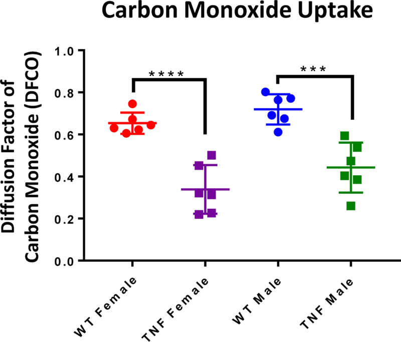
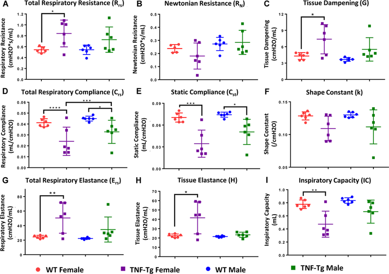
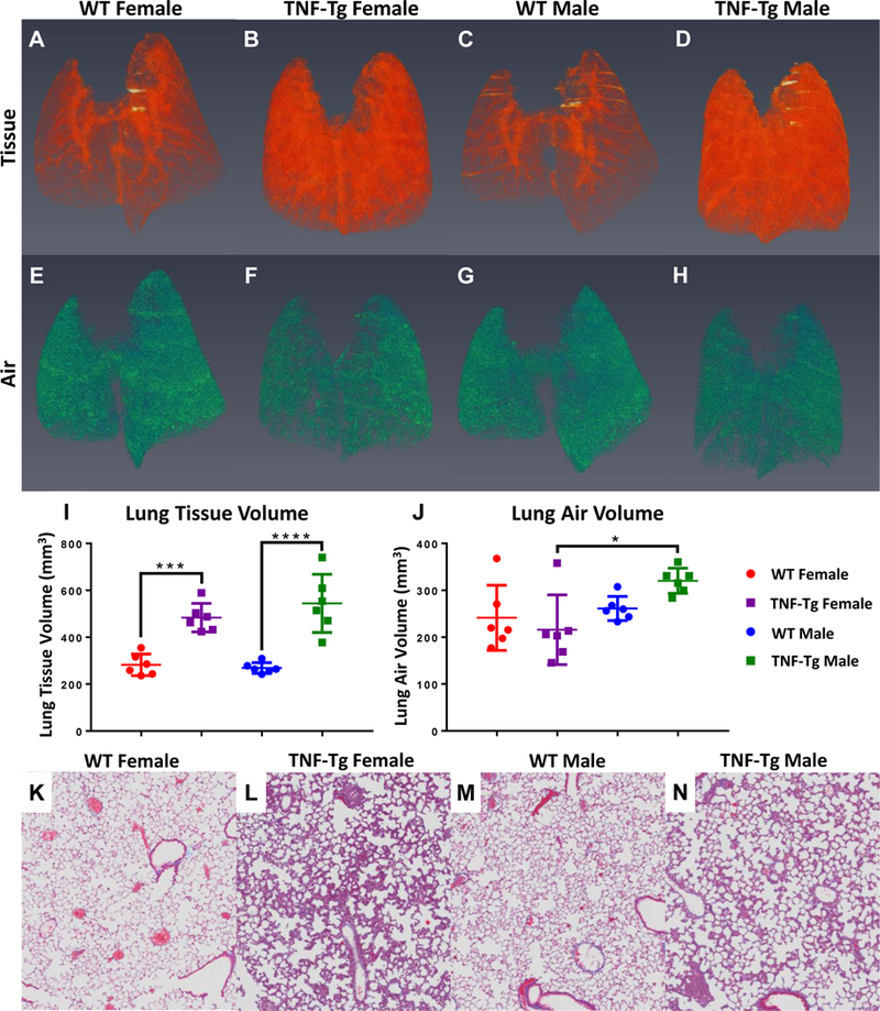
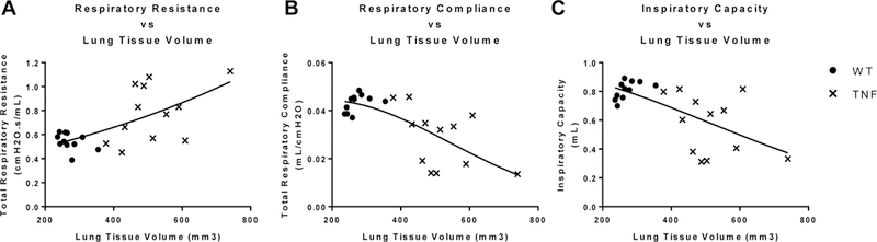
Purpose: Micro-computed tomography (µCT) is increasingly being used on animal models as a minimally-invasive longitudinal outcome measure of pulmonary disease progression. However, while imaging can elucidate macroscopic structural changes over the whole lung, µCT is unable to describe the mechanical changes and functional impairments imposed by progressive disease, which can only be measured via pulmonary function tests (PFTs). The tumor necrosis factor-transgenic (TNF-Tg) mouse model of rheumatoid arthritis (RA) develops pulmonary pathology that mimics many aspects of the inflammatory interstitial lung disease (ILD) seen in a subset of patients with RA. Prior studies using µCT imaging of these mice found increased pulmonary density, characteristic of restrictive disease; however, there have been conflicting reports in the literature regarding the obstructive versus restrictive phenotype of this model. Our study looks to 1) define the functional impairments and 2) characterize the restrictive/obstructive nature of the disease found in this model. Materials and Methods: In this study, we performed PFTs at end-stage ILD, and paired these findings with µCT results, correlating radiology to functional parameters. TNF-Tg and WT littermates of both sexes underwent µCT imaging and PFT testing at 5.5 months-old. Spearman's correlation analyses were performed comparing lung tissue volume (LTV) to PFT parameters of gas exchange and tissue stiffness. Results: Compared to WT, TNF-Tg mice had impaired gas exchange capacity, increased respiratory resistance, and reduced lung compliance, elastance, and inspiratory capacity, indicating increased tissue stiffness and compromised pulmonary function. LTV was also consistently higher in TNF-Tg lungs. Conclusions: These findings demonstrate that: 1) TNF-Tg mice display a restrictive pathology, and 2) in vivo µCT is a valid outcome measure to infer changes in pulmonary mechanical and functional parameters.

Keywords: TNF-transgenic; micro-CT; pulmonary function testing; rheumatoid arthritis-associated interstitial lung disease.


Figures

Figure 1
Figure 1. Gas exchange deficiency in TNF-Tg mice versus WT littermates.
5.5-month-old TNF-Tg male and female mice and their WT littermates underwent carbon monoxide (CO) diffusion testing as described in Materials and Methods. The relative abundance of CO post-expiration is presented as the diffusion factor of CO (DFCO) for individual mice with the mean +/− SD (n=6; ***p<0.001, ****p<0.0001). Analysis of differences between sexes within genotype were analyzed and found to not be significant (female vs male, WT, p=0.4205; TNF-Tg, p=0.1362).
Figure 2
Figure 2. TNF-Tg lungs have a restrictive pulmonary pathology.
The mice described in Figure 1 underwent forced pulmonary manipulation as described in Materials and Methods. 2-way ANOVAs with Sidak’s post hoc multiple comparisons were used to analyze differences in mechanical parameters between cohorts. Data for mechanical parameters: (A) total respiratory resistance, (B) Newtonian resistance, (C) tissue dampening, (D) total respiratory compliance, (E) static compliance, (F) the shape constant, (G) total respiratory elastance, (H) and tissue elastance, are presented for individual mice with mean +/− SD (n=6, *p>0.05,**p<0.01, ***p<0.001, ****p<0.0001). Note that within sex, differences between TNF-Tg and WT mice were found for multiple parameters measuring tissue “stiffness”, including total respiratory resistance, tissue dampening, total respiratory compliance, static compliance, total respiratory elastance, and tissue elastance. Between the sexes, female TNF-Tg mice consistently trended towards increased disease severity compared to their male counterparts over multiple measures, yet only respiratory compliance reached statistical significance.
Figure 3
Figure 3. Radiographic and histologic confirmation of inflammatory ILD in TNF-Tg mice versus WT littermates.
The mice described in Figure 1 underwent in vivo micro-CT and ex vivo histopathology as described in Materials and Methods. Representative 3D renderings of lung tissue volume (A-D) and lung air volume (E-H) are presented with statistical analyses. Note that TNF-Tg mice have increased lung tissue volumes (I), while maintaining a comparable basal post-expiratory air volume (J) (n=6, mean +/− SD, *p>0.05, ***p<0.001, ****p<0.0001). Histologic sections stained with Masson’s trichrome are shown to illustrate the absence of collagen deposition (blue tissue) in the interstitium of all mice (K-N). Histologic analysis demonstrates marked inflammatory ILD in TNF-Tg mice versus their WT littermates (K-N). Note that the TNF-Tg male mice have lower cellular density by percent of total area than females, as determined through histology.
Figure 4
Figure 4. Correlation of pulmonary tissue physiology outcome measures with lung tissue volume.
Spearman’s correlations were performed comparing the PFT outcome data in Figure 2 with the micro-CT lung volume data in Figure 3. The statistical findings are presented in Table 1. Graphs with best fit non-linear regression curves are presented to illustrate the relative correlation between lung volume and: Total Respiratory Resistance (A), Total Respiratory Compliance (B), and Inspiratory Capacity (C). ● = WT, x = TNF-Tg, n=12 per genotype.

References

    1. Assayag D, Lubin M, Lee JS, King TE, Collard HR, Ryerson CJ. Predictors of mortality in rheumatoid arthritis-related interstitial lung disease. Respirology 2014;19(4):493–500. - PubMed
    1. Gabriel SE, Crowson CS, Kremers HM, Doran MF, Turesson C, O’Fallon WM, Matteson EL. Survival in rheumatoid arthritis: A population-based analysis of trends over 40 years. Arthritis Rheum 2003;48(1):54–58. - PubMed
    1. Bongartz T, Nannini C, Medina-Velasquez YF, Achenbach SJ, Crowson CS, Ryu JH, Vassallo R, Gabriel SE, Matteson EL. Incidence and mortality of interstitial lung disease in rheumatoid arthritis: A population-based study. Arthritis Rheum 2010;62(6):1583–1591. - PMC - PubMed
    1. Olson AL, Swigris JJ, Sprunger DB, Fischer A, Fernandez-Perez ER, Solomon J, Murphy J, Cohen M, Raghu G, Brown KK. Rheumatoid arthritis-interstitial lung disease-associated mortality. Am J Respir Crit Care Med 2011;183(3):372–378. - PMC - PubMed
    1. Sparks JA, Chang SC, Liao KP, Lu B, Fine AR, Solomon DH, Costenbader KH, Karlson EW. Rheumatoid arthritis and mortality among women during 36 years of prospective follow-up: Results from the nurses’ health study. Arthritis Care Res (Hoboken) 2016;68(6):753–762. - PMC - PubMed

Publication types

Substances