. 2019 Jul 18;14(7):e0219384.
doi: 10.1371/journal.pone.0219384. eCollection 2019.

Defining the binding interface of Amyloid Precursor Protein (APP) and Contactin3 (CNTN3) by site-directed mutagenesis

Affiliations

Defining the binding interface of Amyloid Precursor Protein (APP) and Contactin3 (CNTN3) by site-directed mutagenesis

Xi Peng et al. PLoS One. .

Abstract

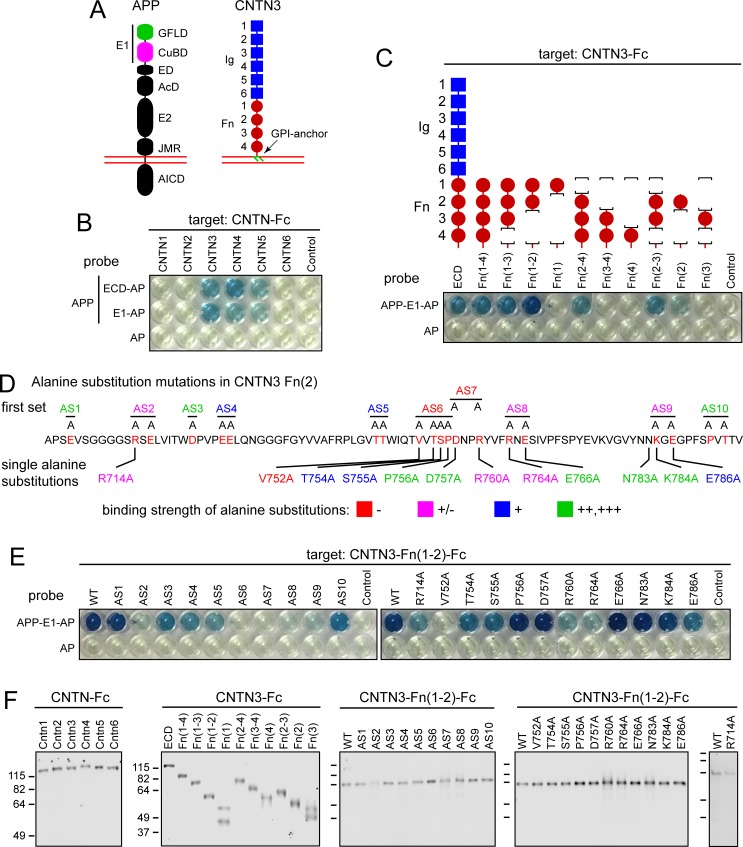
The Amyloid Precursor Protein (APP) and Contactin (CNTN) families of cell-surface proteins have been intensively studied in the context of neural development and neuropsychiatric diseases. Earlier studies demonstrated both genetic and biochemical interactions between the extracellular domains of APP and CNTN3, but their precise binding interfaces were not defined. In the present study, we have used binding assays between APP-alkaline phosphatase (AP) fusion proteins and CNTN-Fc fusion proteins, together with alanine substitution mutagenesis, to show that: (i) the second Fibronectin domain (Fn(2)) in CNTN3 mediates APP binding; (ii) the copper binding domain (CuBD) in APP mediates CNTN3 binding; and (iii) the most important amino acids for APP-CNTN3 binding reside on one face of CNTN3-Fn(2) and on one face of APP-CuBD. These experiments define the regions of direct contact that mediate the binding interaction between APP and CNTN3.


Conflict of interest statement

The authors have declared that no competing interests exist.

Figures

Fig 1
Fig 1. CNTN3 sequences required for binding to APP.
(A) Schematic of APP and CNTN3 full-length proteins, showing their domain structures. Red lines indicate the lipid bilayer. Abbreviations for APP: GFLD, growth factor-like domain; CuBD, copper binding domain; ED, extension domain; AcD, acidic domain; E2, central domain; JMR, juxtamembrane region; AICD, APP intracellular domain [2]. Abbreviations for CNTN3: Ig, immunoglobulin domain; Fn, fibronectin domain. (B) Image of a Protein-G coated 96-well tray showing the AP reaction product (blue). The indicated Fc-fusion proteins (“target”) were immobilized by Protein-G binding and then incubated with the indicated APP-AP fusion proteins (the full ECD or the E1 domain) or with AP alone (“probe”). For each CNTN protein the full ECD (including all Ig and Fn domains) was fused to Fc. The concentrations of the different Fc-fusions were adjusted to provide equivalent amounts, as determined by immunoblotting (S1 Fig). The control wells in these and other AP assays were coated with an irrelevant Fc fusion protein. (C) Domain deletions in the ECD of CNTN3-Fc show that Fn(1–2) contains the principal sites of APP-E1-AP binding. The parental fusion protein (WT) has the full CNTN3 ECD fused to Fc. The first Ig domain is adjacent to the signal peptide. (D) Sequence of CNTN3 Fn(2) showing the locations of alanine substitution mutations targeting groups of mostly polar surface amino acids (“first set”; upper sequence) or single amino acids (lower sequence). AS, alanine substitution. (E) 96-well binding assay showing APP-E1-AP binding to the CNTN3 Fn(2) alanine substitution mutations shown in (D) constructed in the context of CNTN3-Fn(1–2)-Fc. (F) Immunoblots of serum-free conditioned medium containing the indicated WT and mutant CNTN-Fc proteins, visualized with anti-human IgG. Mass in kDa is listed for the protein size standards. These are the same for the right-most four blots. Quantification of AP binding is shown in S1A and S1B Fig and is summarized in S1 Table.
Fig 2
Fig 2. APP sequences required for binding to CNTN3.
(A) Left, 96-well binding assay using the AP fusions with the full APP ECD or its amino-terminal domains as probe and CNTN3-Fn(1–3)-Fc as target. Right, schematic of APP ECD domain structure, showing APP-AP fusions. (B) Sequence of APP CuBD showing the locations of alanine substitution mutations targeting groups of mostly polar surface amino acids (“first set”; upper sequence) or single amino acids (lower sequence). (C) 96-well binding assay showing alanine substitution mutations in the CuBD of APP-CuBD-AP binding to CNTN3-Fn(1–2)-Fc. (D) Immunoblots of serum-free conditioned medium containing the indicated WT and mutant APP-AP proteins, visualized with anti-myc mAb. All AP fusion proteins contain a myc-tag between the APP segment and AP. Mass in kDa is listed for the protein size standards, which are the same for the three left-most blots. Quantification of AP binding is shown in S1C–S1E Fig, and is summarized in S1 Table. The concentrations of the different probes were adjusted to provide equivalent amounts of AP enzyme activity, as determined by colorimetric assay.
Fig 3
Fig 3. Amino acids that are critical for APP-CNTN3 binding displayed on the 3-dimensional structures of CNTN3-Fn(2) and APP-CuBD.
(A) Locations of alanine substitutions in CNTN3-Fn(2) PDB ID 5I99 [25], color coded by their effect on APP-E1-AP binding. (B) Locations of alanine substitutions in APP CuBD PDB ID 2FKL [26], color coded by their effect on CNTN-Fn(1–2)-Fc binding. The cylinders and arrows show the 90 degree rotations that relate the images in adjacent panels. Only data from CNTN-Fn(1–2)-Fc or APP-CuBD-AP fusion proteins that were secreted well are shown. In the leftmost panels, side chains are labeled for those amino acid that are most critical for binding, i.e., those for which alanine substitution severely impairs binding (categories: +/- (magenta) and–(red)).

References

    1. Müller UC, Deller T, Korte M. Not just amyloid: physiological functions of the amyloid precursor protein family. Nat Rev Neurosci. 2017. May;18(5):281–298. 10.1038/nrn.2017.29 - DOI - PubMed
    1. Coburger I, Hoefgen S, Than ME. The structural biology of the amyloid precursor protein APP—a complex puzzle reveals its multi-domain architecture. Biol Chem. 2014. May;395(5):485–98. 10.1515/hsz-2013-0280 - DOI - PubMed
    1. Selkoe DJ, Hardy J. The amyloid hypothesis of Alzheimer's disease at 25 years. EMBO Mol Med. 2016. June 1;8(6):595–608. 10.15252/emmm.201606210 - DOI - PMC - PubMed
    1. Wang P, Yang G, Mosier DR, Chang P, Zaidi T, Gong YD, et al. Defective neuromuscular synapses in mice lacking amyloid precursor protein (APP) and APP-Like protein 2. J Neurosci. 2005. February 2;25(5):1219–25. 10.1523/JNEUROSCI.4660-04.2005 - DOI - PMC - PubMed
    1. Wang Z, Wang B, Yang L, Guo Q, Aithmitti N, Songyang Z, et al. Presynaptic and postsynaptic interaction of the amyloid precursor protein promotes peripheral and central synaptogenesis. J Neurosci. 2009. September 2;29(35):10788–801. 10.1523/JNEUROSCI.2132-09.2009 - DOI - PMC - PubMed

Publication types