. 2020 Mar;145(3):968-981.e14.
doi: 10.1016/j.jaci.2019.07.004. Epub 2019 Jul 15.

B lymphocytes directly contribute to tissue fibrosis in patients with IgG4-related disease

Affiliations

B lymphocytes directly contribute to tissue fibrosis in patients with IgG4-related disease

Emanuel Della-Torre et al. J Allergy Clin Immunol. 2020 Mar.

Abstract

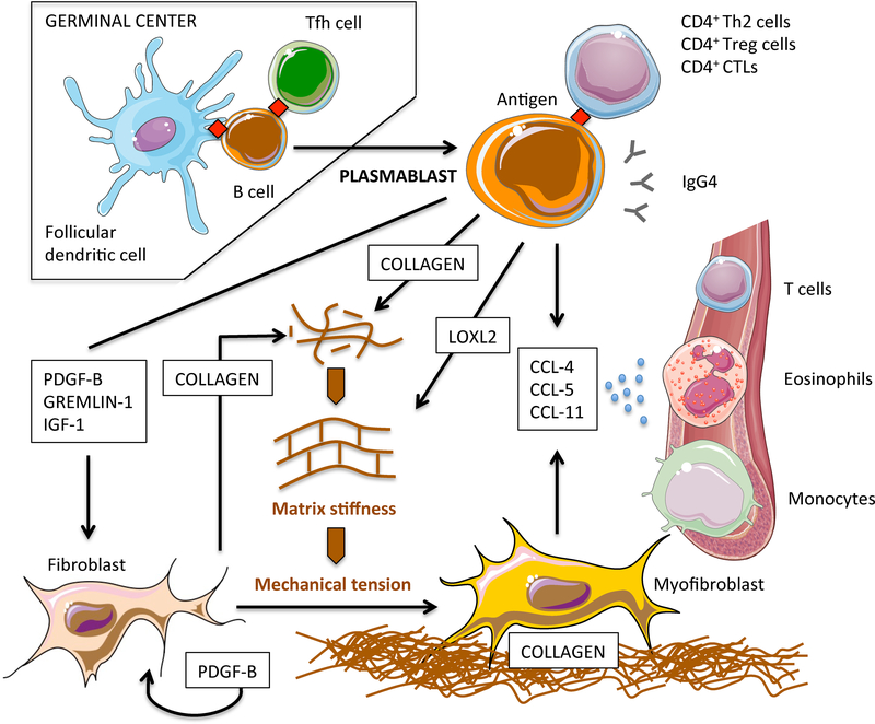
Background: IgG4-related disease (IgG4-RD) is a fibroinflammatory condition marked by rapid clinical improvement after selective depletion of B lymphocytes with rituximab. This feature suggests that B cells might participate in fibrogenesis and wound healing.

Objective: In the present work we aimed to demonstrate that B lymphocytes contribute directly to tissue fibrosis in patients with IgG4-RD.

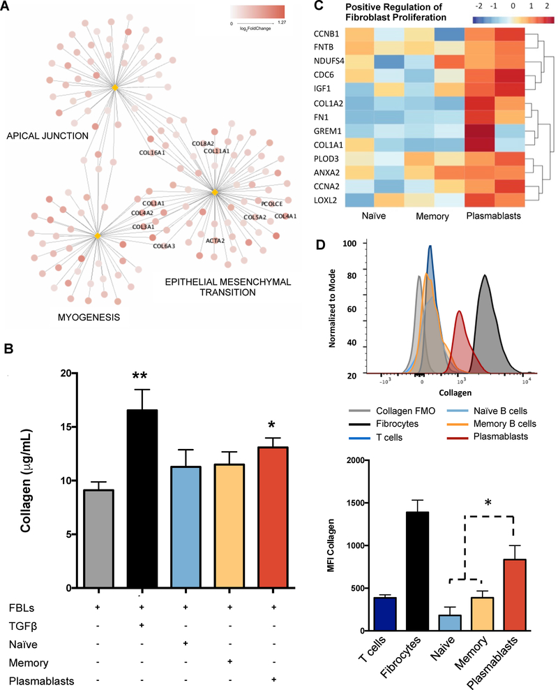
Methods: Total circulating CD19+ B lymphocytes, naive B cells, memory B cells, or plasmablasts from patients with IgG4-RD were cultivated with human fibroblasts. Profibrotic soluble factors and collagen production in cocultures were assessed by using ELISAs and Luminex assays. RNA sequencing and quantitative RT-PCR were used to assess fibroblast activation in the presence of B cells, as well as induction of profibrotic pathways in B-cell subsets. Relevant profibrotic and inflammatory molecules were confirmed in vitro by using functional experiments and on IgG4-RD tissue sections by using multicolor immunofluorescence studies.

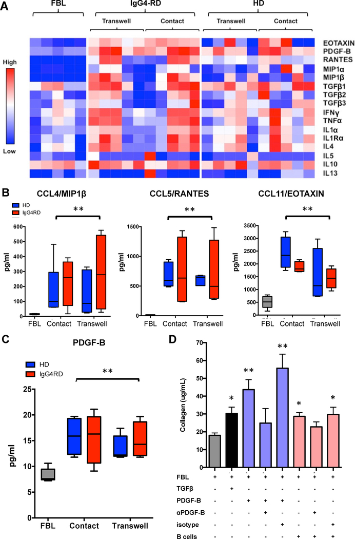
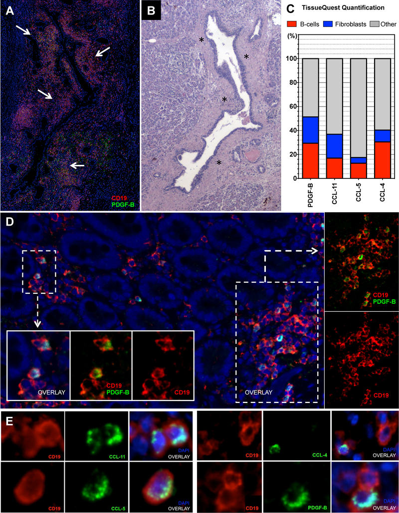
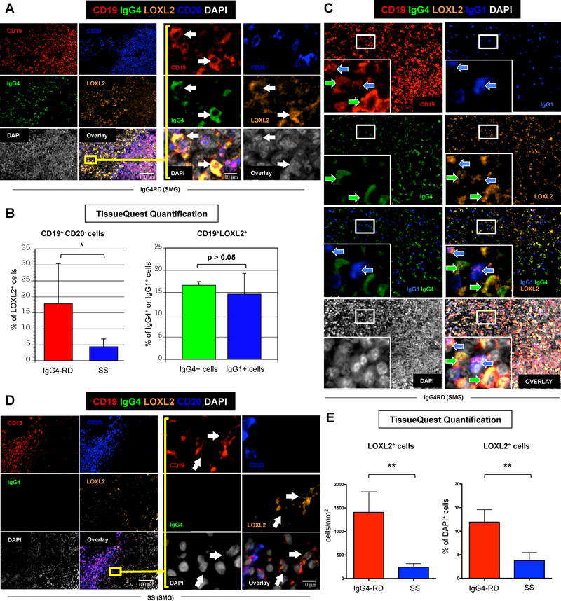
Results: B cells from patients with IgG4-RD (1) produced the profibrotic molecule platelet-derived growth factor B and stimulated collagen production by fibroblasts; (2) expressed enzymes implicated in extracellular matrix remodeling, such as lysyl oxidase homolog 2; (3) produced the chemotactic factors CCL4, CCL5, and CCL11; and (4) induced production of these same chemokines by activated fibroblasts. Plasmablasts expressed sets of genes implicated in fibroblast activation and proliferation and therefore represent cells with intrinsic profibrotic properties.

Conclusion: We have demonstrated that B cells contribute directly to tissue fibrosis in patients with IgG4-RD. These unanticipated profibrotic properties of B lymphocytes, particularly plasmablasts, might be relevant for fibrogenesis in patients with other fibroinflammatory disorders and for wound-healing processes in physiologic conditions.

Keywords: B cells; IgG(4); IgG(4)-related disease; fibroblasts; fibrosis; lysyl oxidase homolog 2; plasmablasts; platelet-derived growth factor; rituximab.


Conflict of interest statement

CONFLICT of INTEREST - Nothing to report.

Figures

Figure 1.
Figure 1.. B lymphocytes drive pro-fibrotic genes expression and collagen production by human pancreatic fibroblasts by means of soluble factors.
(A) Top results of GSEA performed on log2 fold changes between gene expression of fibroblasts co-cultured with B cells from patients with IgG4-RD AIP in the absence of semipermeable membranes and of control fibroblasts at 72hrs (see Online Supplementary Material for details). The network graph shows top scoring enriched gene-sets (FDR q-value <0.01) and their associated leading edge genes (those responsible of the core enrichment of the gene-set). Nodes representing genes are coloured according to their log2 fold change and genes considered of particular interest are labelled. (B) Quantitative PCR showing fold change expression of ACTA2, COL1A1, COL1A2, and COL3A1 genes in pancreatic fibroblasts after 3 days co-culture with B lymphocytes from patients with IgG4-RD AIP (n=5) and from healthy donors (n=5). (C) Collagen concentration in the supernatant of different experimental conditions. Human pancreatic fibroblasts (FBL); FBL treated with TGFβ1 20ng/ml (TGFβ); FBL co-cultured with B cells from healthy controls (blue bars); FBL co-cultured with B cells from patients with IgG4-RD (red bars) in the presence (Transwell) or absence (Contact) of semipermeable membranes. * = p < 0.05 for the comparison with fibroblasts alone; ° = p < 0.05 for the comparison with co-cultures in the presence of B cells from healthy controls. Results are expressed as mean ± SD of 5 experiments.
Figure 2.
Figure 2.. PDGF-B is increased in the co-cultures of fibroblasts with B lymphocytes together with CCL-4, CCL-5, and CCL-11, and mediates collagen production.
(A) Quantification of different cytokines and chemokines by Luminex analysis after 3 days of culture with specific focus on those significantly increased in the supernatant of the co-cultures compared to control fibroblasts: (B) CCL-4, CCL-5, CCL-11, and (C) PDGF-B. Abbreviations: primary human pancreatic fibroblast (FBL); FBL stimulated with 20 ng/ml of recombinant human TGF-β1 (TGFβ); FBL co-cultured with B cells from healthy controls (n=5) (blue bars); FBL co-cultured with B cells from patients with IgG4-RD (n=5) (red bars), in the presence (Transwell) or absence (Contact) of semipermeable membranes. Results are expressed as mean ± SD. (D) PDGF-B inhibition decreases B-cell induced collagen production by primary human skin fibroblasts. Abbreviations: primary human skin fibroblasts (FBL); FBL stimulated with 20 ng/ml of recombinant human TGF-β1 (TGFβ); FBL stimulated with 10 ng/ml of recombinant human PDGF-B (PDGF-B); FBL incubated with 0.5 μg/ml of anti-PDGF-B antibody two hours prior to stimulation with 10 ng/ml of recombinant human PDGF-B (αPDGF-B); FBL incubated with 0.5 μg/ml of IgG isotype antibody two hours prior to stimulation with 10 ng/ml of recombinant human PDGF-B (Isotype); co-cultures with B cells from patients with IgG4-RD (B cells); * = p < 0.05. Results are expressed as mean ± SD of 5 experiments.
Figure 3.
Figure 3.. B-lymphocytes and fibroblasts infiltrating IgG4-RD autoimmune pancreatitis express PDGF-B, CCL-4, CCL-5, and CCL-11.
(A) Immunofluorescence staining of an IgG4-RD AIP tissue, showing PDGF-B expressing B cells (arrows) surrounding pancreatic ducts (CD19 (red), PDGF-B (green), and DAPI (blue)). (B) Sequential matched Hematoxylin/Eosin stain showing a dense fibrosis (asterisks) likely generated as a consequence of the peri-ductal inflammatory infiltrate described in A. (C) Quantification of double-positive cells for CD19+ (red bars) or αSMA+ (blue bars) and CCL-4, CCL-5, CCL-11, or PDGF-B in IgG4-RD AIP expressed in percentage of total CCL-4, CCL-5, CCL-11, or PDGF-B positive cells (i.e. CD19+PDGF-B+ cells/PDGF-B+ cells). Bar length represents the average quantification of 5 tissue slides. (D) PDGF-B expressing B lymphocytes infiltrating the sub-epithelial layer of the duodenum in a case of IgG4-RD AIP of the pancreatic head (low magnification); inserts (high magnification). (E) Immunofluorescence staining of CD19 (red), CCL-4, CCL-5, CCL-11, and PDGF-B (all green), and DAPI (blue) positive cells in representative tissue samples from a patient with IgG4-RD AIP (high magnification).
Figure 4.
Figure 4.. Plasmablasts from IgG4-RD patients induce collagen secretion by human fibroblasts, produce collagenous proteins, and express extracellular-matrix remodelling enzymes.
(A) Top results of GSEA performed on log2 fold changes of gene expression in fibroblasts co-cultured with B cells from patients with IgG4-RD AIP and that in fibroblasts co-cultured with B cells from healthy controls at 72hrs in the presence of semipermeable membranes (see Online Supplementary Material for details). The network has been realized with the same logic described for Figure 1.A (B) Collagen production is increased in co-cultures with plasmablasts compared to control fibroblasts. Primary human skin fibroblasts (FBL); FBL stimulated with 20 ng/ml of recombinant human TGF-β1 (TGFβ); FBL co-cultured with naïve B cells, memory B cells, or circulating plasmablasts. Results are presented as mean ± SD of 5 independent experiments. * = p < 0.05. (C) Heatmap showing up-regulation in circulating plasmablasts of genes significantly mapping to the Gene Ontology “GO:0048146: positive regulation of fibroblasts proliferation”. (D) Flow cytometry showing increased collagen expression in circulating plasmablasts compared to naïve B cells, and memory B cells. Circulating fibrocytes and CD3+ T cells are used as positive and negative controls, respectively. Overlaid histograms represent collagen expression in the different cell types in a representative patient. Histograms represent Mean Fluorescence Intensity (MFI) of intracellular collagen in different cell types. * = p < 0.05. Results are expressed as mean ± SD of 5 experiments.
Figure 5.
Figure 5.. LOXL2 is highly expressed by plasmablasts/plasma cells in IgG4-related sialoadenitis.
(A) Immunofluorescence staining of CD19 (red), IgG4 (green), LOXL-2 (orange), CD20 (blue), and DAPI (white) in salivary glands from a representative patient with IgG4-RD showing abundant infiltrate of LOXL2 expressing IgG4+ plasmablasts (arrows) (inserts: high magnification). (B) Quantification of LOXL2+ plasmablasts (CD19+ CD20 B cells) in salivary glands from patients with IgG4-RD (red bar) and Sjögren’s syndrome (SS) (blue bar) using TissueQuest software. * = p < 0.05 (mean ± SD of n=5 experiments). Quantification of LOXL2+ cells within the total pool IgG4+ (red bar) or IgG1+ (blue bar) plasmablasts in salivary glands from patients with IgG4-RD using TissueQuest software (mean ± SD of n=5 experiments). (C) Immunofluorescence staining of CD19 (red), IgG4 (green), LOXL-2 (orange), IgG1 (blue), DAPI (white) in the salivary gland of a representative patient with IgG4-RD showing LOXL2 expression by both IgG4 (green arrows) and IgG1 (blue arrows) producing B cells (inserts: high magnification). (D) Immunofluorescence staining of CD19 (red), IgG4 (green), LOXL-2 (orange), CD20 (blue), and DAPI (white) in salivary glands from a representative patient with SS showing few LOXL2 expressing IgG4+ B lymphocytes (arrows) (inserts: high magnification). (E) Quantification of LOXL2+ cells in salivary glands from patients with IgG4-RD (red bar) and SS (blue bar) showing a significantly lower number of LOXL2 expressing cells in SS compared to IgG4-RD. ** = p < 0.01 (mean ± SD of n=5 experiments).
Figure 6.
Figure 6.. Revised pathogenetic model of IgG4-RD pathogenesis: B-cell contribution to tissue fibrosis.
After germinal centre reaction, antigen experienced naïve B cells differentiate into class-switched plasmablasts and migrate into inflamed sites where they contribute to tissue fibrosis in different ways. Plasmablasts initiate fibroblast differentiation into myofibroblasts through soluble factors that activate transcriptional programs implicated in mesenchymal transition. Plasmablasts also stimulate fibroblasts to secrete collagen and PDGF-B, thus amplifying fibroblast activation in an autocrine/paracrine loop. Plasmablasts directly contribute to tissue fibrosis by secreting collagenous proteins. Plasmablasts regulate extracellular matrix stiffness by secreting enzymes responsible for the crosslinking of collagen molecules such as LOXL2. Increased extracellular matrix stiffness leads to the activation of mechanoceptors on fibroblasts, thus fostering their full differentiation into myofibroblasts. Plasmablasts attract inflammatory cells with additional fibrotic potential such as CD4+ CTLs, eosinophils, and M2 macrophages, by secreting CCL-4, CCL-5, and CCL-11, and by stimulating fibroblasts to produce these same chemokines. The pathogenic relevance of IgG4 antibodies and of T-cell populations presumably sustained by cognate antigen specific plasmablasts remains to be defined.

Comment in

References

    1. Wynn TA, Ramalingam TR. Mechanisms of fibrosis: therapeutic translation for fibrotic disease.Nat Med. 2012;18:1028–40 - PMC - PubMed
    1. Rockey DC, Bell PD, Hill JA. Fibrosis-a Common Pathway to Organ Injury and Failure. NEJM. 2015;372:1138–49. - PubMed
    1. Ho YY, Lagares D, Tager AM, Kapoor M. Fibrosis-a lethal component of systemic sclerosis. Nat Rev Rheumatol. 2014;10:390–402. - PubMed
    1. Della-Torre E, Lanzillotta M, Doglioni C. Immunology of IgG4-related disease. Clin Exp Immunol 2015;181:191–206. - PMC - PubMed
    1. Carruthers MN, Topazian MD, Khosroshahi A, Witzig TE, Wallace ZS, Hart PA, et al. Rituximab for IgG4-related disease: a prospective, open-label trial. Ann Rheum Dis. 2015;74:1171–7. - PubMed

Publication types