Review
. 2019 Jul 17;11(7):418.
doi: 10.3390/toxins11070418.

Rapid Detection of Botulinum Neurotoxins-A Review

Affiliations
Review

Rapid Detection of Botulinum Neurotoxins-A Review

Robert J Hobbs et al. Toxins (Basel). .

Abstract

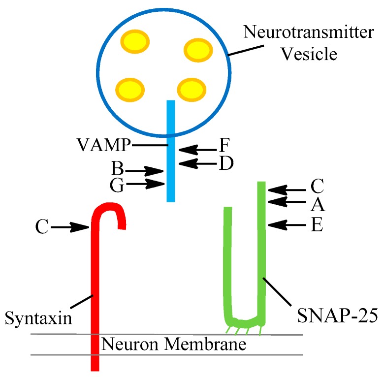
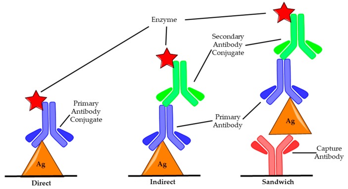
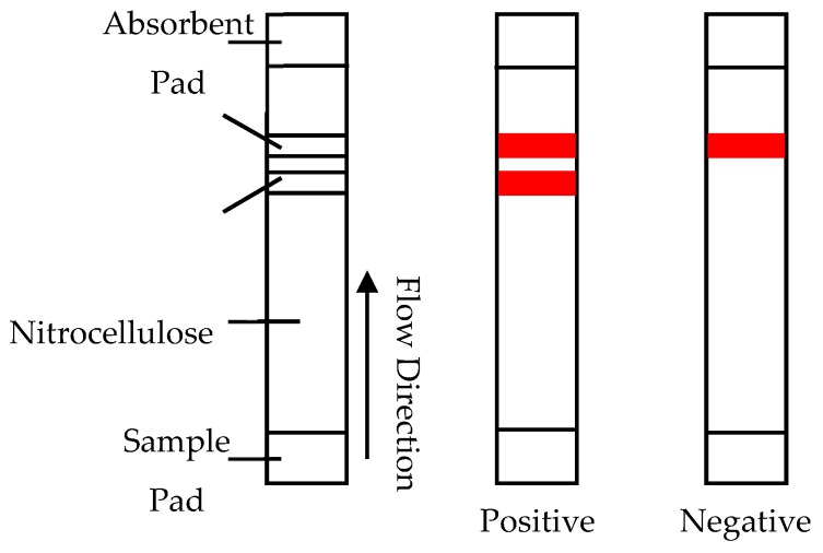
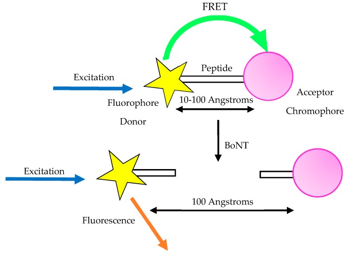
A toxin is a poisonous substance produced within living cells or organisms. One of the most potent groups of toxins currently known are the Botulinum Neurotoxins (BoNTs). These are so deadly that as little as 62 ng could kill an average human; to put this into context that is approximately 200,000 × less than the weight of a grain of sand. The extreme toxicity of BoNTs leads to the need for methods of determining their concentration at very low levels of sensitivity. Currently the mouse bioassay is the most widely used detection method monitoring the activity of the toxin; however, this assay is not only lengthy, it also has both cost and ethical issues due to the use of live animals. This review focuses on detection methods both existing and emerging that remove the need for the use of animals and will look at three areas; speed of detection, sensitivity of detection and finally cost. The assays will have wide reaching interest, ranging from the pharmaceutical/clinical industry for production quality management or as a point of care sensor in suspected cases of botulism, the food industry as a quality control measure, to the military, detecting BoNT that has been potentially used as a bio warfare agent.

Keywords: Botulinum Neurotoxin; Botulism; PoC; Rapid Detection; Sensitivity.


Conflict of interest statement

The authors declare no conflict of interest.

Figures

Figure 1
Figure 1
Diagram showing the proteins that make up the Soluble N-ethylmaleimide-sensitive factor activating protein receptor (SNARE) complex; Syntaxin (Red), SNAP-25 (Green) and Synaptobrevin, also known as vesicle-associated membrane protein (VAMP) (Blue). Additionally, botulinum neurotoxin serotype is listed next to the corresponding protein it is responsible for fragmenting, along with the location on the protein at which it cleaves.
Figure 2
Figure 2
Image of common ELISA set ups: Direct, Indirect and Sandwich. In both direct and indirect ELISA, antigens (Ag) are bound to the microtiter plate first, an antibody specific to the antigen is then introduced. In direct assays this primary antibody (blue) has been modified with an enzyme (red star) such as HRP, which when exposed to a substrate produces a measurable colour change. In indirect ELISA, this enzyme is bound to a secondary antibody (green) that has been modified with an enzyme to facilitate colour change; this secondary antibody binds to the primary antibody. In sandwich ELISA, the surface is treated with a capture antibody (red) specific to a desired antigen before the rest of the assay proceeds in the same manner as the indirect assay.
Figure 3
Figure 3
Diagram of lateral flow assay and examples of positive and negative results [67].
Figure 4
Figure 4
Schematic explaining the action of the FRET assay. Typically, an acceptor chromophore (pink) is linked to a fluorophore donor (yellow) via a peptide; this allows the transfer of energy resulting in FRET being detected. Upon exposure to botulinum neurotoxin, the peptide linker is cleaved and fragmented allowing the chromophore and fluorophore to dissociate. This inhibits the transfer of energy in the system.
Figure 5
Figure 5
Schematic showing how botulinum neurotoxin is detected by measuring the decrease in anodic peak charge observed when BoNT cleaves a section of the self-assembled SNAP-25 monolayer.
Figure 6
Figure 6
Schematic showing how whole and cleaved SNAP-25 differs in blocking the reduction of the redox probe, with the whole SNAP-25 providing a greater blocking ability of the redox probe due to its larger size.
Figure 7
Figure 7
An overview of the endopeptidase mass spectrometry assay, serotype specific antibodies are conjugated to magnetic beads then added to a sample (A); the beads are removed and thoroughly washed before a substrate that mimics the toxins natural target is added (B). The solution is then incubated, and the resulting mixture is analysed by mass spectrophotometry. Both whole substrate and cleaved fragments that result from incubation with BoNT can be detected (C).
Figure 8
Figure 8
UV-visible spectra showing un-modified (left) and modified (right) gold colloids (GC), detailing that upon addition of sodium chloride, the unmodified colloid decreases in stability and undergoes aggregation. The inset images show the visible colour shift [116].
Figure 9
Figure 9
Schematic of cell-based assay for botulinum neurotoxin.
Figure 10
Figure 10
A visual representation of the BoNT detection methods analysed in this review. The assays were compared using the mouse bioassay sensitivity as the baseline (10 pg/mL), an analysis time of <1 h and cost of <£30 per test. Highlighted are a method’s ability to distinguish active toxin and improved sensitivity over MBA (Green), active toxin quantification but poorer LoD than the MBA (Orange) and no active toxin quantification by still-improved sensitivity over MBA (Blue). The two detection methods that fall outside of satisfying the parameters of speed, cost and sensitivity are placed outside of the Venn diagram. Additionally, the following notation are used: (●) combined with endopeptidase activity assay, (▲) with an additional immunoseparation step, (†) cell reporter line coupled with ELISA detection, (1) have been tested in multiplex systems/with multiple serotypes and (2) complex sample matrices tested.

References

    1. Montecucco C., Molgó J. Botulinal neurotoxins: revival of an old killer. Curr. Opin. Pharmacol. 2005;5:274–279. doi: 10.1016/j.coph.2004.12.006. - DOI - PubMed
    1. Shih T.-M., Hulet S.W., McDonough J.H. The effects of repeated low-dose sarin exposure. Toxicol. Appl. Pharmacol. 2006;215:119–134. doi: 10.1016/j.taap.2006.02.003. - DOI - PubMed
    1. Prigent J., Panigai L., Lamourette P., Sauvaire D., Devilliers K., Plaisance M., Volland H., Créminon C., Simon S. Neutralising Antibodies against Ricin Toxin. PLoS ONE. 2011;6:e20166. doi: 10.1371/journal.pone.0020166. - DOI - PMC - PubMed
    1. Chai P.R., Hayes B.D., Erickson T.B., Boyer E.W. Novichok agents: a historical, current, and toxicological perspective. Toxicol. Commun. 2018;2:45–48. doi: 10.1080/24734306.2018.1475151. - DOI - PMC - PubMed
    1. Binz T., Rummel A. Cell entry strategy of clostridial neurotoxins. J. Neurochem. 2009;109:1584–1595. doi: 10.1111/j.1471-4159.2009.06093.x. - DOI - PubMed

Publication types

LinkOut - more resources