Review
. 2019 Jul 17;8(7):733.
doi: 10.3390/cells8070733.

RhoA as a Key Regulator of Innate and Adaptive Immunity

Affiliations
Review

RhoA as a Key Regulator of Innate and Adaptive Immunity

Matthias Bros et al. Cells. .

Abstract

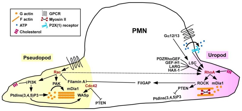
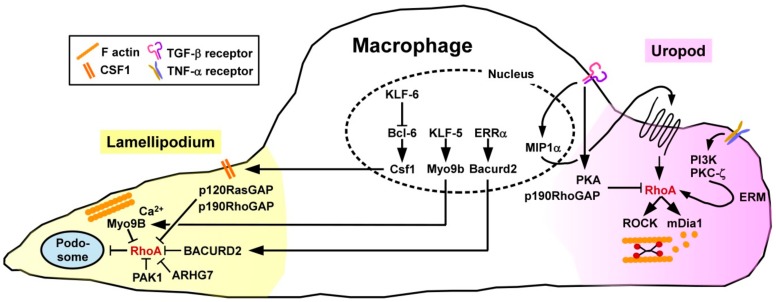
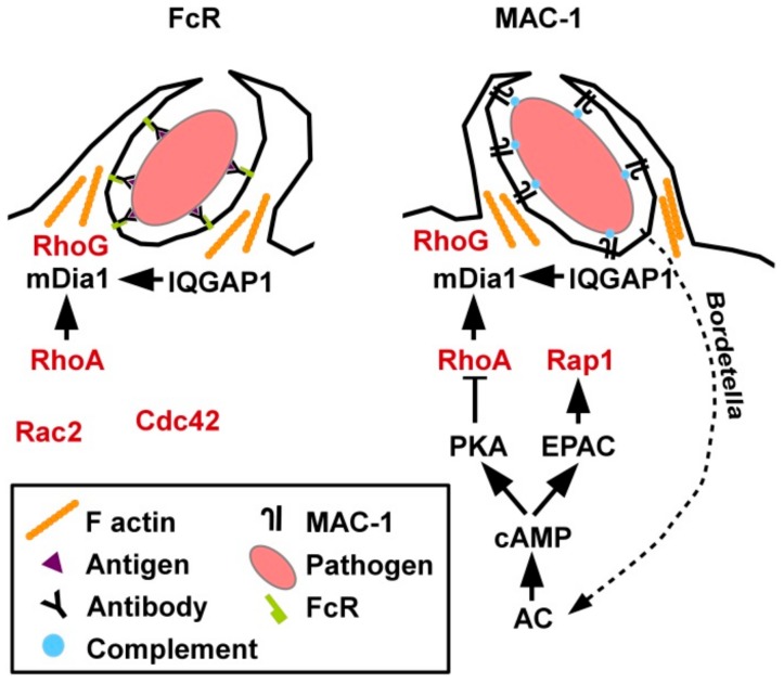
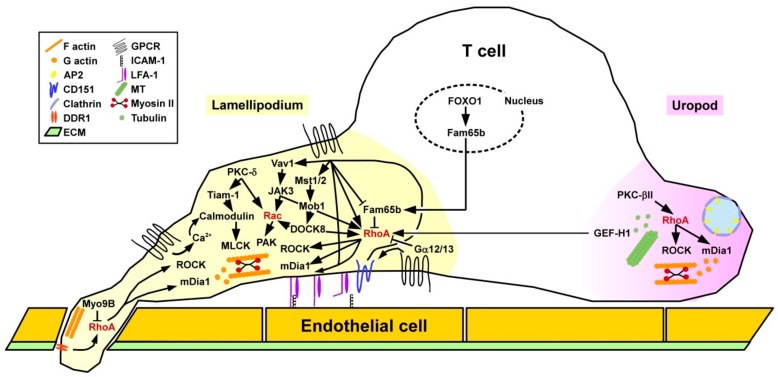
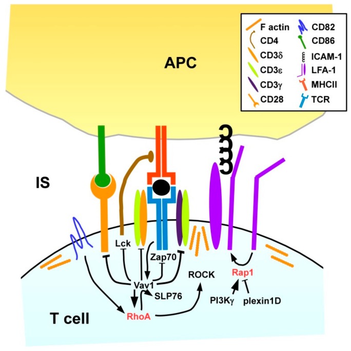
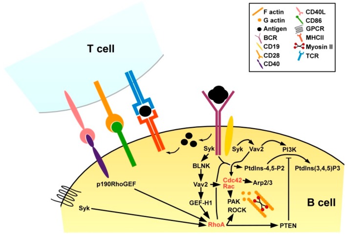
RhoA is a ubiquitously expressed cytoplasmic protein that belongs to the family of small GTPases. RhoA acts as a molecular switch that is activated in response to binding of chemokines, cytokines, and growth factors, and via mDia and the ROCK signaling cascade regulates the activation of cytoskeletal proteins, and other factors. This review aims to summarize our current knowledge on the role of RhoA as a general key regulator of immune cell differentiation and function. The contribution of RhoA for the primary functions of innate immune cell types, namely neutrophils, macrophages, and conventional dendritic cells (DC) to (i) get activated by pathogen-derived and endogenous danger signals, (ii) migrate to sites of infection and inflammation, and (iii) internalize pathogens has been fairly established. In activated DC, which constitute the most potent antigen-presenting cells of the immune system, RhoA is also important for the presentation of pathogen-derived antigen and the formation of an immunological synapse between DC and antigen-specific T cells as a prerequisite to induce adaptive T cell responses. In T cells and B cells as the effector cells of the adaptive immune system Rho signaling is pivotal for activation and migration. More recently, mutations of Rho and Rho-modulating factors have been identified to predispose for autoimmune diseases and as causative for hematopoietic malignancies.

Keywords: RhoA signaling; autoimmune diseases; immune cells; infection; inflammasome; small GTPases.


Conflict of interest statement

The authors declare no conflict of interest.

Figures

Figure 1
Figure 1
Scheme of RhoA signaling. Binding of exogenous ligands via different types of receptors as well as intracellular events trigger activation of RhoA GEF which, in turn, engage membrane-bound RhoA and mediate the exchange of GDP by GTP resulting in RhoA activation. GAP elevate the GTPase activity of RhoA, thereby promoting its inactivation. GDI translocate RhoA from the membrane and keep it in an inactive state. Active RhoA via protein kinases regulates cytoskeletal rearrangements. Active RhoA via ROCK/LIMK negatively regulates cofilin, which is required for F-actin turnover. Additionally, ROCK via inhibition of MLCP confers activation of MLC promoting actomyosin assembly. Active RhoA also promotes mDia activity which, in turn, activates profilin that is also involved in actin remodeling.
Figure 2
Figure 2
Spatially regulated activity of RhoA and other small GTPases is necessary for coordinated PMN migration. Chemoattractants induce Rac/Cdc42 activation at the migration front (pseudopod), and RhoA activty at the cells´ rear end (uropod). Spatial regulation of small GTPase activity is mediated by localized activity of GEF (e.g., LSC, GEF-H1), and GTPase-induced activation/inhibition of GAP (e.g., FilGAP), other adaptor factors, like PTEN, and PI3K signaling.
Figure 3
Figure 3
Migration of MAC requires spatially controlled activation of RhoA. At the migration front various GAP (e.g., Myo9B, p120RasGAP) and other factors (e.g., BACURD2) that inhibit RhoA activity. Endopeptidases concentrated in podosomes mediate dissociation of the ECM which is necessary for MAC migration. Podosome formation is inhibited by RhoA. At the rear end RhoA activity is required for uropod retraction. RhoA is activated, e.g., by TNF-α via PI3K/PKC-.ζ signaling resulting in activation of ERM which, in turn, translocate RhoA to the membrane. TGF-β first promotes RhoA activity via an autocrine MIP-1α stimulation loop, but at later time points inhibits RhoA via PKA signaling and p190RhoGAP.
Figure 4
Figure 4
Phagocytic uptake of pathogens by MAC requires activity of RhoA and other small GTPases. Phagocytosis of opsonized pathogens is conferred by FcR recognizing the constant Fc part of antibodies which bind pathogen-specific surface antigens or by MAC-1 which binds activated complement deposited on the pathogen surface. In both cases partially overlapping sets of small GTPases are involved in phagocytic activity. As an evasion mechanism Bordetella bacteria generate toxins that trigger AC activity which inhibits RhoA.
Figure 5
Figure 5
Dynamic regulation of spatial activity of small GTPases confers DC migration. DC migration requires coordinated activity of Rac/Cdc42 at the front and of RhoA at the rear end as regulated mainly by GEF (e.g., ARHGEF5) and GAP (e.g., Myo9B, SWAP-70). Active RhoA and CYTIP/Cytohesin-1 are necessary to mediate inside-out activation of LFA-1 to enable binding of ICAM and, thereby, cell-cell interaction.
Figure 6
Figure 6
DC interact with T cells via the immunological synapse. Upregulated surface expression of the antigen receptor MHCII is inhibited by active RhoA, whereas RhoB may stimulate MHCII expression. RhoA is also involved in co-stimulatory expression. RhoA activity is regulated by GEF (e.g., p190RhoGEF) and GAP (e.g., SWAP-70, Myo9B), and by tetraspanins that also modulate MHCII/co-stimulator expression, presumably by other mechanisms. The duration of DC/T cell interaction is modulated by LFA-1 activity which is regulated by RhoA and CYTIP/Cytohesin-1.
Figure 7
Figure 7
Migration of T cells is regulated by spatially regulated activity of RhoA and other small GTPases. Chemokine-induced T cell migration is conferred by dynamic regulation of the small GTPases RhoA/Rac at the front and of RhoA at the rear end. The activity of small GTPases is differentially regulated by GEF (e.g., GEF-H1), GAP (e.g., Myo9B), and other factors (e.g., JAK3, Fam65b, PKC). Microtubuli turnover and the presence of clathrin structures contribute to the migratory activity.
Figure 8
Figure 8
Activation of antigen-specific T cells by APC via receptor pairs localized within the IS results in RhoA activation. Recognition of the MHC/antigen complex by the TCR, concomitant co-stimulation via CD86/CD28 and ICAM/LFA-1 dependent adhesion results in activation of the Rho/Rac GEF Vav1. CD82 further promotes Vav1 activation/interaction with SLP76 in a RhoA-dependent manner. Active Vav1 activates small GTPases, and inhibits TCR components and CD28 to prevent excessive T cell stimulation.
Figure 9
Figure 9
BCR activation and concomitant T cell help activate RhoA and other small GTPases in B cells. Binding of a protein antigen by an antigen-specific BCR supported by the co-receptor CD19 triggers Syk which, in turn, activates Vav2 mediating, e.g., via the GEF GEF-H1 activation of RhoA, and of other small GTPases, as well as PI3K activation. RhoA via PTEN negatively regulates PI3K. Antigen-specific CD4+ T cells that bind antigen presenting B cells and receive co-stimulation via CD86/CD28 transiently upregulate CD40L. Binding of CD40 by CD40L activates RhoA via p190RhoGEF. In addition, chemokines may stimulate RhoA.

References

    1. Reiner D.J., Lundquist E.A. Small GTPases. Wormbook Online Rev. C. Elegans Biol. 2018;2018:1–65. doi: 10.1895/wormbook.1.67.2. - DOI - PMC - PubMed
    1. Steffen A., Stradal T.E., Rottner K. Signalling Pathways Controlling Cellular Actin Organization. Handb. Exp. Pharmacol. 2017;235:153–178. doi: 10.1007/164_2016_35. - DOI - PubMed
    1. Narumiya S., Thumkeo D. Rho signaling research: history, current status and future directions. FEBS Lett. 2018;592:1763–1776. doi: 10.1002/1873-3468.13087. - DOI - PMC - PubMed
    1. Durand-Onayli V., Haslauer T., Harzschel A., Hartmann T.N. Rac GTPases in Hematological Malignancies. Int. J. Mol. Sci. 2018;19:4041. doi: 10.3390/ijms19124041. - DOI - PMC - PubMed
    1. Aguilar B.J., Zhou H., Lu Q. Cdc42 Signaling Pathway Inhibition as a Therapeutic Target in Ras- Related Cancers. Curr. Med. Chem. 2017;24:3485–3507. doi: 10.2174/0929867324666170602082956. - DOI - PubMed

Publication types

Substances

LinkOut - more resources