. 2019 Jul 17;9(7):287.
doi: 10.3390/biom9070287.

Caveolin-1 Endows Order in Cholesterol-Rich Detergent Resistant Membranes

Affiliations

Caveolin-1 Endows Order in Cholesterol-Rich Detergent Resistant Membranes

Carla Raggi et al. Biomolecules. .

Abstract

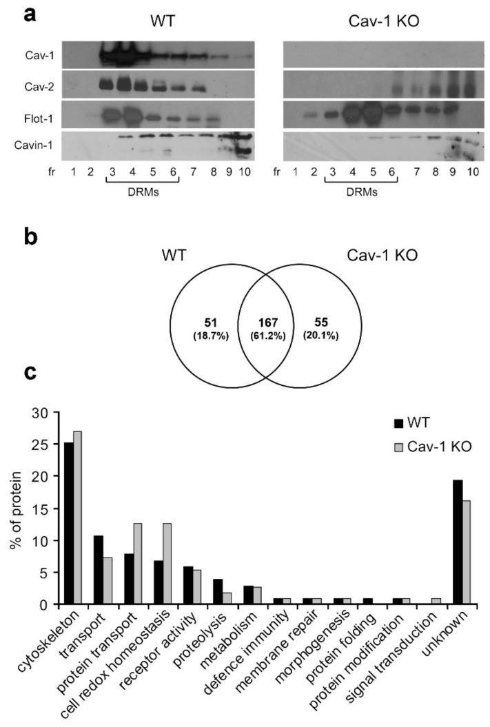
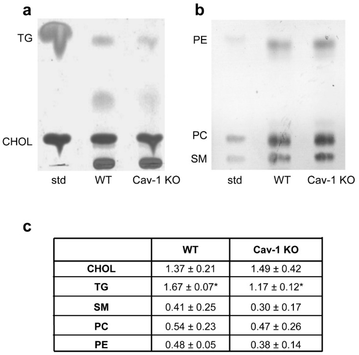
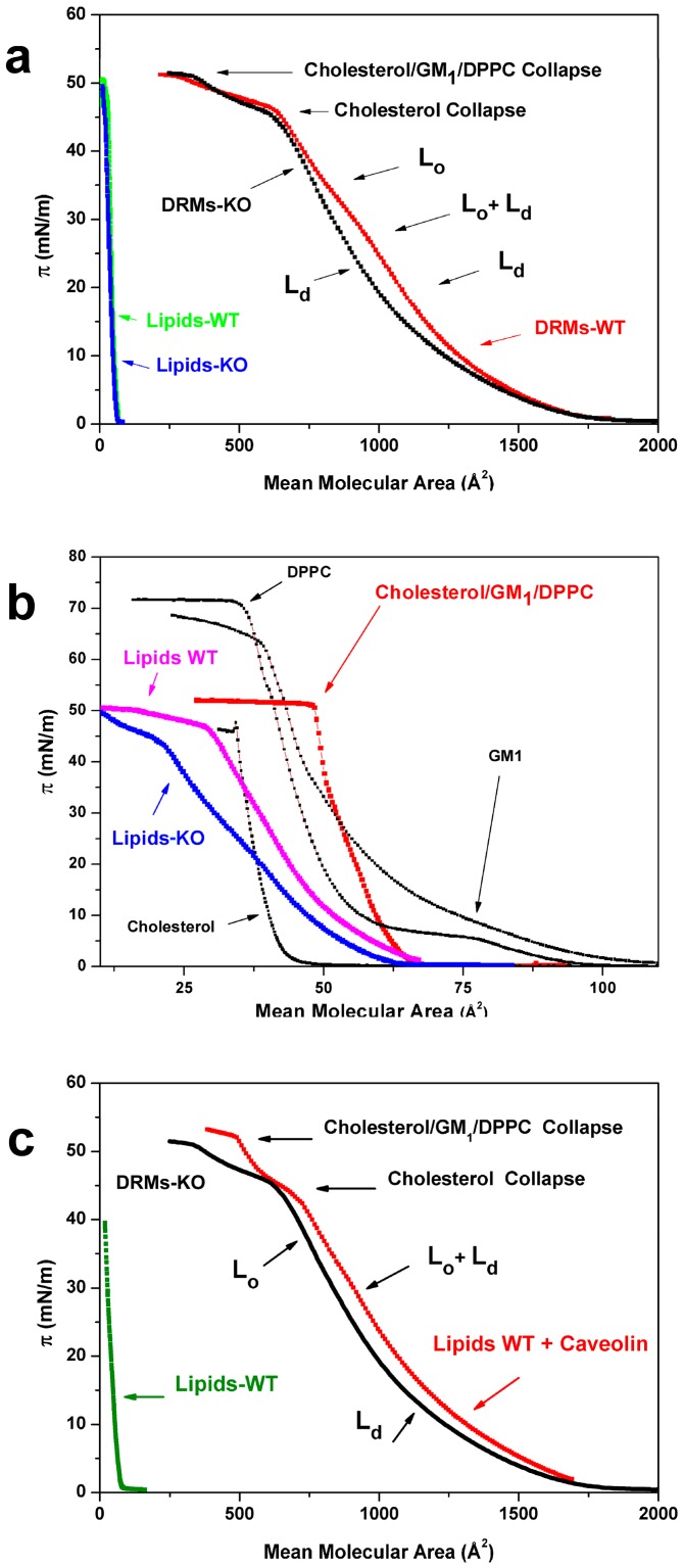
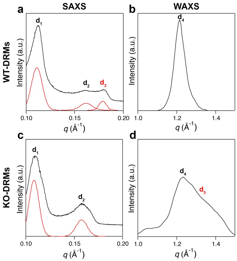
Cholesterol-enriched functional portions of plasma membranes, such as caveolae and rafts, were isolated from lungs of wild-type (WT) and caveolin-1 knockout (Cav-1 KO) mice within detergent resistant membranes (DRMs). To gain insight into their molecular composition we performed proteomic and lipid analysis on WT and Cav-1 KO-DRMs that showed predicted variations of proteomic profiles and negligible differences in lipid composition, while Langmuir monolayer technique and small and wide-angle X-ray scattering (SAXS-WAXS) were here originally introduced to study DRMs biophysical association state. Langmuir analysis of Cav-1 containing DRMs displayed an isotherm with a clear-cut feature, suggesting the coexistence of the liquid-ordered (Lo) phase typical of the raft structure, namely "cholesterol-rich Lo phase," with a phase fully missing in Cav-1 KO that we named "caveolin-induced Lo phase." Furthermore, while the sole lipid component of both WT and KO-DRMs showed qualitatively similar isotherm configuration, the reinsertion of recombinant Cav-1 into WT-DRMs lipids restored the WT-DRM pattern. X-ray diffraction results confirmed that Cav-1 causes the formation of a "caveolin-induced Lo phase," as suggested by Langmuir experiments, allowing us to speculate about a possible structural model. These results show that the unique molecular link between Cav-1 and cholesterol can spur functional order in a lipid bilayer strictly derived from biological sources.

Keywords: Langmuir films; X-ray diffraction; caveolae; cholesterol-rich microdomains; lipid rafts; liquid order; membrane heterogeneity; membranes/fluidity; membranes/model lipid-rafts; membranes/physical chemistry.


Conflict of interest statement

The authors declare no conflicts of interest.

Figures

Figure 1
Figure 1
Characterization of detergent-resistant membranes (DRMs)-associated proteins. (a) Equal volumes of sucrose gradient fractions from lungs of wild-type (WT) and caveolin-1 knockout (Cav-1 KO) mice were subjected to western blot analyses for Cav-1, Cav-2, Flot-1, and Cavin-1. (b) Comparative analysis by reverse phase liquid chromatography-tandem mass spectrometry (RP-LC-MS/MS) of DRMs (pooled fractions 3–6). The Venn diagram shows the number of specific and common WT/Cav-1 KO proteins identified in at least two of the three independent biological replicates. (c) Classification of WT and KO DRM proteins into functional categories based on PANTHER v.12.
Figure 2
Figure 2
Characterization of DRM lipids. Post-nuclear homogenates from WT (a) and Cav-1 KO (b) lung tissue were extracted using 1% TX-100 and fractionated in 5%–30% sucrose gradient. Equal volumes of sucrose gradient fractions were subjected to high performance thin layer chromatography (HPTLC) analysis. Fractions 3–6 corresponding to DRMs showed enrichment of sphingomyelin (SM) and cholesterol (CHOL). (c) HPTLC chromatograms of lipids extracted from DRMs. 10 µg of each lipid standard were spotted on the plate. Cholesterol (CHOL) and triacylglycerols (TG) analysis. Phospholipids (PE; PC) and sphingomyelin (SM) analysis. Lipid concentrations were normalized to DRM protein and calculated against the reference standard. Data are presented as mean ± s.e. (n = 3) (* p < 0.05).
Figure 3
Figure 3
DRMs compression isotherms. (a) Compression isotherms of Langmuir films prepared with DRMs of WT (red) and Cav-1 KO (black) mice and isotherms relative to the same samples after protein removal (green and blue). (b) Protein-free DRMs isotherms (WT pink and KO blue) in comparison to the isotherm of a simplified “raft” model made of cholesterol, monosialotetrahexosylganglioside (GM1), and dipalmitoylphosphatidylcholine (DPPC) (red) and to the single lipid components (black). (c) The reconstitution of Cav-1 in proteoliposomes: Isotherm obtained after reconstitution (red) in comparison with the KO-DRMs isotherm (black) and the protein-free sample obtained from WT-DRMs, used to prepare proteoliposomes (green). Lo, liquid-ordered phase; Ld, liquid-disordered phase; GM1, monosialotetrahexosylganglioside; DPPC, dipalmitoylphosphatidylcholine.
Figure 4
Figure 4
X-ray scattering experiments. (a) Small angle X-ray scattering (SAXS) pattern of lung Cav-1 WT-DRMs. The first two Bragg peaks are the first-order reflections of two lamellar phases with d-spacings of d1 = 57.2 and d2 = 39.9 Å. A third diffraction peak, with lamellar periodicity of d3 = 34.0 Å (symbol in read), is also present. (b) The wide-angle X-ray scattering (WAXS) pattern of lung WT-DRMs shows a sharp diffraction peak, characteristic of an ordered lipid phase with a packing spacing of d4 = 4.2 Å. (c) The SAXS pattern of lung KO-DRMs resembled that of Cav-1 WT-DRMs. However, the main difference is the total absence of the diffraction peak d3. (d) Conversely, in the WAXS scan of lung KO-DRMs another contribution appears at d5 = 3 Å, together with the less intense peak relative to d4 = 4.2 Å. Double peak is characteristic of disordered lipid phases.
Figure 5
Figure 5
Schematic representation of the DRMs bilayer molecular organization. The body of our results, Langmuir isotherm and X-ray scattering experiments, suggest that an ordered phase made of saturated lipids (sphingolipids and gangliosides) and cholesterol is formed (blu), named “cholesterol-rich Lo phase”, together with a more fluid Ld phase made of unsaturated lipids (red). From X-ray scattering results, d1 = 57.2 and d4 = 4.2 Å could be associated with the “cholesterol-rich Lo phase”. A less ordered Ld phase, characterized by d2 = 39.9 and d5 = 3 Å, is also present. Both phases are typical of the lipid-rafts and are present even in the absence of Cav-1 (KO-DRMs). In the presence of Cav-1 a “caveolin-induced Lo phase” can be formed, embedded in the first and characterized by d3 = 34 and d4 = 4.2 Å.

References

    1. Sargiacomo M., Sudol M., Tang Z., Lisanti M.P. Signal transducing molecules and glycosyl-phosphatidylinositol-linked proteins form a caveolin-rich insoluble complex in MDCK cells. J. Cell Biol. 1993;122:789–807. doi: 10.1083/jcb.122.4.789. - DOI - PMC - PubMed
    1. Brown D.A., Rose J.K. Sorting of GPI-anchored proteins to glycolipid-enriched membrane subdomains during transport to the apical cell surface. Cell. 1992;68:533–544. doi: 10.1016/0092-8674(92)90189-J. - DOI - PubMed
    1. Lisanti M.P., Scherer P.E., Vidugiriene J., Tang Z., Hermanowski-Vosatka A., Tu Y.H., Cook R.F., Sargiacomo M. Characterization of caveolin-rich membrane domains isolated from an endothelial-rich source: Implications for human disease. J. Cell Biol. 1994;126:111–126. doi: 10.1083/jcb.126.1.111. - DOI - PMC - PubMed
    1. Lisanti M.P., Tang Z.L., Sargiacomo M. Caveolin forms a hetero-oligomeric protein complex that interacts with an apical GPI-linked protein: Implications for the biogenesis of caveolae. J. Cell Biol. 1993;123:595–604. doi: 10.1083/jcb.123.3.595. - DOI - PMC - PubMed
    1. Simons K., Ikonen E. Functional rafts in cell membranes. Nature. 1997;387:569–572. doi: 10.1038/42408. - DOI - PubMed

Publication types