

Intrathecal Use of Gadobutrol for Glymphatic MR Imaging: Prospective Safety Study of 100 Patients
- PMID: 31320462
- PMCID: PMC7048483
- DOI: 10.3174/ajnr.A6136
Intrathecal Use of Gadobutrol for Glymphatic MR Imaging: Prospective Safety Study of 100 Patients
Abstract
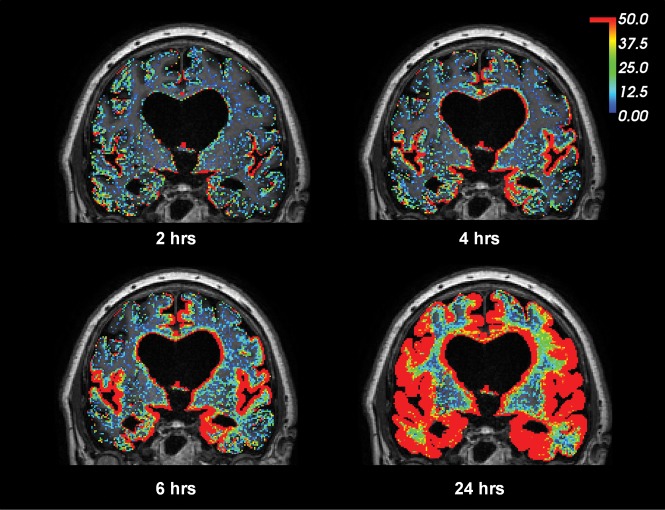
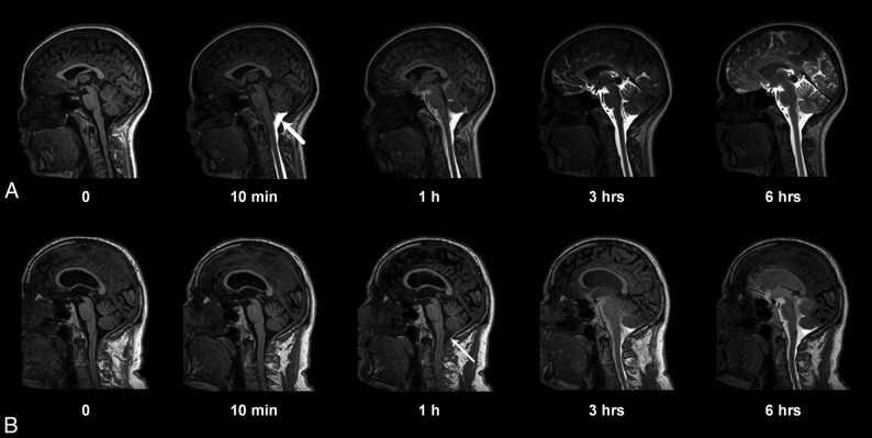
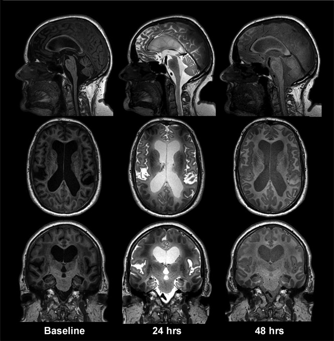
Background and purpose: Intrathecal contrast-enhanced glymphatic MR imaging has shown promise in assessing glymphatic function in patients with dementia. The purpose of this study was to determine the safety profile and feasibility of this new MR imaging technique.
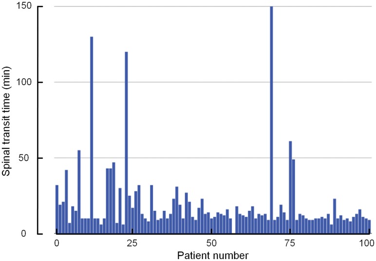
Materials and methods: A prospective safety and feasibility study was performed in 100 consecutive patients (58 women and 42 men, 51 ± 19 years of age) undergoing glymphatic MR imaging from September 2015 to August 2018. Short- and long-term serious and nonserious adverse events were registered clinically and by interview after intrathecal administration of 0.5 mL of gadobutrol (1.0 mmol/mL) along with 3 mL of iodixanol (270 mg I/mL). Adverse events are presented as numbers and percentages.
Results: One serious adverse event (anaphylaxis) occurred in a patient with known allergy to iodine-containing contrast agents (1%). The main nonserious adverse events during the first 1-3 days after contrast injection included severe headache (28%) and severe nausea (34%), though the frequency depended heavily on the diagnosis. After 4 weeks, adverse events had resolved.
Conclusions: Intrathecal administration of gadobutrol in conjunction with iodixanol for glymphatic MR imaging is safe and feasible. We cannot conclude whether short-duration symptoms such as headache and nausea were caused by gadobutrol, iodixanol, the lumbar puncture, or the diagnosis. The safety profile closely resembles that of iodixanol alone.
© 2019 by American Journal of Neuroradiology.
Figures
Similar articles
-
Off-label intrathecal use of gadobutrol: safety study and comparison of administration protocols.Neuroradiology. 2021 Jan;63(1):51-61. doi: 10.1007/s00234-020-02519-4. Epub 2020 Aug 15. Neuroradiology. 2021. PMID: 32803338 Free PMC article.
-
Prospective Safety Study of Intrathecal Gadobutrol in Different Doses.AJNR Am J Neuroradiol. 2023 May;44(5):511-516. doi: 10.3174/ajnr.A7841. Epub 2023 Apr 6. AJNR Am J Neuroradiol. 2023. PMID: 37024308 Free PMC article.
-
Measuring Glymphatic Flow in Man Using Quantitative Contrast-Enhanced MRI.AJNR Am J Neuroradiol. 2019 Apr;40(4):648-651. doi: 10.3174/ajnr.A5931. Epub 2019 Jan 24. AJNR Am J Neuroradiol. 2019. PMID: 30679221 Free PMC article.
-
Neurofluid Dynamics and the Glymphatic System: A Neuroimaging Perspective.Korean J Radiol. 2020 Nov;21(11):1199-1209. doi: 10.3348/kjr.2020.0042. Epub 2020 Aug 11. Korean J Radiol. 2020. PMID: 32783417 Free PMC article. Review.
-
Safety of Intrathecal Administration of Gadolinium-based Contrast Agents: A Systematic Review and Meta-Analysis.Radiology. 2020 Oct;297(1):75-83. doi: 10.1148/radiol.2020191373. Epub 2020 Jul 28. Radiology. 2020. PMID: 32720867
Cited by
-
In vivo assessment of cerebrospinal fluid efflux to nasal mucosa in humans.Sci Rep. 2020 Sep 11;10(1):14974. doi: 10.1038/s41598-020-72031-5. Sci Rep. 2020. PMID: 32917953 Free PMC article. Clinical Trial.
-
Functional analysis of the human perivascular subarachnoid space.Nat Commun. 2024 Mar 5;15(1):2001. doi: 10.1038/s41467-024-46329-1. Nat Commun. 2024. PMID: 38443374 Free PMC article.
-
High-resolution 3D demonstration of regional heterogeneity in the glymphatic system.J Cereb Blood Flow Metab. 2022 Nov;42(11):2017-2031. doi: 10.1177/0271678X221109997. Epub 2022 Jul 3. J Cereb Blood Flow Metab. 2022. PMID: 35786032 Free PMC article.
-
Impaired glymphatic function in idiopathic intracranial hypertension.Brain Commun. 2021 Mar 21;3(2):fcab043. doi: 10.1093/braincomms/fcab043. eCollection 2021. Brain Commun. 2021. PMID: 34235434 Free PMC article.
-
Neurofluids and the glymphatic system: anatomy, physiology, and imaging.Br J Radiol. 2023 Nov;96(1151):20230016. doi: 10.1259/bjr.20230016. Epub 2023 Jun 1. Br J Radiol. 2023. PMID: 37191063 Free PMC article. Review.
References
Publication types
MeSH terms
Substances
LinkOut - more resources
Full Text Sources