. 2019 Sep;31(9):2241-2258.
doi: 10.1105/tpc.19.00155. Epub 2019 Jul 18.

A Photosynthesis-Specific Rubredoxin-Like Protein Is Required for Efficient Association of the D1 and D2 Proteins during the Initial Steps of Photosystem II Assembly

Affiliations

A Photosynthesis-Specific Rubredoxin-Like Protein Is Required for Efficient Association of the D1 and D2 Proteins during the Initial Steps of Photosystem II Assembly

Éva Kiss et al. Plant Cell. 2019 Sep.

Abstract

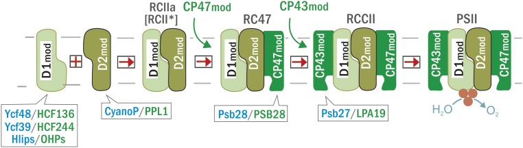
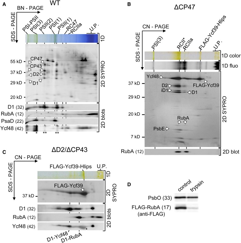
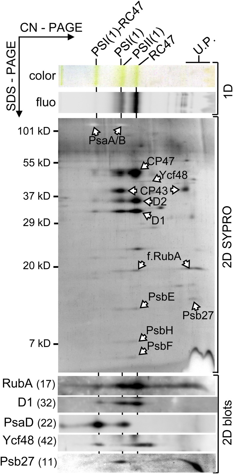
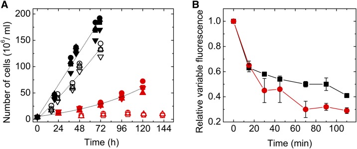
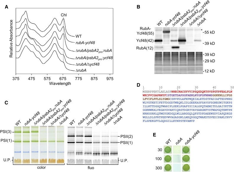
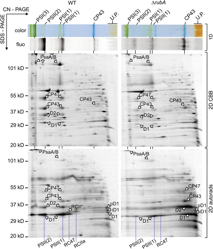
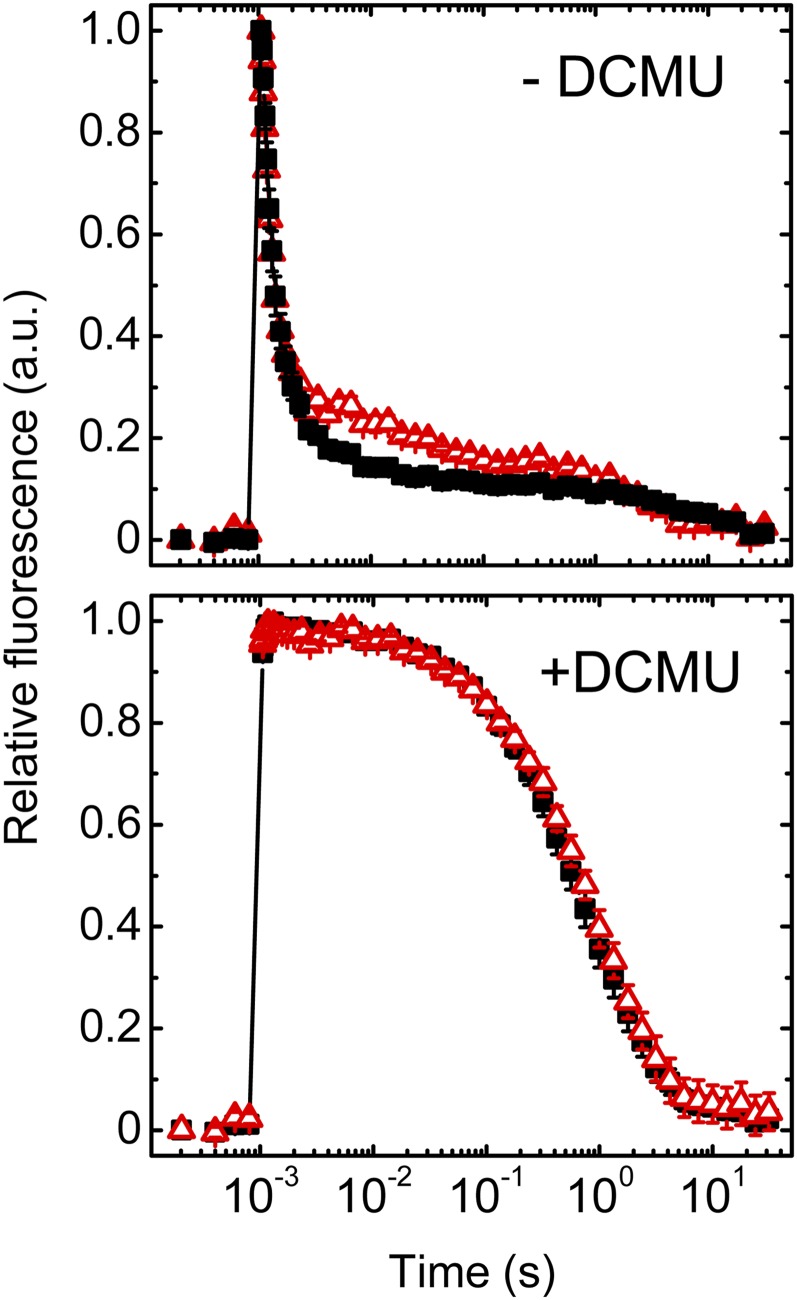
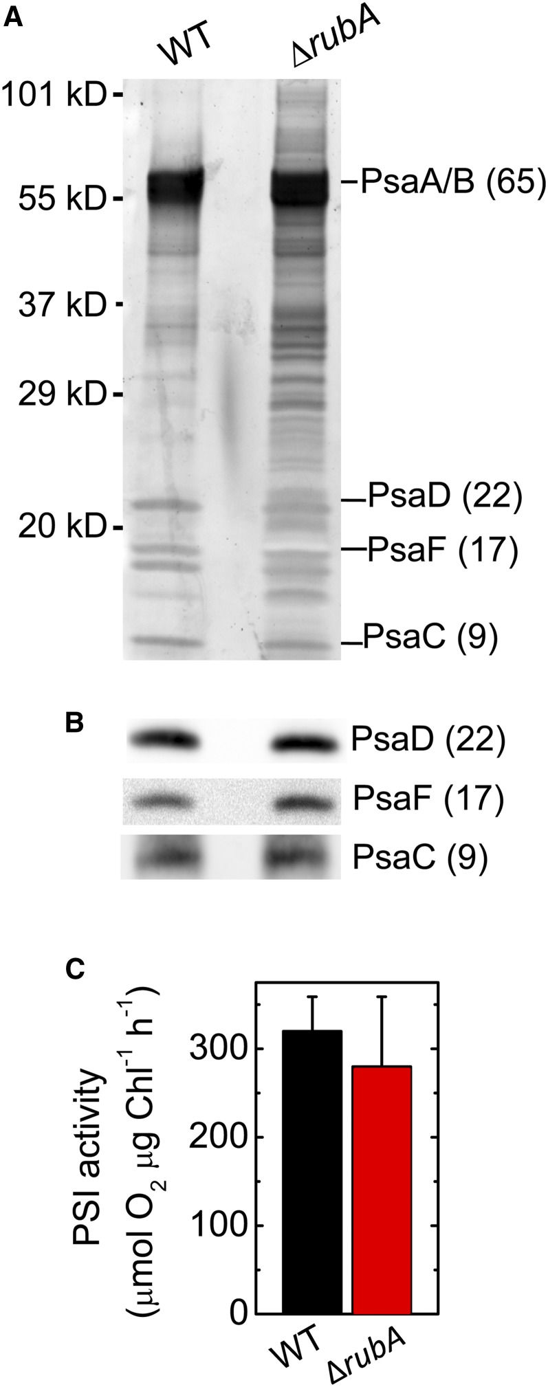
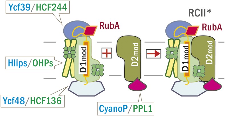
Oxygenic photosynthesis relies on accessory factors to promote the assembly and maintenance of the photosynthetic apparatus in the thylakoid membranes. The highly conserved membrane-bound rubredoxin-like protein RubA has previously been implicated in the accumulation of both PSI and PSII, but its mode of action remains unclear. Here, we show that RubA in the cyanobacterium Synechocystis sp PCC 6803 is required for photoautotrophic growth in fluctuating light and acts early in PSII biogenesis by promoting the formation of the heterodimeric D1/D2 reaction center complex, the site of primary photochemistry. We find that RubA, like the accessory factor Ycf48, is a component of the initial D1 assembly module as well as larger PSII assembly intermediates and that the redox-responsive rubredoxin-like domain is located on the cytoplasmic surface of PSII complexes. Fusion of RubA to Ycf48 still permits normal PSII assembly, suggesting a spatiotemporal proximity of both proteins during their action. RubA is also important for the accumulation of PSI, but this is an indirect effect stemming from the downregulation of light-dependent chlorophyll biosynthesis induced by PSII deficiency. Overall, our data support the involvement of RubA in the redox control of PSII biogenesis.


Figures

Figure 1.
Figure 1.
Simplified Scheme for the Modular Assembly of PSII. Modules containing the D1 and D2 proteins combine to form the RCII complexes RCIIa and RCII*. They bind the CP47 module followed by the CP43 module to obtain a non-oxygen-evolving PSII core complex (RCCII), and finally, the monomeric and dimeric oxygen-evolving PSII complexes (PSII). Cyanobacterial accessory factors associating with the assembly complexes and their plant homologs are indicated in blue and green, respectively. For simplicity, the RCII complexes (RCIIa and RCII*) as well as PSII dimers and monomers are not distinguished in the scheme.
Figure 2.
Figure 2.
Localization and Topology of RubA in the Isolated Synechocystis Membranes. (A) Membrane proteins were separated by blue native (BN) PAGE in the first dimension (1D) and further separated by SDS-PAGE in the second dimension. The 2D gel was stained with SYPRO Orange (2D SYPRO) and electroblotted, and the indicated proteins were immunodetected using specific antibodies (2D blots). The relative molecular masses in kD are shown on the side of the stained gel and in parentheses next to the names of the immunodetected proteins. The loaded sample contained 5 µg of Chl. Designation of complexes: PSI-PSII, supercomplex of PSI and PSII; PSI(3) and PSI(1), trimeric and monomeric PSI; PSII(2) and PSII(1), dimeric and monomeric PSII; RC47 and RCIIa are PSII assembly complexes consisting of the D1/D2 heterodimer with and without the CP47 antenna, respectively; U.P., unassembled proteins; WT, wild type. Asterisks indicate signals of PsaD (*) and D1 (**) present on the blot before the Ycf48 detection. (B) Immunodetection of RubA in RCII complexes. The FLAG-Ycf39 protein with its interacting partners was purified from the CP47-less background that accumulates the RCII complexes. The preparation was analyzed using CN-PAGE. The gel was photographed (1D color), and Chl fluorescence (1D fluo) was detected. The subsequent 2D analysis was performed as described for (A). The complexes are designated as in (A); RCII*, PSII reaction center complex consisting of the D1/D2 heterodimer plus the Ycf39-Hlips complex. U.P., unassembled proteins. (C) The FLAG-Ycf39 protein was immunopurified from a strain that lacks the D2 and CP43 subunits of PSII, so the complex assembly is arrested before the association of D1mod with D2mod. The preparation was analyzed using CN-PAGE in the first dimension (1D) and analyzed in the second dimension as described for (A). The complexes are designated as in (A) and (B). The asterisk on the blot developed after probing with the antibody against Ycf48 indicates a cross-reaction with the strong band of Ycf39. U.P., unassembled proteins. (D) Trypsinization of membranes for the determination of the RubA membrane topology. Right-side-out membrane vesicles from the strain expressing an N-terminally FLAG-tagged RubA (FLAG-RubA) before and after 1 h of trypsinization were analyzed by SDS-PAGE, the gel was electroblotted, and FLAG-RubA as well as PsbO were detected using specific antibodies. Each loaded sample contained 3 µg of Chl.
Figure 3.
Figure 3.
Protein Interacting Partners of RubA. The FLAG-RubA preparation was analyzed by CN-PAGE in the first dimension. The native gel was photographed (1D color) and scanned for Chl fluorescence (1D fluo), and proteins were separated in the second dimension as described for Figure 2A. Designation of complexes is as in Figure 2A; f.RubA, FLAG-RubA; PSI(1)-RC47 is a complex containing monomeric PSI and RC47. U.P., unassembled proteins.
Figure 4.
Figure 4.
Photoautotrophic Growth and High Light-Induced PSII Photoinhibition of Wild-Type and ∆rubA Cells. (A) The relative changes in cell number of the wild type (black symbols) and ∆rubA (red symbols) were assessed during exponential growth in liquid cultures under continuous (solid symbols) or fluctuating (15 min of light/15 min of dark; open symbols) light conditions. The circle, square, and triangle symbols represent three independent cultures. (B) The same amount of wild-type (black) and ∆rubA (red) cells were exposed to 300 µmol photons m−2 s−1. Variable fluorescence was measured and normalized to the initial value (0 min). Mean values and the indicated se are derived from measurements of three independent cultures of each strain. The initial value measured for the ∆rubA mutant represented 20 ± 3% of the wild-type value.
Figure 5.
Figure 5.
Comparative Analysis of the Strains Lacking RubA and/or Ycf48 or Expressing a Fused Version of These Proteins. (A) Whole-cell absorption spectra of the wild type, the strain lacking both RubA and Ycf48 but expressing a RubA-Ycf48 fusion protein (rubA-ycf48), the strain lacking RubA and expressing RubA from the psbA2 promotor (∆rubA/psbA2pro:rubA), the strain lacking RubA and expressing Ycf48 from the psbA2 promotor (∆rubA/psbA2pro:ycf48), the strain lacking both RubA and Ycf48 (∆rubA/∆ycf48), and the strain lacking RubA (∆rubA) were recorded when the photoautotrophic cultures reached an OD of 0.5 at 750 nm; they are shown after shifting for better visibility. The absorption peaks at 679 nm, which reflect Chl content, are designated by a dotted line. (B) Membranes isolated from strains described in (A) were analyzed by SDS-PAGE. Proteins were subsequently blotted onto a PVDF membrane, and RubA as well as Ycf48 were detected using specific antibodies. For each strain, membranes corresponding to the same number of cells (1.4 × 10⁸) were loaded into the lanes. The SYPRO-stained gel is shown to prove the equal loading of the lanes. The asterisk designates an unspecific cross-reaction. WT, wild type. (C) Membranes isolated from strains described in (A) were analyzed by CN-PAGE, and the gel was photographed (color) and scanned for Chl fluorescence (fluo). Designation of complexes is as in Figure 2. For each strain, membranes corresponding to the same number of cells (2.5 × 10⁸) were loaded into the lanes. U.P., unassembled proteins. WT, wild type. (D) Amino acid sequence of the RubA-Ycf48 fusion protein. The RD (red), the rubredoxin linker peptide (purple), the transmembrane helix of RubA (brown), and the Ycf48 part (blue) are shown. (E) Drops containing 1.25 × 10⁴ cells of the wild type, ∆rubA, or the rubA-ycf48 strain were pipetted on a solid agar plate. The plate was photographed after 3 d of autotrophic growth under constant illumination at 30, 100, or 300 µmol photons m−2 s−1. WT, wild type.
Figure 6.
Figure 6.
2D Analysis of Radioactively Labeled Membrane Proteins of the Wild Type and ∆rubA. Membranes isolated from radioactively labeled cells were analyzed by CN-PAGE in the first dimension (1D color and 1D fluo). After SDS-PAGE in the second dimension, the gel was stained (2D CBB), and the radiolabeled proteins were subsequently detected by autoradiography (2D autorads). Designation of complexes is as described in Figure 2; iD1, intermediate D1; pD1, precursor D1. Each loaded sample contained 5 µg of Chl. U.P., unassembled proteins. WT, wild type.
Figure 7.
Figure 7.
Decay Kinetics of Flash-Induced Chl Fluorescence. Equal numbers of wild-type (solid black squares) and ∆rubA (open red triangles) cells were dark adapted for 5 min in the absence (−DCMU) or presence (+DCMU) of DCMU and subsequently excited with single turnover flash at time = 1 ms. The relaxations of the flash-induced Chl fluorescence were recorded and are shown after normalization to the initial amplitude. Mean values (averages) and the indicated se are derived from five independent measurements. a.u., arbitrary units.
Figure 8.
Figure 8.
Determination of the Structural and Functional Integrity of PSI in the ΔrubA Mutant. (A) Electrophoretic analysis of PSI-enriched membranes. After separation of proteins by SDS-PAGE, the proteins were stained using SYPRO Orange. WT, wild type. (B) Immunodetection of PsaD, PsaF, and PsaC subunits in the PSI-enriched membranes. (C) PSI activities of the PSI-enriched membranes isolated from the wild type (black bar) and the ΔrubA mutant (red bar) measured by means of oxygen consumption in the presence of an artificial electron donor (dichlorophenolindophenol) and acceptor (methyl viologen). Each column and error bar represents a mean value (average) and the sd, respectively, for five independent measurements. The same amounts of wild-type and mutant thylakoid particles (estimated according to Chl content) were used for all assays presented in (A) to (C). WT, wild type.
Figure 9.
Figure 9.
Analysis of the Tetrapyrrole Biosynthetic Pathway in ΔrubA and in the DCMU-Treated Wild Type. (A) Levels of selected tetrapyrrole biosynthesis enzymes in the membranes of the wild type, ΔrubA, and the wild type grown in the presence of 0.8 µM DCMU. After 2 weeks of autotrophic cultivation, the cells were harvested. Membranes corresponding to the same amount of cells were analyzed by SDS-PAGE, and separated proteins were electroblotted and immunodetected. ChlG, chlorophyll synthase; ChlH, catalytic subunit of Mg-chelatase; ChlM, Mg-protoporphyrin IX methyltransferase; WT, wild type. (B) Abundance of Chl precursors in the control wild-type (black solid line), ∆rubA (red line), and DCMU-treated wild-type (WT+DCMU; black dotted line) cells. Pigments were extracted by methanol from equal numbers of cells. The signals of the subsequent HPLC analysis were detected by a pair of fluorescence detectors (FLD1 and FLD2) set for different wavelengths to cover all Chl precursors. a.u., arbitrary units. DV, divinyl; MgP, Mg-protophorphyrin IX; MV, monovinyl.
Figure 10.
Figure 10.
Characterization of Photoautotrophic and LAHG-Grown Wild-Type and ΔrubA Cells. (A) Whole cell absorption spectra of the wild type (black line) and ΔrubA (red line) grown for 6 d under LAHG conditions. The spectra are shown after normalization to the OD at 750 nm and are shifted for better visibility. a.u., arbitrary units. (B) Separation of membrane protein complexes from LAHG-grown wild-type and ∆rubA cells using CN-PAGE. The gel was photographed (color) and scanned for Chl fluorescence (fluo). Designation of complexes is as in Figure 5C. a.u., arbitrary units. (C) 77 K Chl fluorescence emission spectra from cells of the wild type (black line) and ∆rubA (red line) grown either photoautotrophically (PAT) or heterotrophically (LAHG). Equal amounts of cells were frozen in liquid nitrogen and excited at 435 nm. Spectra were normalized to the emission peak of the internal standard rhodamine at 570 nm. Curves represent mean values of three independent measurements. WT, wild type. U.P., unassembled proteins. (D) Comparison of the relative cellular contents of Chl and its biosynthetic precursors in LAHG-grown wild-type (black bars) and ∆rubA (red bars) cultures. The Chl content was determined spectroscopically, while the relative abundances of Chl precursors were quantified by HPLC. Abbreviations are as in Figure 9B. The values were normalized to the autotrophically grown wild-type control levels that are taken as 100% and indicated by the dotted line. The columns and error bars represent means ± se, respectively, for at least three independent cultures.
Figure 11.
Figure 11.
Model for the Early Stages of PSII Assembly Showing the Locations of RubA and the Other Cyanobacterial/Plant Accessory Proteins. Ycf48 (HCF136 in plants) and the chlorophyll-containing Ycf39/Hlip complex (HCF244/OHP in plants) assist in the insertion of Chl into D1mod, while CyanoP (PPL1 in plants) stabilizes D2mod. The transmembrane domain of RubA associates with the N-terminal region of D1, positioning the C-terminal tail of RubA in close proximity to the lumenal Ycf48 factor. The linker region of RubA allows the rubredoxin domain to bind at the interface of D1 and D2. All factors remain associated with the modules after they form the RCII* complex.
None

References

    1. Adams N.B.P., Brindley A.A., Hunter C.N., Reid J.D. (2016). The catalytic power of magnesium chelatase: A benchmark for the AAA(+) ATPases. FEBS Lett. 590: 1687–1693. - PMC - PubMed
    1. Anderson S.L., McIntosh L. (1991). Light-activated heterotrophic growth of the cyanobacterium Synechocystis sp. strain PCC 6803: A blue-light-requiring process. J. Bacteriol. 173: 2761–2767. - PMC - PubMed
    1. Bečková M., Gardian Z., Yu J., Koník P., Nixon P.J., Komenda J. (2017). Association of Psb28 and Psb27 proteins with PSII-PSI supercomplexes upon exposure of Synechocystis sp. PCC 6803 to high light. Mol. Plant 10: 62–72. - PubMed
    1. Boehm M., Romero E., Reisinger V., Yu J., Komenda J., Eichacker L.A., Dekker J.P., Nixon P.J. (2011). Investigating the early stages of photosystem II assembly in Synechocystis sp. PCC 6803: Isolation of CP47 and CP43 complexes. J. Biol. Chem. 286: 14812–14819. - PMC - PubMed
    1. Boehm M., Yu J., Reisinger V., Bečkova M., Eichacker L.A., Schlodder E., Komenda J., Nixon P.J. (2012). Subunit composition of CP43-less photosystem II complexes of Synechocystis sp. PCC 6803: Implications for the assembly and repair of photosystem II. Philos. Trans. R. Soc. Lond. B Biol. Sci. 367: 3444–3454. - PMC - PubMed

Publication types

MeSH terms