. 2019 Jul 18;10(8):552.
doi: 10.1038/s41419-019-1763-2.

Increase in HDAC9 suppresses myoblast differentiation via epigenetic regulation of autophagy in hypoxia

Affiliations

Increase in HDAC9 suppresses myoblast differentiation via epigenetic regulation of autophagy in hypoxia

Zhang Zhang et al. Cell Death Dis. .

Abstract

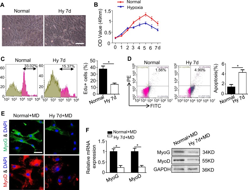
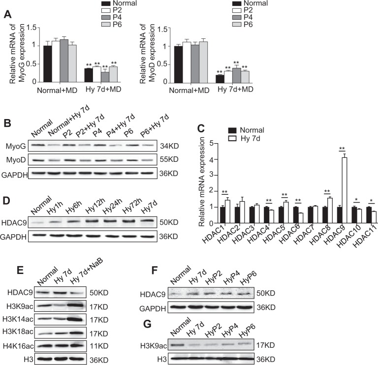
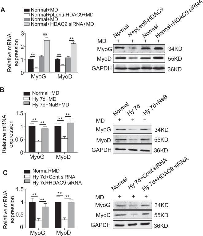
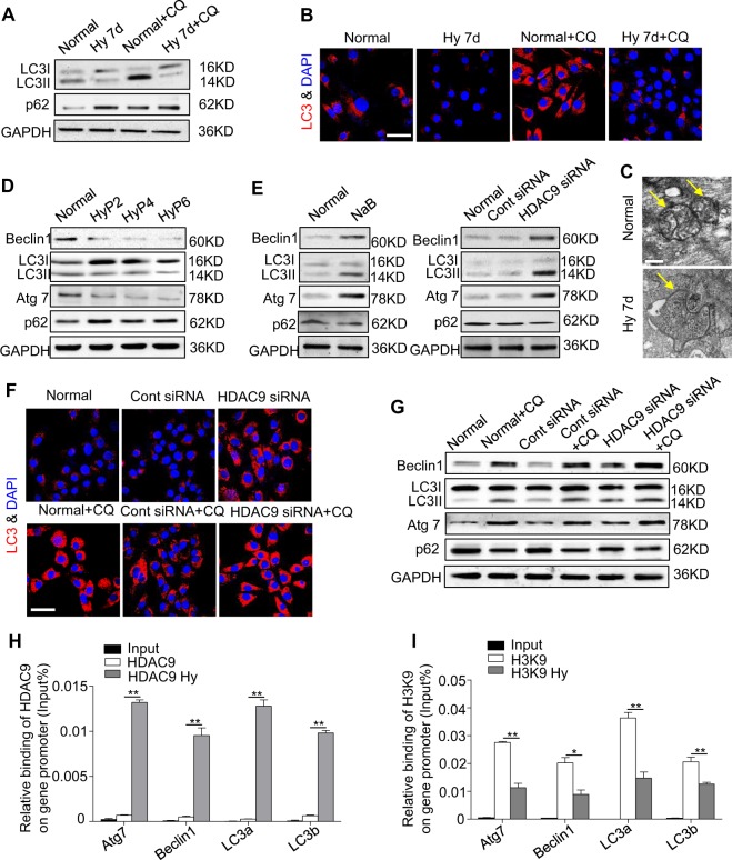
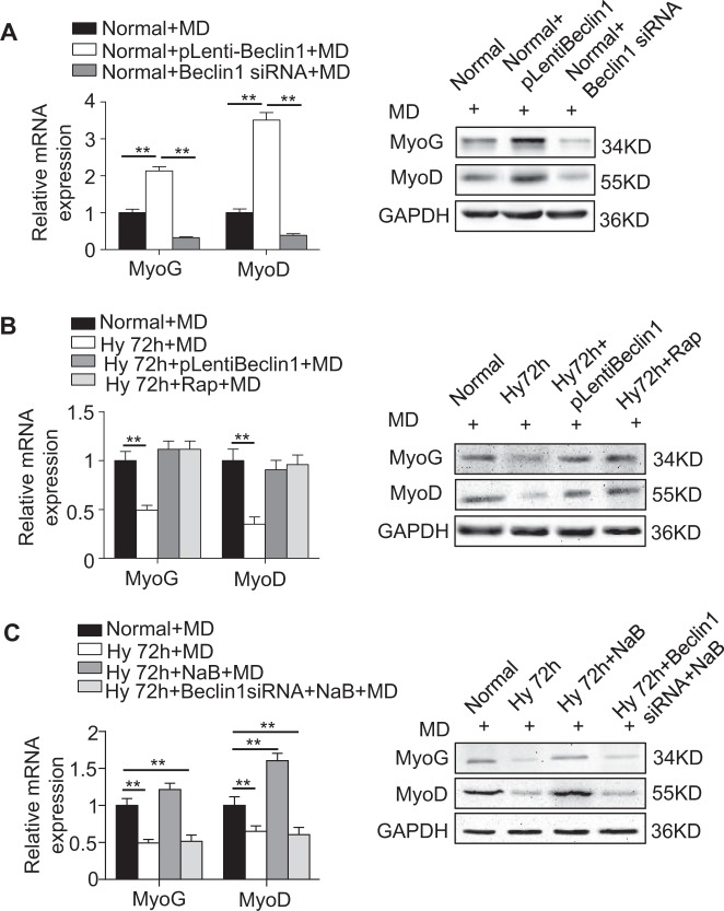
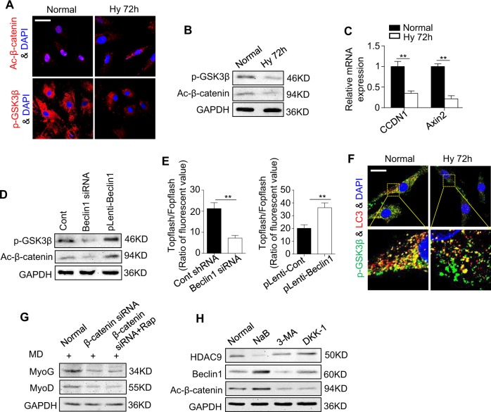
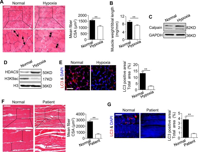
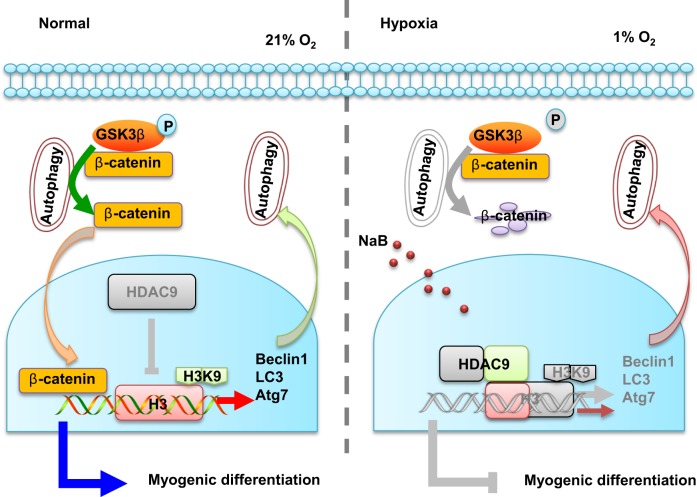
Extremely reduced oxygen (O2) levels are detrimental to myogenic differentiation and multinucleated myotube formation, and chronic exposure to high-altitude hypoxia has been reported to be an important factor in skeletal muscle atrophy. However, how chronic hypoxia causes muscle dysfunction remains unknown. In the present study, we found that severe hypoxia (1% O2) significantly inhibited the function of C2C12 cells (from a myoblast cell line). Importantly, the impairment was continuously manifested even during culture under normoxic conditions for several passages. Mechanistically, we revealed that histone deacetylases 9 (HDAC9), a member of the histone deacetylase family, was significantly increased in C2C12 cells under hypoxic conditions, thereby inhibiting intracellular autophagy levels by directly binding to the promoter regions of Atg7, Beclin1, and LC3. This phenomenon resulted in the sequential dephosphorylation of GSK3β and inactivation of the canonical Wnt pathway, impairing the function of the C2C12 cells. Taken together, our results suggest that hypoxia-induced myoblast dysfunction is due to aberrant epigenetic regulation of autophagy, and our experimental evidence reveals the possible molecular pathogenesis responsible for some muscle diseases caused by chronic hypoxia and suggests a potential therapeutic option.


Conflict of interest statement

The authors declare that they have no conflict of interest.

Figures

Fig. 1
Fig. 1. The properties of C2C12 cells decrease significantly under hypoxia.
C2C12 cells were cultured under a normoxic or hypoxic microenvironment for 7 days. a The morphologies of the C2C12 cells grown under these two conditions were observed using an inverted microscope. Scale bar: 50 μm. b, c The viability and proliferation of the C2C12 cells under the two treatments were measured using MTT (b) and Edu (c) assays. d Apoptosis of the two C2C12 cells was examined by flow cytometry. e The C2C12 cells were cultured in myogenic differentiation medium (MD) under a normoxic or hypoxic microenvironment for 7 days, and MyoG (green)/MyoD (red)/nuclei (blue) were examined by immunofluorescence staining. Scale bar: 50 μm. f The myogenic genes of the C2C12 cells were detected by qRT-PCR and western blotting. The data are presented as the mean ± s.d. of triplicate samples from a representative experiment. *P< 0.05. c, d unpaired two-tailed Student’s t-test. b, f one-way analysis of variance (ANOVA)
Fig. 2
Fig. 2. The effect of hypoxia on the disrupted function of C2C12 cells occurs in an epigenetic-dependent manner.
a, b C2C12 cells were cultured in MD under normoxic or hypoxic conditions for 7 days as Normal (P1) or Hypoxia P1 (Hy), respectively, and then, both were continuously passaged six times under normoxic conditions. The expression of MyoG and MyoD in passages 2, 4, and 6 in the C2C12 cells was analyzed by qRT-PCR (a) and western blotting (b). c The C2C12 cells were cultured under normoxic or hypoxic conditions for 7 days, and the expression of the histone deacetylase family (HDAC1-11) was examined by qRT-PCR. d The expression levels of HDAC9 in the C2C12 cells at different time points after exposure to hypoxic stimuli were examined by western blotting. e The expression levels of HDAC9 and well-known histone targets of HDAC9 in normoxia or hypoxia with or without NaB treatment (the dose of NaB is 200 μM) in the C2C12 cells are shown. f, g The expression levels of HDAC9 and H3K9ac in different passages (normal, Hy7d, HyP2, HyP4, and HyP6) of the C2C12 cells were examined by western blotting. The data are presented as the mean ± s.d. of triplicate samples from a representative experiment. *P< 0.05, **P < 0.01. One-way analysis of variance (ANOVA)
Fig. 3
Fig. 3
Downregulation of the expression of HDAC9 by an inhibitor or siRNA rescues the inhibitory effect of hypoxia on C2C12 cell myogenesis. a The regulation of HDAC9 expression through lentiviral vectors in the C2C12 cells is depicted. The expression of MyoG and MyoD was examined by qRT-PCR and western blotting at day 7 after myogenic induction. b, c To observe the effect of HDAC9 on the myogenesis in the C2C12 cells, C2C12 cells were treated with the HDAC inhibitor sodium butyrate (NaB) (b) or with HDAC9 siRNA (c). After myogenic induction for 7 days, the expression levels of MyoG and MyoD in the C2C12 cells were examined by qRT-PCR and western blotting. The data are presented as the mean ± s.d. of triplicate samples from a representative experiment. *P< 0.05, **P < 0.01. One-way analysis of variance (ANOVA)
Fig. 4
Fig. 4. HDAC9 epigenetically regulates the autophagy level in C2C12 cells.
a The expression levels of the autophagy-related proteins Beclin1, LC3I/II and specific cargo p62 after exposure to hypoxia for 7 days with or without chloroquine (CQ) were examined by western blotting. b The C2C12 cells were cultured under a normoxic or hypoxic microenvironment for 7 days with or without CQ. The LC3 (red)/nuclei (blue) in the C2C12 cells were analyzed by immunofluorescence staining. Scale bar: 50 μm. c To observe the autophagosomes, the C2C12 cells were cultured under normoxic or hypoxic conditions for 7 days and then observed using an electron microscope. Scale bar: 2 μm. d The expression levels of autophagy-related proteins in Hyp 2, Hyp 4, and Hyp 6 of the C2C12 cells were examined by western blotting. e The expression levels of autophagy-related proteins were examined by western blotting after treatment with NaB and HDAC9 siRNA. f, g Downregulation of HDAC9 in the normoxic C2C12 cells with or without CQ is shown. The LC3 (red)/nuclei (blue) in the C2C12 cells was analyzed by immunofluorescence staining, and the autophagy-related genes were analyzed by western blotting. Scale bar: 50 μm. h, i Chromatin was isolated from the C2C12 cells exposed or not to hypoxia and subjected to the chromatin immunoprecipitation assay using acetylated-histone H3K9 (Ac-H3K9) and HDAC9 antibodies. The IgG antibody was included as a control. The data are presented as the mean ± s.d. of triplicate samples from a representative experiment. *P< 0.05, **P < 0.01. One-way analysis of variance (ANOVA)
Fig. 5
Fig. 5. HDAC9 regulates myogenic differentiation of C2C12 cells likely through autophagy.
a The regulation of Beclin1 expression in the C2C12 cells through the lentiviral vector and siRNA is depicted. The myogenesis-related genes MyoG and MyoD were examined by qRT-PCR and western blotting. b After activating autophagy in the hypoxic C2C12 cells by overexpression of Beclin1 or Rapamycin (Rap), the myogenesis-related genes MyoG and MyoD were examined by qRT-PCR and western blotting. c The C2C12 cells were cultured in MD and treated with NaB or Beclin1 siRNA under hypoxia for 72 h. The myogenesis-related genes MyoG and MyoD were examined in the C2C12 cells by qRT-PCR and western blotting. Normoxic C2C12 cells were used as a control. The data are presented as the mean ± s.d. of triplicate samples from a representative experiment. *P< 0.05, **P < 0.01. One-way analysis of variance (ANOVA)
Fig. 6
Fig. 6. Autophagy regulates myogenic differentiation in C2C12 cells through regulation of the canonical Wnt pathway.
ac The expression levels of p-GSK3β and active-β-catenin were examined by immunofluorescence staining (a) and western blotting (b) after the cells were cultured in normoxia or hypoxia for 72 h. The downstream genes of the canonical Wnt pathway were analyzed by qRT-PCR (c). Scale bar: 50 μm. d The expression levels of p-GSK3β and active-β-catenin in the C2C12 cells were examined by western blotting after downregulation of Beclin1. e Activation of the canonical Wnt pathway was examined 48 h after transfection by luciferase assay. f Immunostaining showed overlapping of LC3 (red) and p-GSK3β (green) in the C2C12 cells cultured in normoxia and hypoxia for 72 h. Scale bar: 25 μm. g C2C12 cells were cultured in MD and transfected with control siRNA or β-catenin siRNA, and rapamycin was used to activate autophagy. Myogenesis-related genes were examined by western blotting after treatment for 72 h. h The C2C12 cells were treated with NaB, 3-MA, and DKK-1. The expression levels of HDAC9, Beclin1, and ac-β-catenin were examined by western blotting. The data are presented as the mean ± s.d. of triplicate samples from a representative experiment. *P< 0.05, **P < 0.01. c One-way analysis of variance (ANOVA). e unpaired two-tailed Student’s t-test
Fig. 7
Fig. 7. The autophagy that is epigenetically regulated by HDAC9 is also observed in the femoral artery (FA) ligation model and arteriosclerosis obliterans patients.
We constructed a mouse single FA ligation model and harvested samples from both the control and surgery groups on day 21. a Hematoxylin and eosin staining (h&e) was used for visualizing the tissue from the mouse gastrocnemius muscle of the FA ligation model. The results from the quantitative analysis of the muscle fiber is indicated in the right panel. Scale bar: 200 μm. b, c The ratio of muscle weight/tibial length (b) and calpain expression (c) were analyzed in the control and ligation groups. d The expression levels of HDAC9 and H3K9 in the two groups were examined by western blotting. e The muscles samples were immunostained using anti-LC3 (red) and nuclear staining (blue, DAPI) to reveal the autophagy level. The results from the quantification analysis of the LC3+ cells is shown in the right panel. Scale bar: 100 μm. f H&E staining of the muscle in the normal control and arteriosclerosis obliterans patients is shown. The results from the quantification analysis are shown in the right panel. Scale bar: 200 μm. g The immunostaining analysis showed the expression of LC3 (red) and DAPI (blue) in the distal gastrocnemius muscle. Quantification of the LC3+ cells is indicated in the right panel. Scale bar: 100 μm. n = 8 for the mouse model and n = 2 for the patients. The data are presented as the mean ± s.d. of triplicate samples from a representative experiment. *P< 0.05, **P < 0.01. Unpaired two-tailed Student’s t-test
Fig. 8
Fig. 8. Schematic diagram depicts how hypoxia regulates myogenic differentiation of C2C12 cells and an epigenetics-guided therapeutic method.
In the presence of normal oxygen, intracellular autophagy controls the phosphorylation of GSK3β and then promotes the transfer of β-catenin from the cytoplasm into the nucleus, which subsequently activates myogenesis-related gene expression. Under hypoxia, HDAC9 expression is increased due to the hypoxic microenvironment, and HDAC9 deacetylates H3K9 of autophagy-related genes and inhibits autophagosome formation. Insufficient autophagy subsequently results in dephosphorylation of GSK3β and inactivation of the canonical Wnt pathway, ultimately preventing myogenic differentiation. NaB treatment can partially rescue the impaired myogenic differentiation of the C2C12 cells caused by hypoxia

Similar articles

Cited by

References

    1. Slot IG, Schols AM, De Theije CC, Snepvangers FJ, Gosker HR. Alterations in skeletal muscle oxidative phenotype in mice exposed to 3 weeks of normobaric hypoxia. J. Cell Physiol. 2016;231:377–392. doi: 10.1002/jcp.25083. - DOI - PubMed
    1. Di Carlo A, et al. Hypoxia inhibits myogenic differentiation through accelerated MyoD degradation. J. Biol. Chem. 2004;279:16332–16338. doi: 10.1074/jbc.M313931200. - DOI - PubMed
    1. Jash S, Adhya S. Effects of transient hypoxia versus prolonged hypoxia on satellite cell proliferation and differentiation in vivo. Stem Cells Int. 2015;2015:961307. doi: 10.1155/2015/961307. - DOI - PMC - PubMed
    1. Turan N, et al. A systems biology approach identifies molecular networks defining skeletal muscle abnormalities in chronic obstructive pulmonary disease. PLoS Comput. Biol. 2011;7:e1002129. doi: 10.1371/journal.pcbi.1002129. - DOI - PMC - PubMed
    1. Demonbreun AR, McNally EM. Muscle cell communication in development and repair. Curr. Opin. Pharmacol. 2017;34:7–14. doi: 10.1016/j.coph.2017.03.008. - DOI - PMC - PubMed

Publication types

MeSH terms