. 2019 Jun 19:2019:6919834.
doi: 10.1155/2019/6919834. eCollection 2019.

The Protective Effect of Sonneratia apetala Fruit Extract on Acetaminophen-Induced Liver Injury in Mice

Affiliations

The Protective Effect of Sonneratia apetala Fruit Extract on Acetaminophen-Induced Liver Injury in Mice

Jingjing Liu et al. Evid Based Complement Alternat Med. .

Abstract

Acute liver injury is a common consequence of taking overdose of acetaminophen (APAP). The aim of this study was to evaluate the antioxidant activity and hepatoprotective effect of a mangrove plant Sonneratia apetala fruit extract (SAFE) on APAP-induced liver injury in mice. Mice were orally pretreated with SAFE (100, 200, and 400 mg/kg) daily for one week. The control and APAP groups were intragastrically administered with distilled water, and NAC group was treated with N-Acetyl-L-cysteine (NAC) before APAP exposure. The results manifested that SAFE significantly improved survival rates, attenuated hepatic histological damage, and decreased the alanine aminotransferase (ALT) and aspartate aminotransferase (AST) levels in serum in APAP-exposed mice. SAFE treatment also increased glutathione (GSH) level and glutathione peroxidase (GSH-Px) activity, enhanced catalase (CAT), and total antioxidant capacity (T-AOC), as well as reducing malondialdehyde (MDA) level in liver. In addition, the formation of tumor necrosis factor-alpha (TNF-α), interleukin 6 (IL-6), and elevation of myeloperoxidase (MPO) in APAP-exposed mice were inhibited after SAFE treatment. And SAFE also displayed high DPPH radical scavenging activity and reducing power in vitro. The main bioactive components of SAFE such as total phenol, flavonoid, condensed tannin, and carbohydrate were determined. The current study proved that SAFE exerted potential protective effect against APAP-induced acute liver injury, which might be associated with the antioxidant and anti-inflammatory activities of SAFE.


Figures
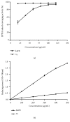
Figure 1
Figure 1
Antioxidant activity of SAFE in vitro. (a) DPPH radical scavenging activity and (b) reducing power. Data are presented as a mean ± SD (n = 3).
Figure 2
Figure 2
SAFE attenuated APAP-induced high mortality rate. The survival rate was expressed as Kaplan-Meier curves followed by the log-rank Mantel-Cox test for comparison among curves (n =15). Survival rate was significantly lower in APAP group mice compared to that in control, 200 mg/kg SAFE and 400 mg/kg SAFE groups (p < 0.01, p < 0.05 and p < 0.01, respectively).
Figure 3
Figure 3
SAFE relieved hepatic histopathological damage in APAP-treated mice. (a) Control; (b) APAP; (c) NAC; (d) 100 mg/kg of SAFE; (e) 200 mg/kg of SAFE; (f) 400 mg/kg of SAFE. Scale bar: 50 μm.
Figure 4
Figure 4
SAFE inhibited ALT (a) and AST (b) levels in serum. Data are presented as a mean ± SD (n = 8), ##p < 0.01 vs. control group; ∗p < 0.05 or ∗∗p < 0.01 vs. APAP group.
Figure 5
Figure 5
SAFE restrained APAP-induced liver oxidative stress in APAP-treated mice. (a) GSH; (b) GSH-Px; (c) CAT; (d) T-AOC; and (e) MDA. Data are presented as a mean ± SD (n = 8), ##p < 0.01 vs. control group; ∗p < 0.05 or ∗∗p < 0.01 vs. APAP group.
Figure 6
Figure 6
SAFE suppressed APAP-induced MPO regulation. Data are presented as a mean ± SD (n = 8), ##p< 0.01 vs. control group; ∗p < 0.05 or ∗∗p < 0.01 vs. APAP group.
Figure 7
Figure 7
SAFE inhibited APAP-induced inflammation in mice. The level of TNF-α (a) and IL-6 (b) was measured. Data are presented as a mean ± SD (n = 8), ##p < 0.01 vs. control group; ∗p < 0.05 or ∗∗p < 0.01 vs. APAP group.

References

    1. Jaeschke H. Acetaminophen: dose-dependent drug hepatotoxicity and acute liver failure in patients. Digestive Diseases. 2015;33(4):464–471. doi: 10.1159/000374090. - DOI - PMC - PubMed
    1. Maes M., Vinken M., Jaeschke H. Experimental models of hepatotoxicity related to acute liver failure. Toxicology and Applied Pharmacology. 2016;290:86–97. doi: 10.1016/j.taap.2015.11.016. - DOI - PMC - PubMed
    1. Jaeschke H., Williams C. D., Ramachandran A., Bajt M. L. Acetaminophen hepatotoxicity and repair: the role of sterile inflammation and innate immunity. Liver International. 2012;32(1):8–20. doi: 10.1111/j.1478-3231.2011.02501.x. - DOI - PMC - PubMed
    1. McGill M. R., Sharpe M. R., Williams C. D., Taha M., Curry S. C., Jaeschke H. The mechanism underlying acetaminophen-induced hepatotoxicity in humans and mice involves mitochondrial damage and nuclear DNA fragmentation. The Journal of Clinical Investigation. 2012;122(4):1574–1583. doi: 10.1172/JCI59755. - DOI - PMC - PubMed
    1. Rhodes K., Braakhuis A. Performance and side effects of supplementation with n-acetylcysteine: a systematic review and meta-analysis. Sports Medicine. 2017;47(8):1619–1636. doi: 10.1007/s40279-017-0677-3. - DOI - PubMed