Review
. 2019 Jun 27:17:862-868.
doi: 10.1016/j.csbj.2019.06.004. eCollection 2019.

Spotting, Transcription and In Situ Synthesis: Three Routes for the Fabrication of RNA Microarrays

Affiliations
Review

Spotting, Transcription and In Situ Synthesis: Three Routes for the Fabrication of RNA Microarrays

Jory Lietard et al. Comput Struct Biotechnol J. .

Abstract

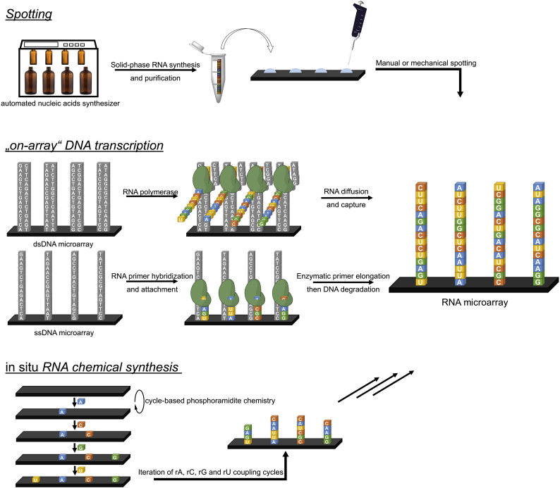
DNA microarrays have become commonplace in the last two decades, but the synthesis of other nucleic acids biochips, most importantly RNA, has only recently been developed to a similar extent. RNA microarrays can be seen as organized surfaces displaying a potentially very large number of unique sequences and are of invaluable help in understanding the complexity of RNA structure and function as they allow the probing and treatment of each of the many different sequences simultaneously. Three approaches have emerged for the fabrication of RNA microarrays. The earliest examples used a direct, manual or mechanical, deposition of pre-synthesized, purified RNA oligonucleotides onto the surface in a process called spotting. In a second approach, pre-spotted or in situ-synthesized DNA microarrays are employed as templates for the transcription of RNA, subsequently or immediately captured on the surface. Finally, a third approach attempts to mirror the phosphoramidite-based protocols for in situ synthesis of high-density DNA arrays in order to produce in situ synthesized RNA microarrays. In this mini-review, we describe the chemistry and the engineering behind the fabrications methods, underlining the advantages and shortcomings of each, and illustrate how versatile these platforms can be by presenting some of their applications.

Keywords: Microarray; Phosphoramidites; Photolithography; RNA; Solid-phase synthesis; Spotting; Transcription; in situ RNA synthesis.


Figures

Fig. 1
Fig. 1
Schematic representation of the three different approaches to the fabrication of RNA microarrays: spotting, transcription of DNA microarrays, and in situ RNA synthesis using phosphoramidite chemistry. DNA transcription can be performed from ssDNA or dsDNA. In ssDNA, a small RNA primer must first hybridize and be covalently bound to the surface or the DNA template, and primer elongation leads to the synthesis of ssRNA. In dsDNA, the transcribed RNA is initially in the solution phase, but can diffuse and be captured in a different region of the array surface, or in a separate surface altogether. DNA nucleotides are shown in grey, and RNA nucleotides in colour. DNA and RNA sequences are arbitrary and serve to illustrate the fabrication processes.
Fig. 2
Fig. 2
Two of the main spotting approaches for RNA microarrays (the given sequence is purely illustrative): left, immobilization of the RNA molecule through formation of a strong biotin-streptavidin bond, right, thiol or thiotriphosphate-containing RNA can be spotted either directly onto the gold layer or indirectly attached to gold by reacting with a maleimide functional group. (For interpretation of the references to colour in this figure legend, the reader is referred to the web version of this article.)
Fig. 3
Fig. 3
Left: an illustration of the photolithography process for RNA array fabrication. Incoming UV light (purple arrows) reflects onto the surface only when the corresponding micromirrors are properly tilted in the “ON” position. The reflected UV light triggers the deprotection of the NPPOC group (black ball) at the 5′ end of the growing oligonucleotide strand. Right: chemical coupling cycle of an RNA phosphoramidite. After UV light deprotection as shown in the left part of the figure, the amidite couples to the 5′-OH oligonucleotides using dicyanoimidazole as a coupling agent, followed by drying of the surface, and oxidation of the phosphite triesters into phosphotriesters. Microarray synthesis then enters a new cycle with the photodeprotection of selected NPPOC groups. After synthesis, RNA oligonucleotides are deprotected in two steps under mildly basic conditions. (For interpretation of the references to colour in this figure legend, the reader is referred to the web version of this article.)

References

    1. Sharp P.A. The centrality of RNA. Cell. 2009;136:577–580. - PubMed
    1. Breaker R.R., Joyce G.F. The expanding view of RNA and DNA function. Chem Biol. 2014;21:1059–1065. - PMC - PubMed
    1. Wilson T.J., Liu Y.J., Lilley D.M.J. Ribozymes and the mechanisms that underlie RNA catalysis. Front Chem Sci Eng. 2016;10:178–185.
    1. Serganov A., Nudler E. A decade of riboswitches. Cell. 2013;152:17–24. - PMC - PubMed
    1. Hentze M.W., Castello A., Schwarzl T., Preiss T. A brave new world of RNA-binding proteins. Nat Rev Mol Cell Biol. 2018;19:327–341. - PubMed

LinkOut - more resources