Review
. 2019 Aug;11(4):533-544.
doi: 10.1111/os.12468. Epub 2019 Jul 18.

Clinical Applications of 3-Dimensional Printing Technology in Hip Joint

Affiliations
Review

Clinical Applications of 3-Dimensional Printing Technology in Hip Joint

Run-Zhi Xia et al. Orthop Surg. 2019 Aug.

Abstract

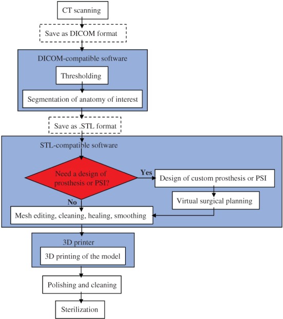
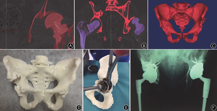
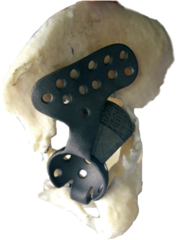
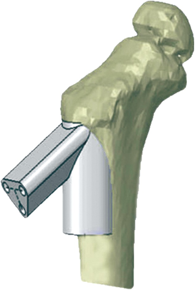
Three-dimensional (3D) printing is a digital rapid prototyping technology based on a discrete and heap-forming principle. We identified 53 articles from PubMed by searching "Hip" and "Printing, Three-Dimensional"; 52 of the articles were published from 2015 onwards and were, therefore, initially considered and discussed. Clinical application of the 3D printing technique in the hip joint mainly includes three aspects: a 3D-printed bony 1:1 scale model, a custom prosthesis, and patient-specific instruments (PSI). Compared with 2-dimensional image, the shape of bone can be obtained more directly from a 1:1 scale model, which may be beneficial for preoperative evaluation and surgical planning. Custom prostheses can be devised on the basis of radiological images, to not only eliminate the fissure between the prosthesis and the patient's bone but also potentially resulting in the 3D-printed prosthesis functioning better. As an alternative support to intraoperative computer navigation, PSI can anchor to a specially appointed position on the patient's bone to make accurate bone cuts during surgery following a precise design preoperatively. The 3D printing technique could improve the surgeon's efficiency in the operating room, shorten operative times, and reduce exposure to radiation. Well known for its customization, 3D printing technology presents new potential for treating complex hip joint disease.

Keywords: Hip Joint; Patient-Specific Modeling; Printing, Three-Dimensional; Prosthesis Design.


Figures

Figure 1
Figure 1
Whole process of three‐dimensional (3D) printing. CT scan obtained from the patients should be saved as digital imaging and communications in medicine (DICOM) format and imported into DICOM‐compatible software to conduct thresholding and segmentation of anatomy of interest. Then, the file should be saved in stereolithographic (.STL) format to be communicated to the STL‐compatible postprocessing software for further mesh editing, cleaning, healing, and smoothing. We can also design custom prostheses or patient‐specific instruments (PSI) for virtual surgical planning if necessary. Finally, the. STL file is imported into a 3D printer to result in the formation of 3D structures.
Figure 2
Figure 2
A, Image thresholding was performed by using software, which allowed for bone to be differentiated from surrounding soft tissue based on bone and soft tissue densities on the CT scan; B, Using the region growing process, both femurs were digitally segmented from their corresponding pelvis. The red pelvis will be retained, while the purple femurs will be removed; C, Once both femurs were erased, the 3D isolated image of pelvis (namely, the anatomy of interest) was created; D, The final life‐size 3D‐printed pelvis model, providing the surgeon with visual and tactile appreciation of the defects; E, Acetabular cup, augment, and buttress sizes, as well as cage dimensions were selected and trialed in preoperative surgical stimulation using a 3D‐printed pelvis; F, Postoperative anteroposterior pelvic plain film radiographs showed satisfactory revision total hip arthroplasty in situ 17.
Figure 3
Figure 3
A custom cage with an iliac braid to ensure enough screws could be used for firm fixation and a 3D‐printed augment to the superior surface of the cage for stable support51.
Figure 4
Figure 4
After reduction of the femoral neck fracture through software simulation, a navigation instrument was designed by the computer to conform to the proximal femur. The instrument provided optimal screw path and screw length for guiding pins and screws, which could be used to fix the Locking Compression Pediatric Hip Plate onto the fractured femoral neck during surgery56.
Figure 5
Figure 5
A, A 3D‐printed patient‐specific instrument (PSI) has a cutting slit that matches the planned resection planes. The black region represents the tumor. The K‐wire holes can stabilize PSI to the bone61; B, A 3D‐printed PSI, which can be used as an osteotomy guide plate. The black region represents the tumor. The flanges of the PSI allow a unique position on the bone surface; the K‐wire holes on the flanges can stabilize PSI on the bone 47.
Figure 6
Figure 6
Numbers of published articles by content of the study.
Figure 7
Figure 7
A graphic history of the published studies addressing both 3D printing and hip joint disease in the past 5 years.

References

    1. Ulrich SD, Seyler TM, Bennett D, et al Total hip arthroplasties: what are the reasons for revision?. Int Orthop, 2008, 32: 597–604. - PMC - PubMed
    1. Bagaria V, Rasalkar D, Bagaria SJ, Ilyas J. Medical Applications of Rapid Prototyping—a New Horizon. London, UK: IntechOpen Limited, 2011. 10.5772/20058. - DOI
    1. Gross BC, Erkal JL, Lockwood SY, Chen C, Spence DM. Evaluation of 3D printing and its potential impact on biotechnology and the chemical sciences. Anal Chem, 2014, 86: 3240–3253. - PubMed
    1. Zerr J, Chatzinoff Y, Chopra R, Estrera K, Chhabra A. Three‐dimensional printing for preoperative planning of total hip arthroplasty revision: case report. Skeletal Radiol, 2016, 45: 1431–1435. - PubMed
    1. Zeng CJ, Huang WH, Huang HJ, Wu ZL. Laparoscopic acetabular fracture fixation after three‐dimensional modelling and printing. Indian J Orthop, 2017, 51: 620–623. - PMC - PubMed