

The embryology of persistent cloaca and urogenital sinus malformations
- PMID: 31322137
- PMCID: PMC7155797
- DOI: 10.4103/aja.aja_72_19
The embryology of persistent cloaca and urogenital sinus malformations
Abstract
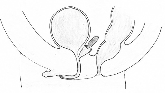
Cloacal malformations are characterized by the confluence of the lower urinary tract, the female reproductive tract, and the rectum to create a common channel with a single opening on the perineum. The presence of a cloaca is a normal phase of early human embryological development. Between the 4th and 7th weeks of gestation, the cloaca undergoes subdivision to form the hindgut and urogenital sinus. Failure of this process results in the congenital anomaly termed persistent cloaca (PC). The term urorectal septum malformation sequence (URSMS) is also used to describe this anomaly. The classic description of this process which is still cited in many standard textbooks dates from the 19th century. However, this has been increasingly called into question by the findings of studies using modern scientific methodology. Urogenital sinus anomalies are defined by the confluence of the urethra and vagina to form a common channel of varying length with a single perineal opening. In this condition, the anorectal canal opens separately on the perineum. The presence of a urogenital sinus represents a transient phase of the normal development of the lower genital tract in the female fetus. However, the form of urogenital sinus most commonly encountered in the developed world is a feature of disordered sexual differentiation and does not arise simply from the persistence of the anatomical structure which is a feature of normal fetal development.
Keywords: cloaca; embryology; urogenital sinus.
Conflict of interest statement
None
Figures

References
-
- Tennant PW, Glinianaia SV, Wellesley D, Draper ES, Kurinczuk JJ, et al. Epidemiology of partial urorectal septum malformation sequence (or 'persistent cloaca'): a population-based study in seven regions of England and Wales, 1985-2010. Arch Dis Child Fetal Neonatal Ed. 2014;99:413–8. - PubMed
-
- Sheldon C, Defoor W. Anorectal malformation and cloaca The Kelalis-King-Belman Textbook of Clinical Pediatric Urology. 5th ed. London: Informa; 2007. pp. 1161–86.
-
- Levitt MA, Peña A. Cloacal malformations: lessons learned from 490 cases. Semin Pediatr Surg. 2010;19:128–38. - PubMed
Publication types
MeSH terms
LinkOut - more resources
Full Text Sources
