Review
. 2019 Nov-Dec;1862(11-12):194405.
doi: 10.1016/j.bbagrm.2019.07.006. Epub 2019 Jul 16.

Repeat-associated RNA structure and aberrant splicing

Affiliations
Review

Repeat-associated RNA structure and aberrant splicing

Melissa A Hale et al. Biochim Biophys Acta Gene Regul Mech. 2019 Nov-Dec.

Abstract

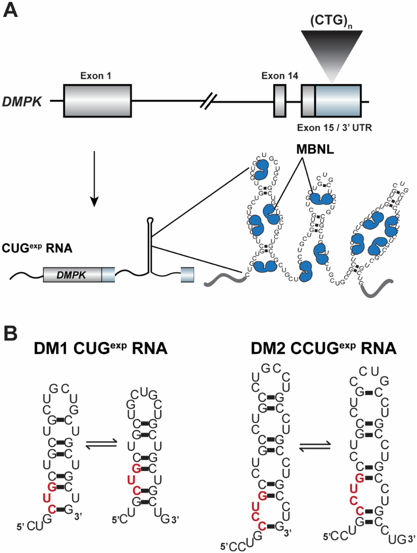
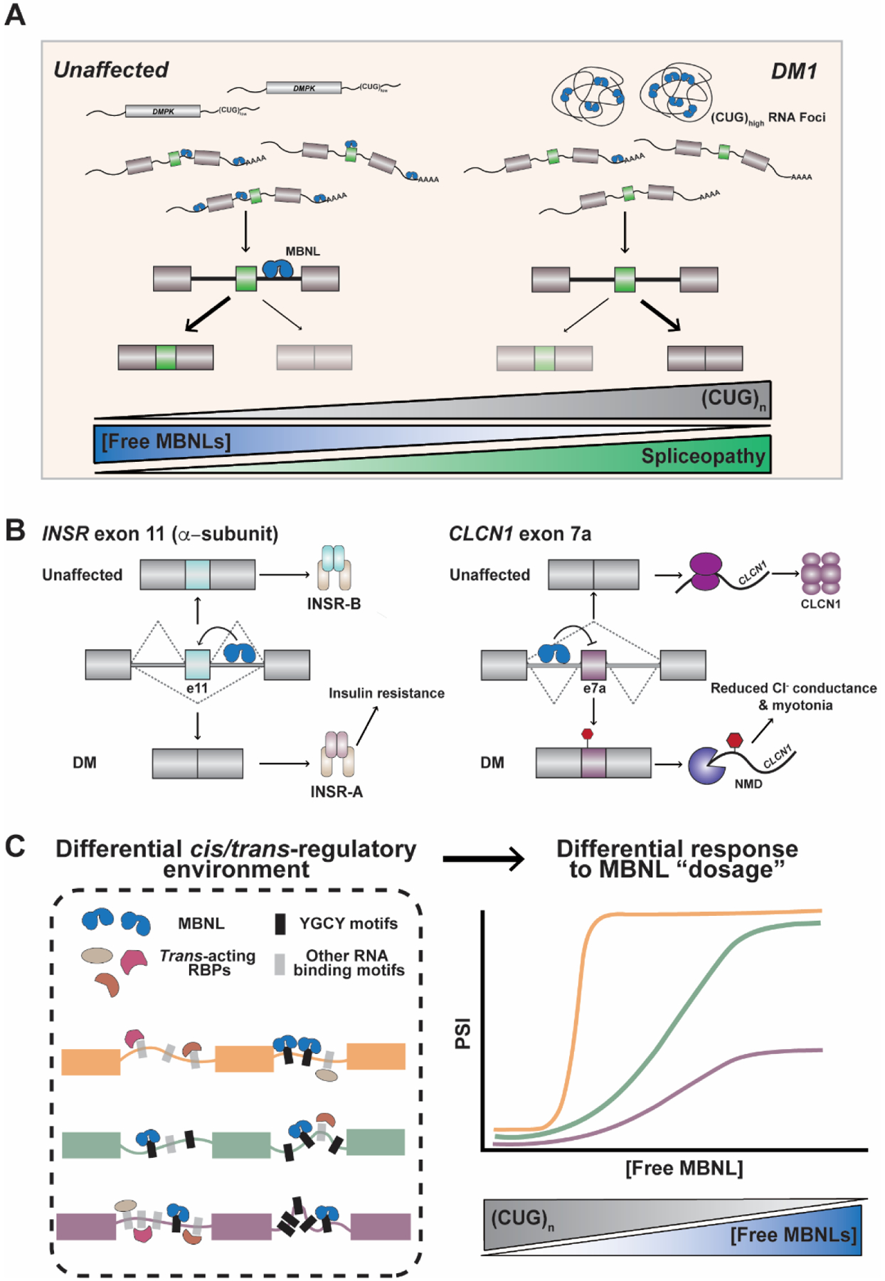
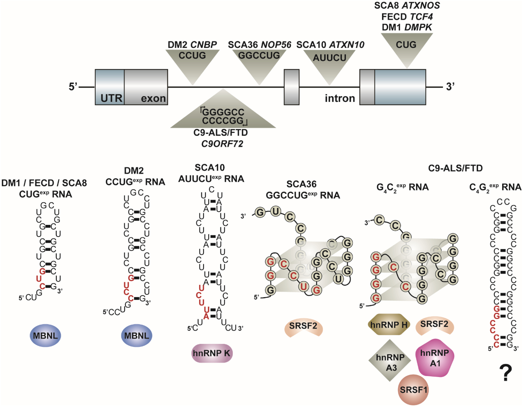
Over 30 hereditary disorders attributed to the expansion of microsatellite repeats have been identified. Despite variant nucleotide content, number of consecutive repeats, and different locations in the genome, many of these diseases have pathogenic RNA gain-of-function mechanisms. The repeat-containing RNAs can form structures in vitro predicted to contribute to the disease through assembly of intracellular RNA aggregates termed foci. The expanded repeat RNAs within these foci sequester RNA binding proteins (RBPs) with important roles in the regulation of RNA metabolism, most notably alternative splicing (AS). These deleterious interactions lead to downstream alterations in transcriptome-wide AS directly linked with disease symptoms. This review summarizes existing knowledge about the association between the repeat RNA structures and RBPs as well as the resulting aberrant AS patterns, specifically in the context of myotonic dystrophy. The connection between toxic, structured RNAs and dysregulation of AS in other repeat expansion diseases is also discussed. This article is part of a Special Issue entitled: RNA structure and splicing regulation edited by Francisco Baralle, Ravindra Singh and Stefan Stamm.

Keywords: Alternative splicing; Muscleblind-like (MBNL); Myotonic dystrophy; RNA binding proteins; RNA structure; Repeat expansion diseases.


Figures

Figure 1:
Figure 1:
The CUGexp / CCUGexp toxic RNAs that cause myotonic dystrophy type 1 and 2 (DM1 and DM2), respectively, have been shown to form “slippery hairpins” in vitro that capture and sequester muscleblind-like (MBNL) proteins. A) The expanded CTG repeat in the DMPK 3’ UTR (colored light blue, coding sequence in grey) is transcribed into CUGexp RNAs that consist of consecutive 5’-UGCU-3’ MBNL RNA binding motifs. MBNL proteins bind to and are sequestered by these RNAs in ribonuclear aggregates known as foci. B) Structural studies have revealed that the CUGexp / CCUGexp RNAs form “slippery hairpins” in vitro in which the CUG repeats dynamically shift and realign. These experimental observations suggest that these RNAs likely fold into metastable, dynamic secondary structures in vivo, although an outstanding question in the field is what structure(s) the CUG / CCUG repeats adopt in vivo. A single repeat unit is highlighted in red in each model RNA structure.
Figure 2:
Figure 2:
Sequestration of MBNL proteins by the toxic DM1 CUGexp RNA leads to transcriptome-wide, MBNL dose-dependent changes in alternative splicing. A) As the CUG repeat number increases, the concentration of free, functional MBNL proteins is progressively reduced as these AS factors become sequestered within RNA foci. Greater sequestration of MBNL leads to enhanced mis-splicing. B) Mis-splicing of MBNL-dependent mRNAs including INSR and CLCN1 leads to disease symptoms in DM patients. MBNL drives exon 11 inclusion within the INSR pre-mRNA by binding to a downstream intronic splicing enhancer. Inclusion of this exon results in the INSR-B protein isoform. When functional MBNL concentrations are reduced in DM, exon 11 is excluded and the less active INSR-A isoform is produced, leading to impaired glucose metabolism and insulin insensitivity. In the context of CLCN1, MBNL proteins drive exclusion of exon 7a. This exon contains a pre-mature stop codon and degradation of the mRNA transcript occurs via nonsense-mediated decay (NMD) when included in the final mRNA product as a result of the sequestration of MBNL. As such, total levels of CLCN1 are reduced and patients experience myotonia as a consequence of reduced chloride ion conductance in skeletal muscle. C) Organization of binding motifs in pre-mRNAs (cis-acting) and the complement of RBPs (trans-acting) within a specific tissue results in differential MBNL dose-dependent splicing regulation of events as measured by exon inclusion, or percent spliced in (PSI). More specifically, splicing events require different MBNL concentrations to reach cell and tissue-specific appropriate responses, making some events markedly sensitive to changes in MBNL concentration as a consequence of CUGexp RNA expression.
Figure 3:
Figure 3:
Expanded repeat RNAs transcribed from many microsatellite repeat expansions associated with genetic disease form secondary structures in vitro predicted to sequester RBPs that regulate alternative splicing. These expanded repeats are located within different regions of genomic architecture and can be transcribed bi-directionally (see C9-ALS/FTD). In vitro studies indicate that these repeat RNAs form secondary structures that classify into two general groups – A-form RNA helices or G-quadruplexes – as shown by these model RNA structures. A single repeat unit is highlighted in red in each model RNA structure. These toxic RNAs are found within ribonuclear foci and have been shown to co-localize with several RBPs that act as alternative splicing (AS) factors as listed below each RNA structure. The sequestration of these RBPs leads to disease-associated mis-splicing of multiple target pre-mRNAs. Outstanding questions in the field include (1) what structure(s) these repeat RNAs adopt in vivo and (2) the role of RNA secondary structure in modulating the interaction of RBPs with the toxic RNA and subsequent downstream dysregulation of alternative splicing and other RNA metabolic processes.

References

    1. Gerstein MB, Rozowsky J, Yan K-K, Wang D, Cheng C, Brown JB, et al., Comparative analysis of the transcriptome across distant species, Nature. 512 (2014) 445–448. doi:10.1038/nature13424. - DOI - PMC - PubMed
    1. Wang ET, Sandberg R, Luo S, Khrebtukova I, Zhang L, Mayr C, et al., Alternative isoform regulation in human tissue transcriptomes, Nature. 456 (2008) 470–476. doi:10.1038/nature07509. - DOI - PMC - PubMed
    1. Pan Q, Shai O, Lee LJ, Frey BJ, Blencowe BJ, Deep surveying of alternative splicing complexity in the human transcriptome by high-throughput sequencing, Nat. Genet 40 (2008) 1413–1415. doi:10.1038/ng.259. - DOI - PubMed
    1. ENCODE Project Consortium, An integrated encyclopedia of DNA elements in the human genome, Nature. 489 (2012) 57–74. doi:10.1038/nature11247. - DOI - PMC - PubMed
    1. Baralle FE, Giudice J, Alternative splicing as a regulator of development and tissue identity, Nat. Rev. Mol. Cell Biol 18 (2017) 437–451. doi:10.1038/nrm.2017.27. - DOI - PMC - PubMed

Publication types