. 2020 Jan;45(2):374-383.
doi: 10.1038/s41386-019-0466-9. Epub 2019 Jul 19.

Chronic adolescent exposure to ∆9-tetrahydrocannabinol decreases NMDA current and extrasynaptic plasmalemmal density of NMDA GluN1 subunits in the prelimbic cortex of adult male mice

Affiliations

Chronic adolescent exposure to ∆9-tetrahydrocannabinol decreases NMDA current and extrasynaptic plasmalemmal density of NMDA GluN1 subunits in the prelimbic cortex of adult male mice

Virginia M Pickel et al. Neuropsychopharmacology. 2020 Jan.

Abstract

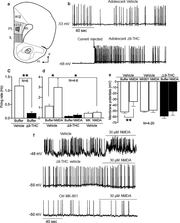
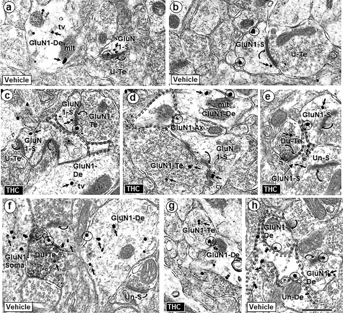
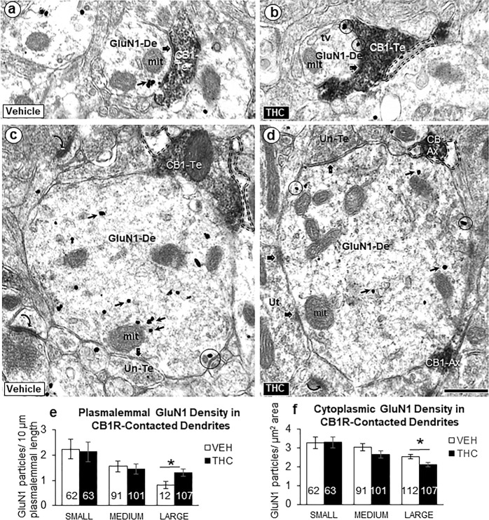
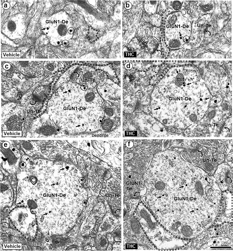
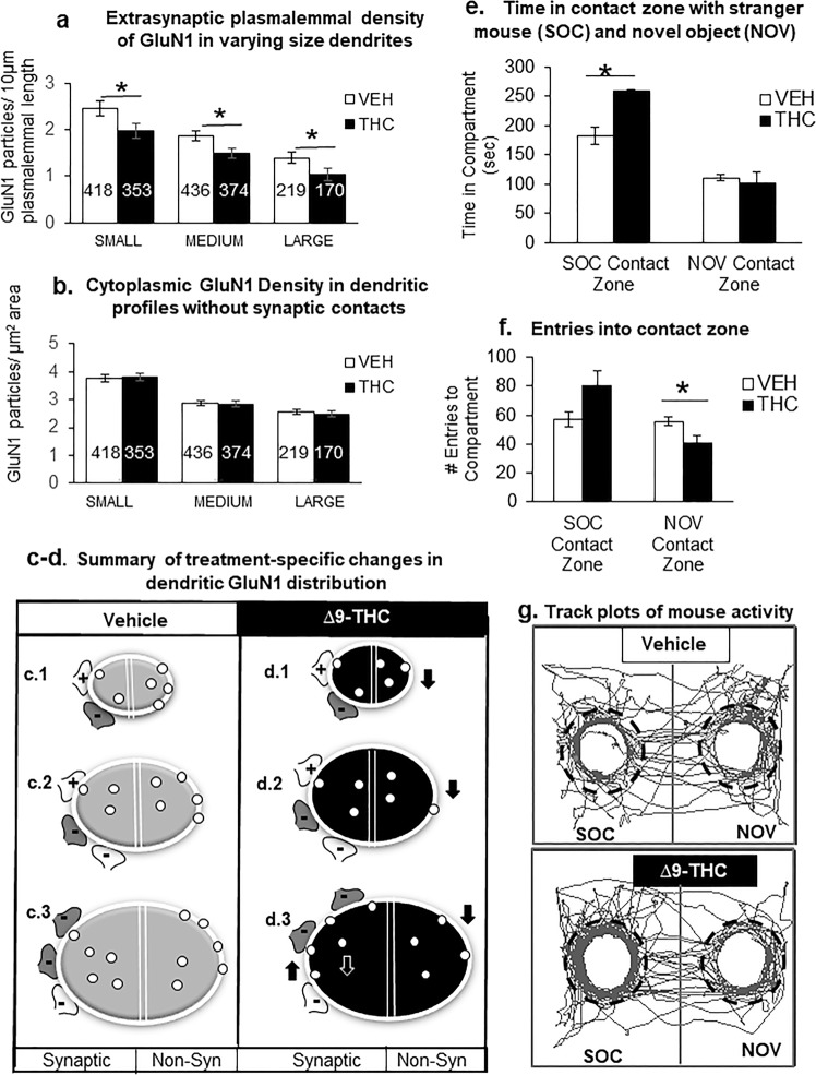
Adolescence is a vulnerable period of development when limbic connection of the prefrontal cortex (PFC) involved in emotional processing may be rendered dysfunctional by chronic exposure to delta-9-tetrahydrocannabinol (∆9-THC), the major psychoactive compound in marijuana. Cannabinoid-1 receptors (CB1Rs) largely mediate the central neural effects of ∆9-THC and endocannabinoids that regulate NMDA receptor-dependent synaptic plasticity of glutamatergic synapses in the prelimbic prefrontal cortex (PL-PFC). Thus, chronic occupancy of CB1Rs by ∆9-THC during adolescence may competitively decrease the functional expression and activity of NMDA receptors in the mature PL-PFC. We used a multidisciplinary approach to test this hypothesis in adult C57BL/6J male mice that received vehicle or ∆9-THC in escalating doses (2.5-10 mg/kg/ip) through adolescence (postnatal day 29-43). In comparison with vehicle, the mice receiving ∆9-THC showed a hyperpolarized resting membrane potential, decreased spontaneous firing rate, increased current-induced firing threshold, and decreased depolarizing response to NMDA in deep-layer PL-PFC neurons analyzed by current-clamp recordings. Electron microscopic immunolabeling in the PL-PFC of adult mice that had received Δ9-THC only during adolescence showed a significant (1) decrease in the extrasynaptic plasmalemmal density of obligatory GluN1-NMDA subunits in dendrites of all sizes and (2) a shift from cytoplasmic to plasmalemmal distribution of GluN1 in large dendrites receiving mainly inhibitory-type synapses from CB1R-labeled terminals. From these results and concomitant behavioral studies, we conclude that social dysfunctions resulting from excessive intake of ∆9-THC in the increasingly available marijuana products used by male teens may largely reflect circuit defects in PL-PFC networks communicating through endocannabinoid-regulated NMDA receptors.


Figures

Fig. 1
Fig. 1
Adolescent exposure to ∆9-THC decreases activity of PL-PFC neurons. a Schematic diagram of the prelimbic (PL) region of the PFC, which was used for current-clamp recording in layer 5 and electron microscopic analysis (trapezoid) in layers 2/3 of the PL-PFC. Other identified brain structures include anterior cingulate cortex (acg), infralimbic cortex (IL), forceps minor corpus callosum (fmi), and anterior commissure (ac). Arrows point medial (m) and dorsal (d) in this modified drawing of a half coronal section at 1.98 mm anterior to Bregma as seen in the mouse brain atlas [19]. b Representative traces of the membrane potential and spontaneous discharge in patch-clamp recordings of deep layer PL-PFC neurons from adult mice receiving vehicle (upper trace) or ∆9-THC (lower trace) during adolescence. The spontaneous spikes seen in slices from vehicle-injected adolescent mice are absent in recordings from a PL-PFC neuron in a mouse receiving ∆9-THC in adolescence, which shows a firing pattern that is dependent on the injected current. c Bar graphs summarize the changes in spontaneous firing rate in PL-PFC neurons of the vehicle vs. the ∆9-THC groups (from 1.18 ± 0.17 Hz of vehicle to 0.19 ± 0.05 Hz of ∆9-THC, P < 0.01, N = 6). d The presence of the NMDA receptor antagonist MK-801 (10 µM) blocked NMDA (30 µM)-induced increase in firing rate (from 3.0 Hz ± 0.86 Hz of vehicle to 0.57 ± 0.11 Hz of ∆9-THC, P < 0.05, N = 4–5). e Bar graphs summarize changes in RMP in PL-PFC neurons in vehicle compared with ∆9-THC-injected mice. NMDA-induced depolarization in mice receiving vehicle injections during adolescence is abolished in mice receiving ∆9-THC or in the presence of MK-801 (from 3.0 ± 0.86 Hz of vehicle to 0.48 ± 0.12 Hz of ∆9-THC, P < 0.05, N = 4–5). The presence of the NMDA (100 µM) induce depolarization in the vehicle groups (from −52.2 ± 2.1 mV of vehicle to −34.4 ± 7.8 mV of NMDA, P < 0.05, N = 4–5). However, there is no depolarization induced by NMDA in ∆9-THC or MK-801-treated groups (P > 0.05, N = 4–12). f Representative traces from PL-PFC neurons from mice receiving ∆9-THC (middle trace) does not result in depolarizing responses to NMDA as seen in mice receiving vehicle (upper trace). The presence of MK-801 inhibited the NMDA-induced increase in firing rate and depolarization (lower trace). N = number of mice per treatment group. Unpaired t-test *p < 0.05
Fig. 2
Fig. 2
Similarities of GluN1-immunogold distribution in neuronal profiles contacted by other neuronal and glial structures in the PL-PFC of adult mice that received vehicle or Δ9-THC. GluN1 is seen within the cytoplasm (small arrows) and on plasma membranes (circles) of selective neuronal (dendritic and axonal) and perisynaptic glial profiles in the PL-PFC of adult mice that received vehicle (a, b) or ∆9-THC (c, d) during adolescence. In these images GluN1-immunogold particles are seen in dendritic spines (GluN1-s) receiving asymmetric synapses (curved arrows) from unlabeled terminals or GluN1-labeled terminals. GluN1 immunogold is also respectively localized together (Du-Te) or separate (GluN1-Te) from dense CB1R-immunoperoxidase labeling in axon terminals forming symmetric synapses (block arrows) with GluN1-labeled somatodendritic profiles (GluN1 Soma) and dendrites (GluN1-De). e Dual labeled terminals (Du-te) contain sparse gold GluN1 and CB1-immunoperoxidase labeling (chevron). In f, unlabeled axon terminals form asymmetric synapses (curved arrows) with a dendritic spine that is contacted by the dual labeled terminal (upper left) and an unlabeled dendritic spine (Un-s) opposed to the GluN1-De. tv = tubulovesicles (tv), mit = mitochondrion, U-Te = unlabeled terminal; Un-S = unlabeled spine; scale bar = 500 nm. A GluN1-immunogold labeled dendrite (GluN1-De) is opposed to a GluN1-labeled terminal (GluN1-Te) and to a perisynaptic glial process (double dashed line) in g and h, respectively
Fig. 3
Fig. 3
Postsynaptic GluN1-immunogold labeling in dendrites receiving synaptic contacts from CB1R-labeled terminals. Electron microscopic images (a–d) and bar graphs (e, f) showing size-dependent changes in the plasmalemmal and cytoplasmic GluN1-immunogold distribution in dendrites recipient to inputs from small axons or terminals that are unlabeled or contain immunoperoxidase labeling for CB1Rs in PL-PFC of adult mice receiving ∆9-THC compared with vehicle. GluN1 immunogold is seen on the plasma membrane (circles) and in the cytoplasm (small arrows) of small (a, b) and large (c, d) dendritic profiles (GluN1-De). These profiles receive symmetric synapses (block arrows) from axon terminals that are unlabeled (Un-Te) or contain dense immunoperoxidase labeling for the CB1R (CB1-Te). The GluN1-labeled dendrites are opposed to glial processes (profiles outlined with dashed line) in bd and to a CB1R-labeled axon (CB1-Ax) in d. Curved arrows = asymmetric excitatory-type axospinous synapses in the neuropil of panel c. Scale bar = 500 nm. Bar graphs of e and f show the number of GluN1-labeled dendritic shafts in 21,170 µm2 of PL-PFC tissue collected equally from ~70 microscopic images in two vibratome sections from five THC and five VEH-injected adult mice. ANOVA *p < 0.05
Fig. 4
Fig. 4
Extrasynaptic GluN1-immunogold labeling in dendritic shafts in PL-PFC adult mice receiving vehicle or ∆9-THC during adolescence. The gold particles are located on the plasma membrane (circles) and in the cytoplasm (small arrows) of dendritic shafts (GluN1-De) having small (a, b), medium (c, d), and large (e, f) mean diameters. Many of these dendrites are opposed to glial processes (profiles outlined with dashed line) and to unlabeled (Un-Te) or GluN1-immunogold-labeled terminals (GluN1-Te) forming asymmetric synapses (curved arrows) on nearby dendritic spines. Scale bar = 500 nm
Fig. 5
Fig. 5
Adult mice chronically exposed to ∆9-THC during adolescents differ from vehicle controls in their extrasynaptic GluN1 distribution in varying sized dendrites within the PL-PFC (a–d) and interactions with a stranger mouse (SOC) vs. novel object (NOV) (e–g). In a and b, the bar graphs respectively show that adolescent ∆9-THC produces a significant decrease in extrasynaptic plasmalemmal GluN1 in small, medium and large dendrites, without effect on GluN1 cytoplasmic distribution in the PL-PFC dendrites of adult mice. In a and b, ANOVA *p < 0.05 and numbers = number of GluN1-labeled dendrites. Schematic drawings in c and d respectively summarize the GluN1-immunogold distribution in PL-PFC dendrites of adult mice receiving ∆9-THC or vehicle during adolescents. In each treatment group, transverse sections through small (c.1), medium (c.2), and large (c.3) dendrites are represented by oval shapes with a white line drawn around the perimeter to indicate the plasmalemma that encloses the cytoplasmic compartment. Each of these dendritic profiles is bisected by a double line to separate portions that are contacted by excitatory (+) and inhibitory (−) axon terminals (i.e., synaptic) or without axonal contact (i.e., non-synaptic (non-Syn)). CB1R labeling is shown by dark shading in a subgroup of the inhibitory-type terminals. Data from bar graphs in Fig. 3e, f are schematically shown by an increase (up arrow) in plasmalemmal density and the corresponding decrease (down white arrow) in the cytoplasmic density of GluN1-immunogold particles (small circles) exclusively in large dendrites of mice receiving ∆9-THC compared with vehicle. In contrast, data summarized from bar graphs in this figure show a decrease (down arrow) in GluN1-immunogold density on the extrasynaptic plasma membrane of all dendrites without synaptic contact (i.e., non-synaptic). Bar graphs in e and f respectively show that administration of ∆9-THC compared with vehicle during adolescence significantly increases the time spent in portions of the chamber in contact with a stranger mouse and decreases the entries into the contact zone surrounding a novel object. ANOVA shows a statistically significant treatment effect on (ambulatory time: (F(1,10) = 12.07, p = 0.006) and number of entries: (F(1,10) = 11.03, p = 0.008). g Representative tracking of adult mice that received vehicle or ∆9-THC during adolescence shows that ∆9-THC enhances the mobilization to portions of the two interconnected compartments within which dashed lines indicate the perimeter of the contact zone around the stranger mouse (SOC) and novel object (NOV)

References

    1. Hashimotodani Y, Ohno-Shosaku T, Kano M. Endocannabinoids and synaptic function in the CNS. Neuroscientist. 2007;13:127–37. - PubMed
    1. Fortin DA, Levine ES. Differential effects of endocannabinoids on glutamatergic and GABAergic inputs to layer 5 pyramidal neurons. Cereb Cortex. 2007;17:163–74. - PubMed
    1. Auclair N, Otani S, Soubrie P, Crepel F. Cannabinoids modulate synaptic strength and plasticity at glutamatergic synapses of rat prefrontal cortex pyramidal neurons. J Neurophysiol. 2000;83:3287–93. - PubMed
    1. Murphy M, Mills S, Winstone J, Leishman E, Wager-Miller J, Bradshaw H, et al. Chronic adolescent delta(9)-tetrahydrocannabinol treatment of male mice leads to long-term cognitive and behavioral dysfunction, which are prevented by concurrent cannabidiol treatment. Cannabis Cannabinoid Res. 2017;2:235–46. - PMC - PubMed
    1. Dow-Edwards D, Silva L. Endocannabinoids in brain plasticity: cortical maturation, HPA axis function and behavior. Brain Res. 2017;1654(Pt B):157–64. - PubMed

Publication types

MeSH terms