. 2019 Jul 18;20(14):3520.
doi: 10.3390/ijms20143520.

Suppression of Brown Adipocyte Autophagy Improves Energy Metabolism by Regulating Mitochondrial Turnover

Affiliations

Suppression of Brown Adipocyte Autophagy Improves Energy Metabolism by Regulating Mitochondrial Turnover

Donghwan Kim et al. Int J Mol Sci. .

Abstract

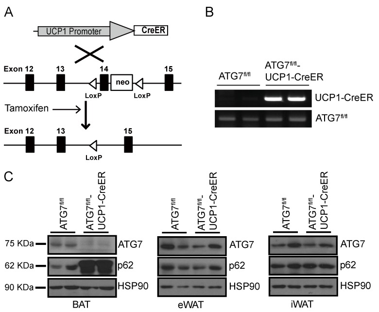
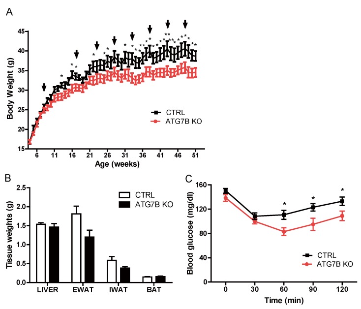
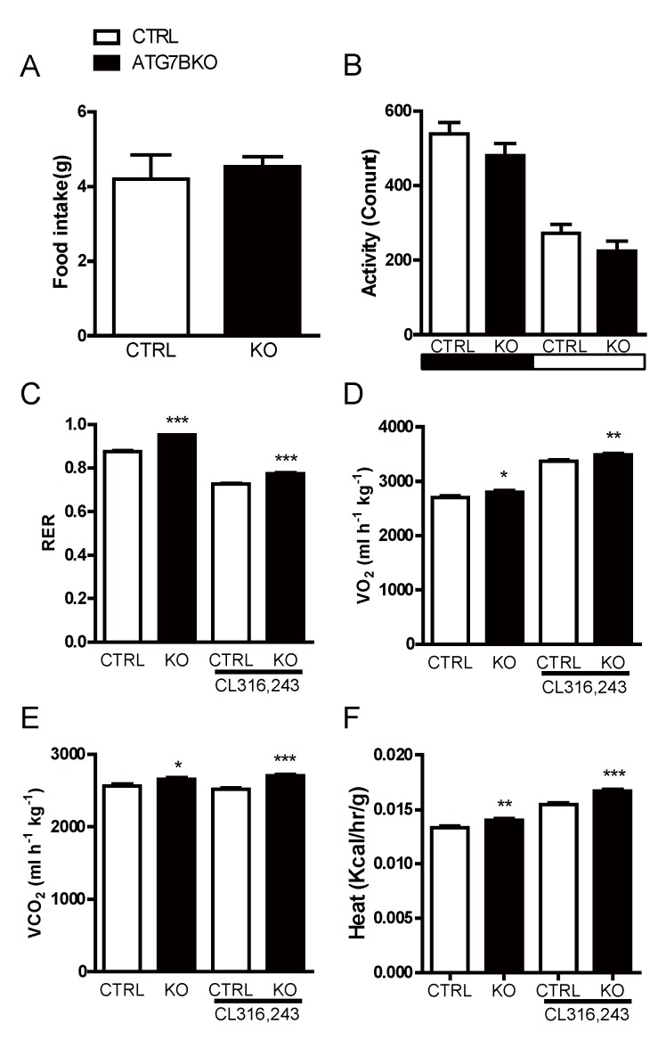
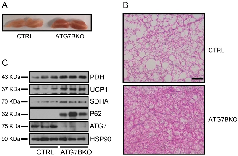
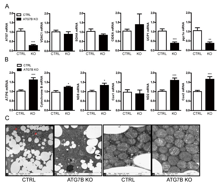
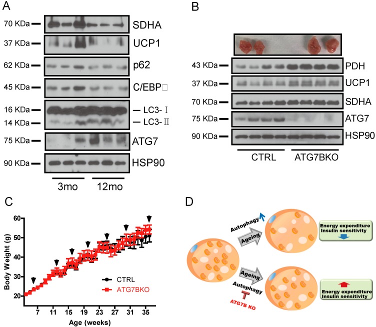
The high abundance of mitochondria and the expression of mitochondrial uncoupling protein 1 (UCP1) confer upon brown adipose tissue (BAT) the unique capacity to convert chemical energy into heat at the expense of ATP synthesis. It was long believed that BAT is present only in infants, and so, it was not considered as a potential therapeutic target for metabolic syndrome; however, the discovery of metabolically active BAT in adult humans has re-stimulated interest in the contributions of BAT metabolic regulation and dysfunction to health and disease. Here we demonstrate that brown adipocyte autophagy plays a critical role in the regulation BAT activity and systemic energy metabolism. Mice deficient in brown adipocyte autophagy due to BAT-specific deletion of Atg7-a gene essential for autophagosome generation-maintained higher mitochondrial content due to suppression of mitochondrial clearance and exhibited improved insulin sensitivity and energy metabolism. Autophagy was upregulated in BAT of older mice compared to younger mice, suggesting its involvement in the age-dependent decline of BAT activity and metabolic rate. These findings suggest that brown adipocyte autophagy plays a crucial role in metabolism and that targeting this pathway may be a potential therapeutic strategy for metabolic syndrome.

Keywords: aging; autophagy; brown adipose tissues; energy homeostasis; mitophagy.


Conflict of interest statement

The authors declare no conflict of interest

Figures

Figure 1
Figure 1
Generation of brown adipocyte-specific Atg7 knockout (ATG7B KO) mice. (A). Schematic illustration of the strategy for establishing brown adipocyte-specific conditional autophagy-deficient mice. Atg7fl/fl mice were crossed with uncoupling protein 1 (UCP1-CreER+/) transgenic mice, and tamoxifen was administered to induce the nuclear migration of CreER. (B). Genotyping results for Atg7fl/fl (control) mice and Atg7fl/fl-UCP1-CreER+/ (ATG7B KO) mice. The top panel shows the genotyping results for the UCP1-CreER allele and the bottom panel shows the genotyping results for the Atg7fl/fl allele. (C). Western blot analysis of ATG7 and p62 expression in brown adipose tissue (BAT) (left panel), epididymal white adipose tissue (eWAT; middle panel) and inguinal white adipose tissue (iWAT; right panel) of control and ATG7B KO mice.
Figure 2
Figure 2
Brown adipocyte-specific Atg7 knockout mice exhibit reduced body weight and improved insulin sensitivity. (A). Weekly body weight chart of control (n = 7) and ATG7B KO mice (n = 7), maintained on a normal chow diet. Arrows indicate weeks that control and ATG7B KO mice were treated with tamoxifen. (B). Weights of liver, eWAT, iWAT, and BAT from 1-year-old control (n = 7) and ATG7B KO mice (n = 7). (C). Insulin tolerance test (ITT) of normal chow-fed control (n = 7) and ATG7B KO mice (n = 7). Statistical significance was determined by Mann–Whitney test. * and ** represent p < 0.05 and p < 0.01 respectively.
Figure 3
Figure 3
Increased energy expenditure in ATG7B KO mice. Shown are comparisons of (A). 24-h food intake and (B). physical activity of control (n = 3) and ATG7B KO mice (n = 5) (C). Respiratory exchange ratio (RER), (D). relative oxygen consumption, (E). carbon dioxide production, and (F). energy expenditure of night-time basal and CL316,243 treated control (n = 4) and ATG7B KO mice (n = 4), maintained on a normal chow diet were analyzed using indirect calorimetry. Statistical significance was determined by the Mann–Whitney test. *, ** and *** represent p < 0.5, p < 0.01 and p < 0.001 respectively.
Figure 4
Figure 4
Increased mitochondrial content in ATG7B KO mice. (A). Appearance of BAT depots dissected from control and ATG7B KO mice. (B). Histological analysis of hematoxylin and eosin (H & E) stained sections of BAT from control and ATG7B KO mice. The scale bar represents 200 μm. (C). Immunoblotting for UCP1 and mitochondria-resident proteins PDH and SDHA in BAT depots of control and ATG7B KO mice.
Figure 5
Figure 5
Suppression of mitochondrial turnover in BAT of ATG7B KO mice. Relative mRNA levels of (A). ATG7, PGC1α and the chromosome-encoded mitochondrial proteins VDAC1, SDHA and CIDEA, and (B). the mitochondrial-encoded proteins ATP6, cytochrome b, ND2, ND5, Cox1, and Cox2 from BAT of control and ATG7B KO mice as quantified by quantitative reverse PCR (qRT-PCR). Statistical significance was determined by Mann–Whitney test. *, ** and *** represent p < 0.05, p < 0.01, and p < 0.001 respectively. (C). Transmission electron microscopy images of the BAT from control and ATG7B KO mice. Left two panels (3000×), right two panels (20,000×).
Figure 6
Figure 6
Age-associated changes of BAT autophagy and mitochondrial content. (A). The relative protein levels of mitochondrial (SDHA, UCP1) and autophagy (ATG7, p62) markers in BAT of young (3-month-old) and aged (12-month-old) mice (B). Appearance of BAT depots and immunoblotting results for ATG7, UCP1 and mitochondrial-resident proteins from BAT of control and ATG7B KO mice maintained on a 60% high-fat diet (HFD). (C). Body weight chart of control (n = 7) and ATG7B KO mice (n = 9) fed 60% HFD. Arrows indicate weeks that control and ATG7B KO mice were treated with tamoxifen. (D). Schematic illustration proposing the role of brown adipocyte autophagy in age-associated decline of BAT activity.

Similar articles

Cited by

References

    1. Cannon B., Nedergaard J. Brown adipose tissue: Function and physiological significance. Physiol. Rev. 2004;84:277–359. doi: 10.1152/physrev.00015.2003. - DOI - PubMed
    1. Smith R.E., Roberts J.C., Hittelman K.J. Nonphosphorylating respiration of mitochondria from brown adipose tissue of rats. Science. 1966;154:653–654. doi: 10.1126/science.154.3749.653. - DOI - PubMed
    1. Aquila H., Link T.A., Klingenberg M. The uncoupling protein from brown fat mitochondria is related to the mitochondrial ADP/ATP carrier. Analysis of sequence homologies and of folding of the protein in the membrane. EMBO J. 1985;4:2369–2376. doi: 10.1002/j.1460-2075.1985.tb03941.x. - DOI - PMC - PubMed
    1. Echtay K.S., Bienengraeber M., Mayinger P., Heimpel S., Winkler E., Druhmann D., Frischmuth K., Kamp F., Huang S.G. Uncoupling proteins: Martin Klingenberg’s contributions for 40 years. Arch. Biochem. Biophys. 2018;657:41–55. doi: 10.1016/j.abb.2018.09.006. - DOI - PubMed
    1. Chouchani E.T., Kazak L., Spiegelman B.M. New Advances in Adaptive Thermogenesis: UCP1 and Beyond. Cell Metab. 2019;29:27–37. doi: 10.1016/j.cmet.2018.11.002. - DOI - PubMed

Substances