. 2019 Jul 18;7(3):52.
doi: 10.3390/biomedicines7030052.

Differential Roles of Dendritic Cells in Expanding CD4 T Cells in Sepsis

Affiliations

Differential Roles of Dendritic Cells in Expanding CD4 T Cells in Sepsis

Samuel Darkwah et al. Biomedicines. .

Abstract

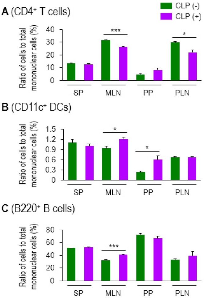
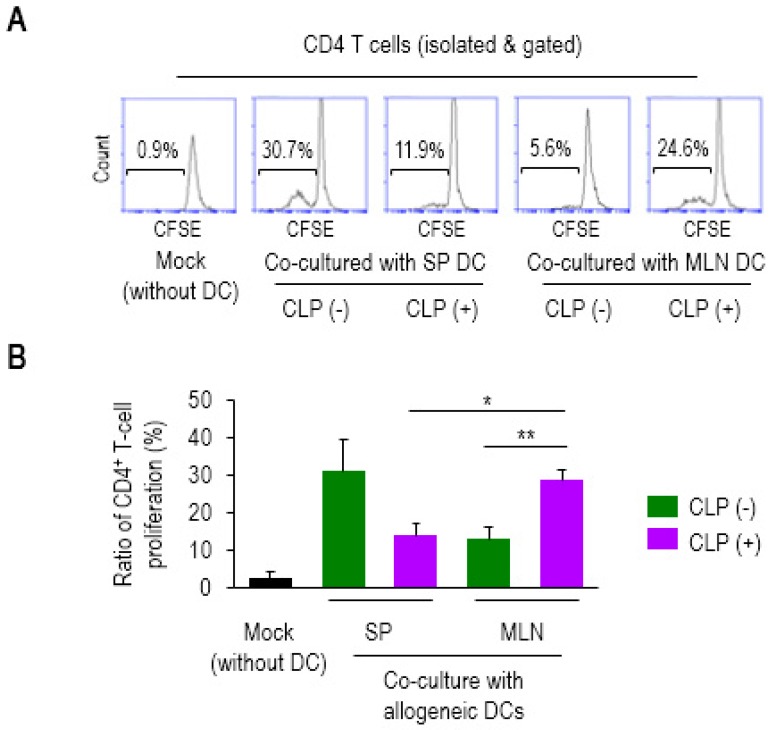
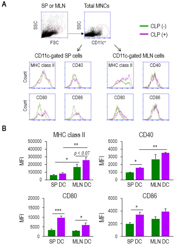
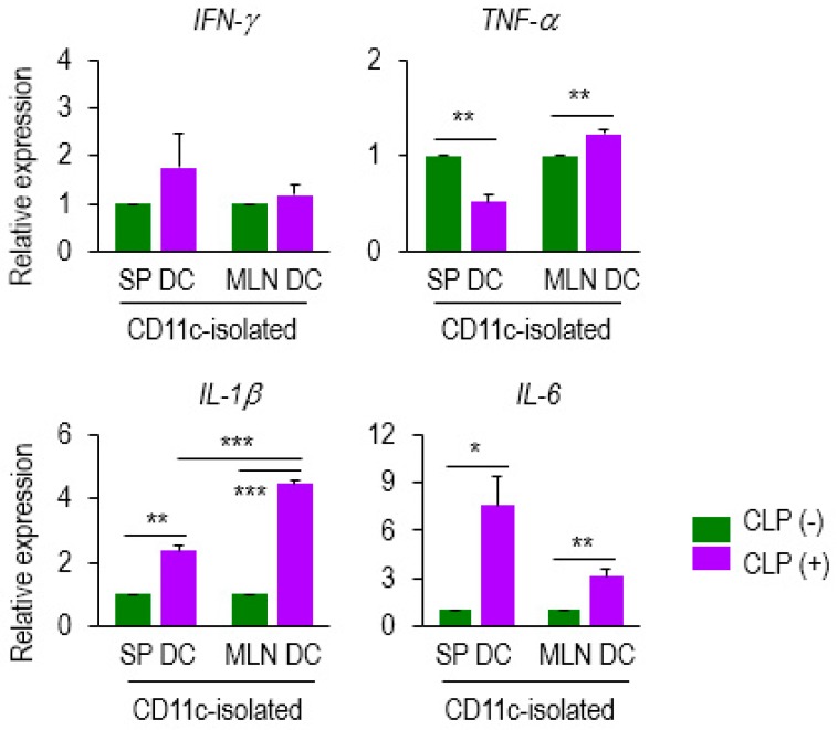
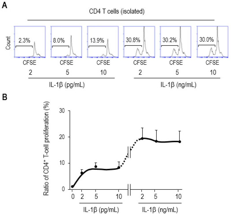
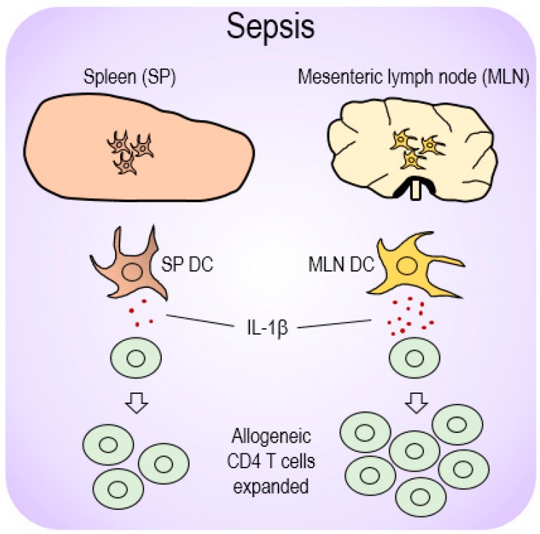
Sepsis is a systemically dysregulated inflammatory syndrome, in which dendritic cells (DCs) play a critical role in coordinating aberrant immunity. The aim of this study is to shed light on the differential roles played by systemic versus mucosal DCs in regulating immune responses in sepsis. We identified a differential impact of the systemic and mucosal DCs on proliferating allogenic CD4 T cells in a mouse model of sepsis. Despite the fact that the frequency of CD4 T cells was reduced in septic mice, septic mesenteric lymph node (MLN) DCs proved superior to septic spleen (SP) DCs in expanding allogeneic CD4 T cells. Moreover, septic MLN DCs markedly augmented the surface expression of MHC class II and CD40, as well as the messaging of interleukin-1β (IL-1β). Interestingly, IL-1β-treated CD4 T cells expanded in a dose-dependent manner, suggesting that this cytokine acts as a key mediator of MLN DCs in promoting septic inflammation. Thus, mucosal and systemic DCs were found to be functionally different in the way CD4 T cells respond during sepsis. Our study provides a molecular basis for DC activity, which can be differential in nature depending on location, whereby it induces septic inflammation or immune-paralysis.

Keywords: CD4 T cells; IL-1β; dendritic cells; mesenteric lymph nodes; sepsis; spleen.


Conflict of interest statement

The authors declare that they have no conflicts of interest with respect to the contents of this article. The funding sponsors had no role in the design of the study, collection, analyses, interpretation of data, writing of the manuscript, or the decision to publish the data.

Figures

Figure 1
Figure 1
Sepsis induced lymphopenia in CD4 T cells and elevated levels of CD11c+ DCs in lymphoid tissues. Ratio of CD4+ T cells (A), CD11c+ DCs (B), and B220+ B cells (C) to total mononuclear cells in spleen (SP), mesenteric lymph nodes (MLNs), Peyer’s patches (PPs), or peripheral lymph nodes (PLNs) was determined by using flow cytometry. For the PLN, inguinal LNs were isolated and used in this assay. CLP (−) indicates control healthy mice and CLP (+) represents CLP mice with moderate sepsis (see Methods section). Bar graphs represent the mean ± SEM obtained from 3 to 4 mice per group. Data are representative of at least three separate experiments. * 0.01 < p < 0.05, *** p < 0.001.
Figure 2
Figure 2
Mesenteric lymph node (MLN) dendritic cells (DCs) in sepsis enhanced the proliferation ratio of allogeneic CD4+ T cells. The CD4 T cells were isolated from SP of Balb/c mice, fluorescently labeled with CFSE and co-cultured with mock (without DC), SP (SP DC), or MLN (MLN DC) (isolated from the tissues of C57BL/6J mice) at a ratio of 4:1 for 7 days. The proliferation ratios were determined via measuring diluted fluorescent intensity of a histogram in flow cytometry in which the numbers inside squares represent the percentages of bracketed regions (A). Bar graphs represent the mean ± SEM obtained from 4 to 5 mice per group (B). Data are representative of at least three independent experiments. * 0.01 < p < 0.05, ** 0.01 < p < 0.001.
Figure 3
Figure 3
MLN DCs in sepsis elevated the surface expression of activation markers. (A) Mononuclear cells (MNCs) were isolated from single-cell suspensions of SP and MLNs. CD11c+ population was gated among total MNCs stained to determine the expression levels of APC markers including MHC class II, CD40, CD80, and CD86. Flow cytometry histograms show sepsis-induced change in their expression. (B) The bar graphs represent the mean ± SEM for MFI obtained from 4 to 5 mice per group, in which green and purple bars indicate without (−) and with (+) CLP, respectively. MFI, mean fluorescence intensity. Data are representative of at least three independent experiments. * 0.01 < p < 0.05, ** 0.01 < p < 0.001, *** p < 0.001.
Figure 4
Figure 4
DC expression of several mediators was altered at the mRNA level during sepsis. Expression of several genes (IFN-γ, TNF-α, IL-1β, and IL-6) was examined on CD11c+ DCs isolated from SP DCs and MLN DCs of either CLP (−) or (+) mice. Data from qPCR analysis were normalized to those controls that represented samples from CLP (−) mice after being normalized to a reference β-actin gene (2−ddCT). Bar graphs represent the mean ± SEM obtained from 4 to 5 mice per group. Data are representative of at least three separate experiments. * 0.01 < p < 0.05, ** 0.01 < p < 0.001, *** p < 0.001.
Figure 5
Figure 5
IL-1β augments CD4 T-cell proliferation. The CD4 T cells were isolated from single-cell suspensions of SP of Balb/c mice, fluorescently labeled with CFSE, treated with recombinant mouse IL-1β (rmIL-1β) at the indicated concentrations, and further incubated for 3 days. (A) Flow-cytometry histograms show representative results. The proliferation ratios were determined via measuring diluted fluorescent intensity of a histogram in flow cytometry in which the numbers inside squares represent the percentages of bracketed regions. (B) These results were shown as a line graph connected by the points that indicate different concentration of rmIL-1β used in the assay. The concentrations of exogenously treated IL-1β used in this study were similar to those in previous proof-of-the-principle studies involving ED50 (2–10 pg/mL) [45] (as also described in the datasheet provided by the manufacturer, R&D Systems). The 1000-times higher concentration (2–10 ng/mL) was included in this assay as well. Data are representative of at least two separate experiments.
Figure 6
Figure 6
A proposed model for differential DC-mediated immune responses depending on location during sepsis. Sepsis initiated upregulation of the pro-inflammatory cytokine, IL-1β, from those DCs present in mucosal lymphoid organs (MLN DCs) and subsequently elevated allogeneic CD4 T cells during MLR. However, SP DCs that produced relatively low levels of IL-1β restricted CD4 T-cell proliferation, and thereby presumably caused an immunosuppressive effect during sepsis.

Similar articles

Cited by

References

    1. Boomer J.S., To K., Chang K.C., Takasu O., Osborne D.F., Walton A.H., Bricker T.L., Jarman S.D., 2nd, Kreisel D., Krupnick A.S., et al. Immunosuppression in patients who die of sepsis and multiple organ failure. JAMA. 2011;306:2594–2605. doi: 10.1001/jama.2011.1829. - DOI - PMC - PubMed
    1. Angus D.C., van der Poll T. Severe sepsis and septic shock. N. Engl. J. Med. 2013;369:840–851. doi: 10.1056/NEJMra1208623. - DOI - PubMed
    1. Shimaoka M., Park E.J. Advances in understanding sepsis. Eur. J. Anaesthesiol. Suppl. 2008;42:146–153. doi: 10.1017/S0265021507003389. - DOI - PMC - PubMed
    1. Hotchkiss R.S., Monneret G., Payen D. Sepsis-induced immunosuppression: From cellular dysfunctions to immunotherapy. Nat. Rev. Immunol. 2013;13:862–874. doi: 10.1038/nri3552. - DOI - PMC - PubMed
    1. Delano M.J., Ward P.A. Sepsis-induced immune dysfunction: Can immune therapies reduce mortality? J. Clin. Investig. 2016;126:23–31. doi: 10.1172/JCI82224. - DOI - PMC - PubMed