. 2019 Jul 18;7(3):68.
doi: 10.3390/vaccines7030068.

Towards the Generation of an ASFV-pA104R DISC Mutant and a Complementary Cell Line-A Potential Methodology for the Production of a Vaccine Candidate

Affiliations

Towards the Generation of an ASFV-pA104R DISC Mutant and a Complementary Cell Line-A Potential Methodology for the Production of a Vaccine Candidate

Ferdinando B Freitas et al. Vaccines (Basel). .

Abstract

African swine fever (ASF) is a fatal viral disease of domestic swine and wild boar, considered one of the main threats for global pig husbandry. Despite enormous efforts, to date, neither the classical vaccine formulations nor the use of protein subunits proved to be efficient to prevent this disease. Under this scenario, new strategies have been proposed including the development of disabled infectious single cycle (DISC) or replication-defective mutants as potential immunizing agents against the ASF virus (ASFV). In this study, we describe the methodology to generate an ASFV-DISC mutant by homologous recombination, lacking the A104R gene, which was replaced by the selection marker (GUS gene). The recombinant viruses were identified when the infected cells acquired a blue color in the presence of X-Gluc (100 µg/mL), which is the substrate for the GUS gene. Since these viral particles result from loss-of-function mutations, being unable to replicate, helper-cell lines expressing the viral pA104R protein were produced. Vero and COS-1 cell lines were transfected by different methods, both physical and chemical, in order to stably express the ASFV-pA104R. Best results were obtained by using Lipofectamine 2000 and Nucleofection methodology of Vero with the pIRESneo vector and by using Flp-FRT site-directed recombination technology system in Flp-In CV-1 cells (transformed COS-1 cells with a single integration site in a transcriptional active region). In order to ensure an efficient and stable integration of the viral ORF on the host cellular genome, the maintenance of the insert was verified by PCR and its expression by immunofluorescence and immunoblot analysis. Although the isolation of the recombinant virus was not achieved, the confirmation of ASFV-ΔA104R sequence, and the detection of the recombinant mutant through three passages, suggest that this approach is feasible and could be a potential strategy to generate safe and efficient DISC vaccine candidates.

Keywords: ASFV; DISC; helper cell line; pA104R; vaccine.


Conflict of interest statement

The authors declare no conflict of interest, financial or otherwise.

Figures
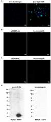
Figure A1
Figure A1
(A) Immunofluorescence studies showed ASFV-pA104R in the cytoplasm and nucleus of COS-1-pA104R cells (right image, green staining), in contrast with COS-1 wild type cells (left image). (B) Transformed Vero cells incubated with the in-house anti-pA104R antibody (left image) and with a goat anti-mouse secondary antibody (right image). (C) Transformed COS-1 cells incubated with the in-house anti-pA104R antibody (left image) and with a goat anti-mouse secondary antibody (right image). (D) Whole cell extracts of Vero cells non-infected and infected with Ba71V isolate (MOI = 2; 18 hpi) probed against the primary in-house antibody (anti-pA104R) and the secondary antibody (anti-mouse horseradish peroxidase-HRP).
Figure 1
Figure 1
Schematic overview to generate helper cell lines expressing pA104R. Vero/COS-1 cells were transfected with the linearized pIRESneo_ASFV-A104R vector and then were subjected to antibiotic selective pressure. The surviving clones were expanded and the Vero-pA104R/COS-1-pA104R cell lines were established.
Figure 2
Figure 2
Schematic representation of the methodology used to generate the deletion plasmid vector. For the deletion of the A104R gene from the genome of Ba71V isolate, a plasmid vector pΔA104R_GUS containing the B646L (p72) promoter and GUS gene flanked by the homology arms of the A104R gene (L arm and R arm) was constructed.
Figure 3
Figure 3
Schematic representation of the adopted strategy to generate the recombinant African swine fever (ASF) viruses. The target A104R gene was replaced by the reporter gene (GUS). The recombinant virus was obtained by homologous recombination between the left and right sequence regions (L arm and R arm) of the A104R gene on wild-type ASFV-Ba71V genome and recombination vector, resulting in the deletion of A104R gene and insertion of GUS gene under control of the strong viral promoter of the ASFV-VP72 gene.
Figure 4
Figure 4
Open reading frame (ORF) A104R was successfully integrated in transfected Vero and COS-1 cells. ORF A104R was amplified by PCR (287 bp) from the genome of Vero-pA104R clones, cultured under antibiotic selective pressure (G418). Line 1: Molecular marker (NZYDNA Ladder III); Line 2: Vero wild type (wt, negative control); Line 3: pIRESneo A104R (positive control); Lines 4–6: three distinct Vero-pA104R clones, transfected with linearized plasmid and Lipofectamine 2000; Lines 7,8: two distinct Vero-pA104R clones, transfected with circular plasmid and Lipofectamine 2000; Line 9: COS-1 wt cells (negative control); Line 10: COS-1 cells transfected by Lipofectamine 2000 with the A104R construct; Line 11: Flp-In wt cells; Line 12: Flp-In cells transformed with the A104R construct; Lines 13–15: COS-1 cells transfected with Lipofectamine LTX (16 weeks); Line 16: FRT/A104R plasmid (positive control); Lines 17–20: Flp-In clones sustaining the selective pressure.
Figure 5
Figure 5
Helper Vero cells expressing viral pA104R. (A) Immunofluorescence studies detected African swine fever virus (ASFV)-pA104R in the cytoplasm and nucleus of Vero-pA104R cells. pA104R and DAPI (4′,6-diamidino-2-phenylindole) staining is shown in green and blue, respectively. (B) Transformed Vero cells non-infected and infected with Ba71V isolate (multiplicities of infection (MOI) = 2; 18 h post infection (hpi)) were re-suspended into buffer containing 0.1% Tx-100 and fractionated. pA104R was only detected in the Triton X-100-insoluble fraction, as expected, both in non-infected and infected transfected cells. β-actin was used as loading control.
Figure 6
Figure 6
Isolation of recombinant virus using the plaques method. (A) Supernatants of the recombination step were used to infect new Vero cells expressing pA104R, which were covered by agarose with X-Gluc. The blue plaques formed were picked and used to infect new cell cultures. (B) Isolation of recombinant virus using serial dilutions. (A–E) Supernatants of the recombination step were diluted (1:10–1:100,000) in Dulbecco’s Modified Eagle’s minimal essential medium (DMEM) with X-Gluc and used to infect new Vero cells expressing pA104R. (F–H) The blue-colored supernatants were collected, diluted and used to infect new cells.

References

    1. Sánchez-Vizcaíno J.M., Mur L., Gomez-Villamandos J.C., Carrasco L. An update on the epidemiology and pathology of African swine fever. J. Comp. Pathol. 2015;152:9–21. doi: 10.1016/j.jcpa.2014.09.003. - DOI - PubMed
    1. Gulenkin V.M., Korennoy F.I., Karaulov A.K., Dudnikov S.A. Cartographical analysis of African swine fever outbreaks in the territory of the Russian Federation and computer modeling of the basic reproduction ratio. Prev. Vet. Med. 2011;102:167–174. doi: 10.1016/j.prevetmed.2011.07.004. - DOI - PubMed
    1. WAHID WAHID WAHID Database. DISEASE Information. [(accessed on 1 March 2019)]; Available online: https://www.oie.int/wahis_2/public/wahid.php/Diseaseinformation/Immsummary.
    1. More S., Miranda M.A., Bicout D., Bøtner A., Butterworth A., Calistri P., Edwards S., Garin-Bastuji B., Good M., Michel V., et al. African swine fever in wild boar. EFSA J. 2018;16:5344. - PMC - PubMed
    1. Ge S., Li J., Fan X., Liu F., Li L., Wang Q., Ren W., Bao J., Liu C., Wang H., et al. Molecular characterization of African swine fever virus, China, 2018. Emerg. Infect. Dis. 2018;24:2131–2133. doi: 10.3201/eid2411.181274. - DOI - PMC - PubMed