Review
. 2019 Jul 9;20(13):3365.
doi: 10.3390/ijms20133365.

Short Tandem Repeat Expansions and RNA-Mediated Pathogenesis in Myotonic Dystrophy

Affiliations
Review

Short Tandem Repeat Expansions and RNA-Mediated Pathogenesis in Myotonic Dystrophy

Łukasz J Sznajder et al. Int J Mol Sci. .

Abstract

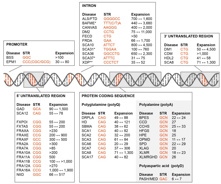
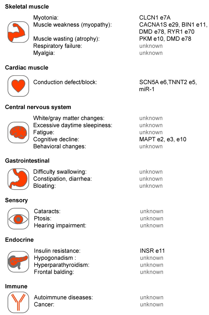
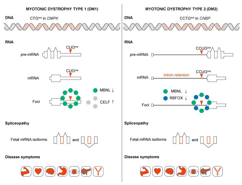
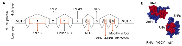
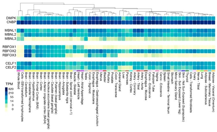
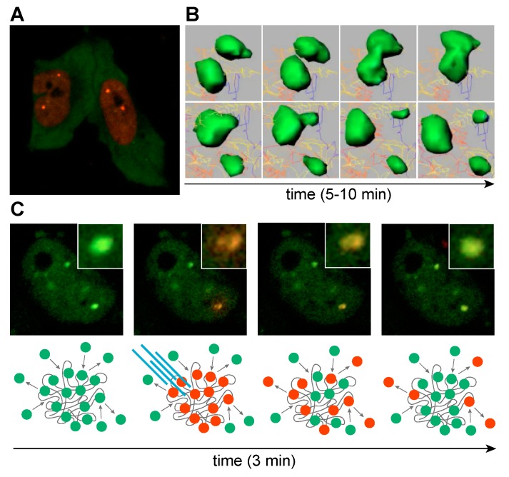
Short tandem repeat (STR) or microsatellite, expansions underlie more than 50 hereditary neurological, neuromuscular and other diseases, including myotonic dystrophy types 1 (DM1) and 2 (DM2). Current disease models for DM1 and DM2 propose a common pathomechanism, whereby the transcription of mutant DMPK (DM1) and CNBP (DM2) genes results in the synthesis of CUG and CCUG repeat expansion (CUGexp, CCUGexp) RNAs, respectively. These CUGexp and CCUGexp RNAs are toxic since they promote the assembly of ribonucleoprotein (RNP) complexes or RNA foci, leading to sequestration of Muscleblind-like (MBNL) proteins in the nucleus and global dysregulation of the processing, localization and stability of MBNL target RNAs. STR expansion RNAs also form phase-separated gel-like droplets both in vitro and in transiently transfected cells, implicating RNA-RNA multivalent interactions as drivers of RNA foci formation. Importantly, the nucleation and growth of these nuclear foci and transcript misprocessing are reversible processes and thus amenable to therapeutic intervention. In this review, we provide an overview of potential DM1 and DM2 pathomechanisms, followed by a discussion of MBNL functions in RNA processing and how multivalent interactions between expanded STR RNAs and RNA-binding proteins (RBPs) promote RNA foci assembly.

Keywords: ALS/FTD; CELF; MBNL; RBFOX; STR; alternative splicing; foci; microsatellite expansion; myotonic dystrophy; phase separation.


Conflict of interest statement

M.S.S. is a member of the scientific advisory board of Locana Bio, Inc. Ł.J.S. declares no conflicts of interest.

Figures

Figure 1
Figure 1
Short tandem repeat (STR) expansion disorders. Diseases caused by STR (orange font) expansions in the promoter, 5’ untranslated region (5’UTR), introns, coding region and 3’ untranslated region (3’UTR) are shown together with the disease acronym and pathogenic expansion range (black font). Some of these mutations are not classical expansions but are insertions due to replication/recombination/duplication (Intron, SCA31, SCA37, BAFME, *; Protein Coding Sequence, Polyalanine and Polyaspartic acid, light grey box) or retrotransposon (Intron, XDP, **) events. Disease-associated STR locations include the: • Promoter. Baratela-Scott Syndrome (BSS) linked to XYLT1 gene [6], Progressive Myoclonus Epilepsy (EPM1)–CSTB [7]; • 5’UTR. Glutaminase Deficiency (GAD)–GLS [8]; Spinocerebellar Ataxia (SCA) Type 12 (SCA12)–PPP2R2B [9]; Fragile X-Associated Primary Ovarian Insufficiency (FXPOI), Fragile X-Associated Tremor/Ataxia Syndrome (FXTAS) and Fragile XA Syndrome (FRAXA or FXS)–FMR1 [10,11]; Fragile XE Syndrome (FRAXE)–AFF2 [12]; Fragile XF Syndrome (FRAXF)–TMEM185A [13]; Folate-sensitive fragile sites (FSFS) FRA2A–AFF3 [14]; FSFS FRA7A–ZNF713 [15]; FSFS FRA10A–FRA10AC1 [16]; FSFS FRA11A–C11orf80 [17]; SFSF FRA11B–CBL2 [18]; FSFS FRA12A–DIP2B [19]; SFSF FRA16A–LOC109617027 [20]; Neuronal Intranuclear Inclusion Disease (NIID)–NOTCH2NLC [21]; • Intron. Amyotrophic Lateral Sclerosis and Frontotemporal Dementia (ALS/FTD)–C9orf72 [22,23]; Benign Adult Familial Myoclonic Epilepsy (BAFME)–SAMD12, TNRC6A and RAPGEF2 [24]; Cerebellar Ataxia, Neuropathy, Vestibular Areflexia Syndrome (CANVAS)–RFC1 [25]; Myotonic Dystrophy Type 2 (DM2)–CNBP [26]; Fuchs Endothelial Corneal Dystrophy (FECD)–TCF4 [27]; Friedrich’s Ataxia (FRDA)–FXN [28]; SCA type 10 (SCA10)–ATXN10 [29]; SCA type 31 (SCA31)–BEAN1/TK2 [30]; SCA36–NOP56 [31]; SCA37–DAB1 [32]; X-Linked Dystonia-Parkinsonism (XDP)–TAF1 [33]. • Coding region (polyglutamine). Dentatorubral-Pallidoluysian Atrophy (DRPLA)–ATN1 [34]; Huntington Disease (HD)–HTT [35]; Spinal and Bulbar Muscular Atrophy (SBMA)–AR [36]; SCA type 1 (SCA1)–ATXN1 [37], SCA type 2 (SCA2)–ATXN2 [38]; SCA type 3 (SCA3)–ATXN3 [39]; SCA type 6 (SCA6)–CACNA1A [40]; SCA type 7 (SCA7)–ATXN7 [41]; SCA type 8 (SCA8)–ATXN8 [42]; SCA type 17 (SCA17)–TBP [43]; • Coding region (polyalanine). Blepharophimosis Syndrome (BPES)–FOXL2 [44]; Cleidocranial Dysplasia (CCD)–RUNX2 [45]; Congenital Central Hypoventilation Syndrome (CCHS)–PHOX2B [46]; Hand-Foot-Genital Syndrome (HFGS)–HOXA13 [47]; Holoprosencephaly (HPE)–ZIC2 [48]; Oculopharyngeal Muscular Dystrophy (OPMD)–PABPN1 [49]; Synpolydactyly Syndrome (SPD)–HOXD3 [50]; X-linked Mental Retardation and Abnormal Genitalia (XLAG) and X-linked Mental Retardation (XLMR)–ARX [51,52]; XLMR and Growth Hormone Deficit (XLMRGHD)–SOX3 [53]; • Coding region (polyaspartic acid). Pseudoachondroplasia and Multiple Epiphyseal Dysplasia (PSACH/MED)–COMP [54]. • 3’UTR. Myotonic Dystrophy Type 1 (DM1) and Congenital Myotonic Dystrophy (CDM)–DMPK [55]; Huntington Disease-Like 2 (HDL2)–JPH3 [56]; SCA8–ATXN8OS [42].
Figure 2
Figure 2
Multi-system involvement in Myotonic Dystrophy types 1 and 2 (DM1 and DM2). Several tissue systems are shown with associated phenotypes together with proposed RNA mis-processing events. These events include: chloride voltage-gated channel 1 (CLCN1) exon (e)7A [81,82,83], calcium voltage-gated channel subunit alpha1 S (CACNA1S) e29 [84], bridging integrator 1 (BIN1) e11 [85], dystrophin (DMD) e78 [86], ryanodine receptor 1 (RYR1) e70 [87], pyruvate kinase isozymes M1/M2 (PKM1/M2) e10 [88], sodium voltage-gated channel alpha subunit 5 (SCN5A) e6 [89,90], troponin T2, cardiac type (TNNT2) e5 [91], microtubule associated protein tau (MAPT) e2, e3, e10 [92,93], insulin receptor (INSR) e11 [94,95] and microRNA-1 (miR-1) [96].
Figure 3
Figure 3
Models of DM1 and DM2 disease mechanisms. CTGexp and CCTGexp in the DMPK and CNBP genes produce pre-mRNA transcripts containing expanded CUG and CCUG repeats. DMPK pre-mRNA is correctly spliced whereas CCUGexp triggers CNBP intron 1 retention. mRNAs with C(C)UGexp sequester Muscleblind-like (MBNL) and RBFOX (in DM2) alternative splicing factors. In addition, CUGexp increase CUGBP Elav-Like Family Member 1 (CELF1) splicing factor stability through protein kinase C (PKC)-mediated hyperphosphorylation. All these changes in the bioavailability of splicing factors cause an imbalance in alternative splicing and enhanced fetal mRNA isoform production in adult tissues. As a result, inappropriate protein expression patterns lead to a variety of DM symptoms.
Figure 4
Figure 4
MBNL functions in RNA biogenesis, localization and stability. MBNL regulates alternative (orange boxes) splicing events, including cassette (e) exon, 5′ splice site, 3′ splice site, mutually exclusive exons, (i) intron retention [121,129] and alternative 3′ end formation by alternative cleavage and polyadenylation (pA) [122]. MBNL also regulates microRNA (miRNA) biogenesis [96], circular RNA (circRNA) formation [123], mRNA localization (horizontal arrow) [121] and increases mRNA stability (vertical arrow) [124]. All examples represent MBNL-mediated events and representative targeted RNAs are indicated.
Figure 5
Figure 5
MBNL gene and ZnF structures. (A) Exonic structure and function of MBNL paralogs [135,148,149,150]. Exon enumeration is derived from previous studies [151]. ZnF1/2 and ZnF3/4–zinc finger domain pairs 1/2 and 3/4 respectively. NLS–nuclear localization signal. MBNL protein levels are shown as Low (bottom) and High (top) which trigger the indicated splicing events (black lines). (B) The structural model of MBNL1 zinc fingers (ZnF; blue) in complex with RNA (red) [152].
Figure 6
Figure 6
Expression of DM1 and DM2 relevant genes in human tissues. Heat map showing DMPK, CNBP, MBNL1-3, RBFOX1-3 and CELF1-2 gene expression in 53 unaffected tissues from >700 individuals. Data were obtained from the Genotype-Tissue Expression (GTEx) project website (gtexportal.org) and the heat map was generated using the Multi-Gene Query function (TPM, transcripts per million).
Figure 7
Figure 7
MBNL sequestration on expanded CUGexp transcripts. (A) Life cell imaging of cells transfected with CUGexp, mCherry-MBNL1 and GFP-MBNL3. MBNL1 localizes primarily in nucleus, whereas MBNL3 is predominately cytoplasmic. (B) Time-lapse sequence images of CUGexp-GFP-MBNL1 complexes (green). Raw data were processed with Imaris software. (C) Time-lapse sequence images of a cell transiently transfected with CUGexp and MBNL1 fused with Dendra2 fluorescence protein. MBNL1 accumulated in two distinct nuclear foci. Laser stimulated Dendra2-MBNL photoconversion from green (emission 507 nm) to red (emission 573 nm) in a single focus reveals dynamic exchange of MBNL between the focus and surrounding nucleoplasm in the course of minutes. Yellowish center of focus core might suggest that MBNL proteins localized in the focus core are less prone to exchange. Below each image, a scheme representing the experimental scheme. (B,C) Images adapted from Reference [135].

References

    1. Bourque G., Burns K.H., Gehring M., Gorbunova V., Seluanov A., Hammell M., Imbeault M., Izsvak Z., Levin H.L., Macfarlan T.S., et al. Ten things you should know about transposable elements. Genome. Biol. 2018;19:199. doi: 10.1186/s13059-018-1577-z. - DOI - PMC - PubMed
    1. Padeken J., Zeller P., Gasser S.M. Repeat DNA in genome organization and stability. Curr. Opin. Genet. Dev. 2015;31:12–19. doi: 10.1016/j.gde.2015.03.009. - DOI - PubMed
    1. McGinty R.J., Mirkin S.M. Cis- and trans-modifiers of repeat expansions: Blending model systems with human genetics. Trends Genet. 2018;34:448–465. doi: 10.1016/j.tig.2018.02.005. - DOI - PMC - PubMed
    1. López Castel A., Cleary J.D., Pearson C.E. Repeat instability as the basis for human diseases and as a potential target for therapy. Nat. Rev. Mol. Cell Biol. 2010;11:165–170. doi: 10.1038/nrm2854. - DOI - PubMed
    1. Iyer R.R., Pluciennik A., Napierala M., Wells R.D. DNA triplet repeat expansion and mismatch repair. Annu Rev. Biochem. 2015;84:199–226. doi: 10.1146/annurev-biochem-060614-034010. - DOI - PMC - PubMed

LinkOut - more resources