. 2019 Jul 19;7(1):190.
doi: 10.1186/s40425-019-0666-1.

Ovarian cancer stem cells and macrophages reciprocally interact through the WNT pathway to promote pro-tumoral and malignant phenotypes in 3D engineered microenvironments

Affiliations

Ovarian cancer stem cells and macrophages reciprocally interact through the WNT pathway to promote pro-tumoral and malignant phenotypes in 3D engineered microenvironments

Shreya Raghavan et al. J Immunother Cancer. .

Abstract

Background: Innate immune cells such as macrophages are abundantly present within malignant ascites, where they share the microenvironment with ovarian cancer stem cells (CSC).

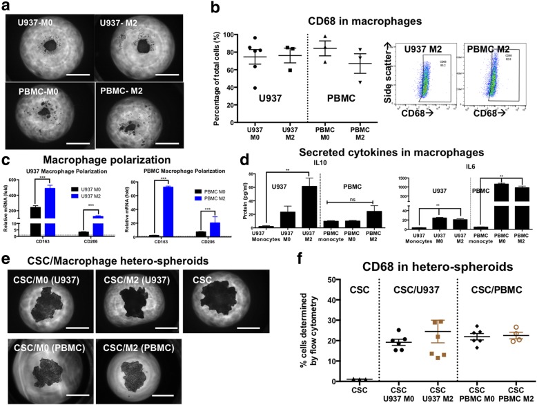
Methods: To mimic this malignant ascites microenvironment, we created a hanging-drop hetero-spheroid model to bring CSCs and macrophages in close association. Within these hetero-spheroids, CD68+ macrophages (derived from U937 or peripheral blood monocytes) make up ~ 20% of the population, while the rest are ovarian cancer cells and ovarian cancer stem cells (derived from the high grade serous ovarian cancer cell line, OVCAR3).

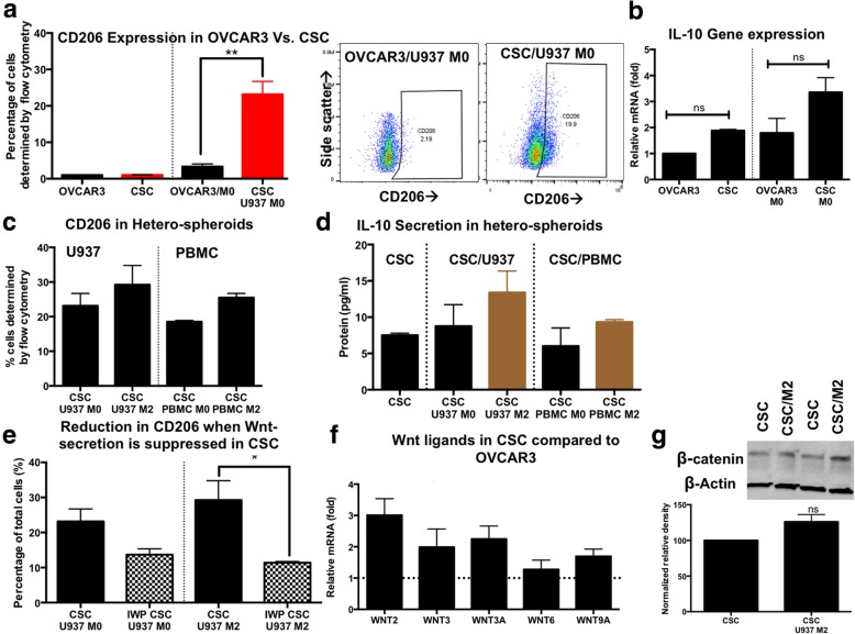
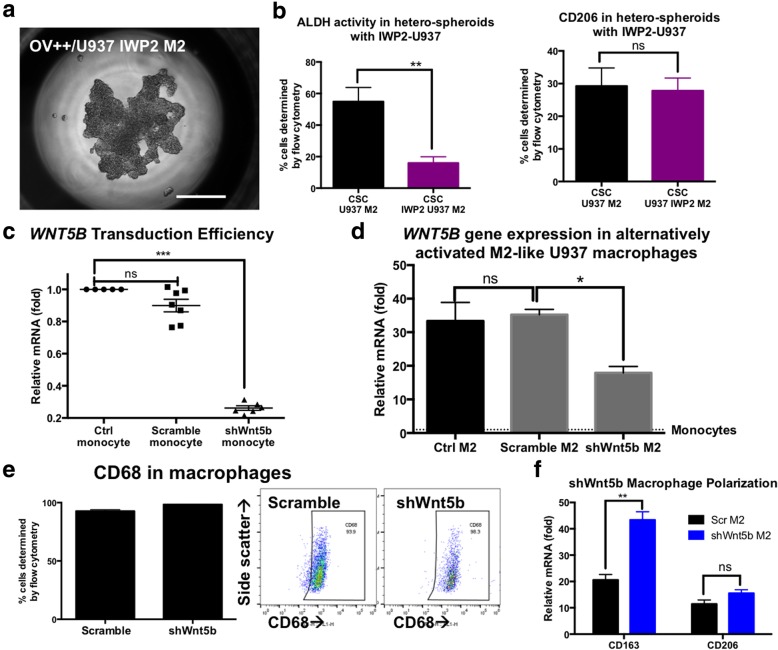
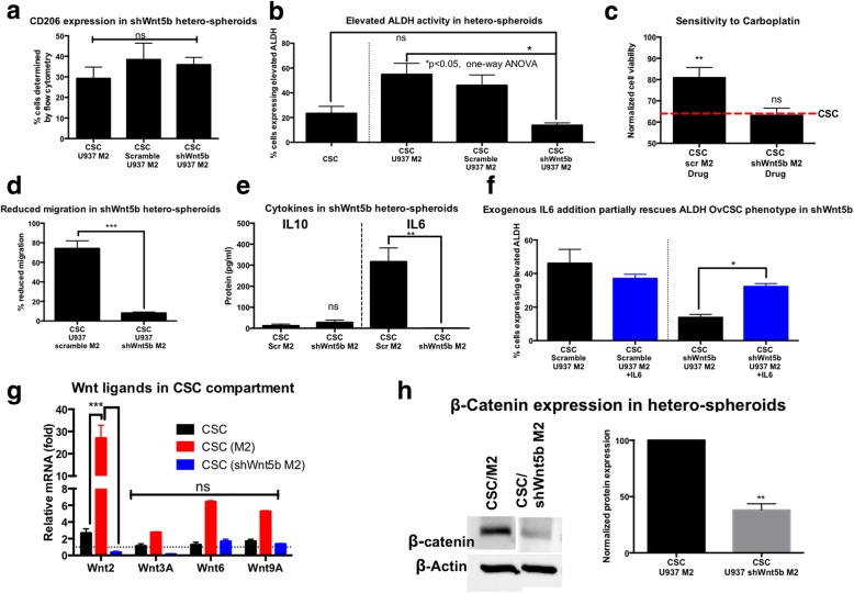
Results: Our results indicate that CSCs drive the upregulation of M2 macrophage marker CD206 within hetero-spheroids, compared to bulk ovarian cancer cells, implying an inherently more immuno-suppressive program. Moreover, an increased maintenance of elevated aldehyde dehydrogenase (ALDH) activity is noted within hetero-spheroids that include pre-polarized CD206+ M2 macrophages, implying a reciprocal interaction that drives pro-tumoral activation as well as CSC self-renewal. Consistent with enriched CSCs, we also observe increased levels of pro-tumoral IL-10 and IL-6 cytokines in the CSC/M2-macrophage hetero-spheroids. CSC/M2-macrophage hetero-spheroids are also less sensitive to the chemotherapeutic agent carboplatin and are subsequently more invasive in transwell assays. Using inhibitors of WNT secretion in both CSCs and macrophages, we found that CSC-derived WNT ligands drove CD206+ M2 macrophage activation, and that, conversely, macrophage-derived WNT ligands enriched ALDH+ cells within the CSC compartment of hetero-spheroids. Upon examination of specific WNT ligand expression within the monocyte-derived macrophage system, we observed a significant elevation in gene expression for WNT5B. In CSCs co-cultured with macrophages within hetero-spheroids, increases in several WNT ligands were observed, and this increase was significantly inhibited when WNT5B was knocked down in macrophages.

Conclusions: Our data implies that macrophage- initiated WNT signaling could play a significant role in the maintenance of stemness, and the resulting phenotypes of chemoresistance and invasiveness. Our results indicate paracrine WNT activation during CSC/M2 macrophages interaction constitutes a positive feedback loop that likely contributes to the more aggressive phenotype, which makes the WNT pathway a potential target to reduce the CSC and M2 macrophage compartments in the tumor microenvironment.


Conflict of interest statement

The authors declare that they have no competing interests.

Figures

Fig. 1
Fig. 1
Hetero-spheroids derived from monocyte-derived macrophages and ovarian cancer stem cells. a Monocytes from the U937 cell line or peripheral blood monocytes (PBMC) were plated into hanging drop arrays, and differentiated to M0 macrophages with phorbol ester treatment, or activated with IL-4 and MCSF treatment. Differentiated and activated macrophages formed compact spheroid-like aggregates. b Differentiated M0 and M2 macrophages all expressed pan macrophage marker, CD68 indicating 75–80% differentiation efficiency from monocytes to macrophages. CD68 expression was evaluated using flow cytometry analysis, with representative plots. c Polarization was assessed using qPCR analysis for two M2 genes, CD163 and CD206. Both U937 and PBMC macrophages expressed significantly higher levels of CD163 and CD206 genes, compared to untreated undifferentiated monocytes. d M2 differentiated macrophages secreted higher amounts of the immuno-suppressive cytokine IL-10, and the pro-tumoral cytokine IL-6. e CSCs were derived from the OVCAR3 cell line based on ALDH+ CD133+ expression. Hetero-spheroids were generated using differentiated U937 or PBMC M0 macrophages and CSCs or activated U937 or PBMC M2 macrophages and CSCs. Representative phase contrast images of hetero-spheroids seen at Day 5 following formation indicate compact spheroids, similar in size to CSC mono-spheroids generated from the same number of CSCs/spheroid. f Hetero-spheroids retain ~ 20% CD68 expression, indicating that at day 5, CD68+ macrophages constitute 20% of the population of cells. Scale bar = 200 μm
Fig. 2
Fig. 2
Hetero-spheroids drive CD206 polarization in monocyte-derived macrophages. a Hetero-spheroids were generated from bulk unsorted OVCAR3 cells and M0 macrophages (OVCAR3/U937 M0), and CSC/U937 M0. Flow analysis for the macrophage polarization marker CD206 indicated that while OVCAR3 and CSCs by themselves do not express CD206, CSCs drive CD206 expression in previously CD206 M0 macrophages within hetero-spheroids at significantly (**p < 0.001, one-way ANOVA) higher levels than OVCAR3. Representative flow analysis plots are shown. b IL10 gene expression was evaluated using qPCR in mono- and hetero-spheroids, indicating that CSCs had a 2-fold increased gene expression of IL10, likely driving CD206 expression in M0 macrophages to higher extents than OVCAR3. c CD206 expression was maintained at elevated levels (18.5–29.24%) in all CSC hetero-spheroid conditions, indicating that the activated M2 phenotype was maintained in macrophages within hetero-spheroids. d Levels of secreted IL-10 were mildly elevated in CSC/M2 hetero-spheroids, but were also similar across all CSC/macrophage hetero-spheroids, indicating the presence of the immuno-suppressive cytokine. e When CSCs were pre-treated with the Wnt secretion inhibitor, IWP-2, significantly lower (*p < 0.05,one-way ANOVA) CD206 expression was observed in CSC/M2 hetero-spheroids, implying the importance of CSC-derived Wnt ligands in the maintenance of M2 activation in macrophages. f qPCR analysis revealed that gene expression of several Wnt ligands were elevated in CSC compared to bulk OVCAR3 spheroids (dotted line = no change); (g) Densitometric measurements of immunoblots for β-catenin compared to the loading control β-actin indicated that CSC/M2 spheroids had a 26% increase in β-catenin expression, compared to CSC mono-spheroids, indicating higher Wnt/β-catenin activity in CSC/M2 hetero-spheroids
Fig. 3
Fig. 3
M2-polarized macrophages increase ALDH+ in CSC/M2 hetero-spheroids. a Elevated ALDH activity was assessed using the ALDEFLUOR flow cytometric analysis assay, with representative flow plots demonstrated. CSC/M2 hetero-spheroids, whether U937 or PBMC, have significantly (**p < 0.001, one-way ANOVA) elevated ALDH+ expression (1.8–2.4 fold). b Secreted pro-tumoral cytokine IL6 is elevated in hetero-spheroids compared to CSC mono-spheroids. c We suppressed signaling through the IL6/STAT3 axis using two inhibitors Ruxolitinib and sc144 in hetero-spheroids, and observed that this suppression also significantly (**p < 0.001, one-way ANOVA) decreased the ALDH+ enrichment within CSC/M2 hetero-spheroids. d Concomitant with the ALDH expression within hetero-spheroids, sensitivity to carboplatin was evaluated using the MTS assay. Normalized absorbance values indicated that CSC/M2 hetero-spheroids were significantly (*p < 0.05, one-way ANOVA) more resistant to carboplatin treatment compared to CSC mono-spheroids made with the same number of CSCs. e Representative phase contrast images indicate the loss of the tight spheroid boundaries in the CSC mono-spheroids in response to carboplatin treatment, indicative of cell death. The extent of the loss of boundary integrity is much lower in the CSC/M2 hetero-spheroids. Scale bar = 100 μm. f Quantification of the number of migrated cells in the lower chamber of a transwell system indicated that CSC/M2 hetero-spheroids were significantly (*p < 0.05, one-way ANOVA) more migratory compared to CSC mono-spheroids
Fig. 4
Fig. 4
Inhibition of macrophage Wnt-secretion reduces ALDH enrichment in hetero-spheroids. a Representative phase contrast image of a hetero-spheroid generated from CSCs and IWP-2 treated U937 M2 macrophages, indicates the formation of aggregated spheroids. Scale bar = 200 μm. b Flow analysis revealed that hetero-spheroids with IWP-2 treated M2 macrophages had a significantly diminished ALDH+ enrichment (**p < 0.001, one-way ANOVA) compared to control untreated CSC/M2 hetero-spheroids. However, no change in CD206 expression was observed in hetero-spheroids. c Transduction efficiency of monocytes with shRNA directed against WNT5B indicated a > 75% efficiency in knockdown of WNT5B gene expression in the shWnt5b targeted construct, and minimal changes in WNT5B gene expression in the scramble non-targeted lentiviral construct. d We utilized shWnt5b monocytes to differentiate and polarize M2 macrophages, and found that there was a 52% reduction in WNT5B gene expression in shWnt5b M2 macrophages, compared to scramble or control untreated M2 macrophages, indicating the knockdown of the Wnt5b gene. e No changes were observed in CD68 expression in monocytes generated from scramble or shWNT5B treated monocytes, demonstrated by flow analysis and representative plots. f Similarly, qPCR analysis of M2 gene expression markers CD163 and CD206 indicated increases in both genes, indicative of activation into the M2 program even in shWnt5b macrophages
Fig. 5
Fig. 5
Macrophage knockdown of Wnt5b suppresses ALDH enrichment, with no change in CD206, and increases chemosensitivity of hetero-spheroids. a Flow analysis of CD206 indicated no significant differences in CSC/M2 and CSC/Scramble M2 and CSC/shWnt5b M2 hetero-spheroids, indicating that CSCs were still capable of maintaining a robust immunosuppressive program in hetero-spheroids, regardless of WNT5B. b ALDH elevation however was significantly (*p < 0.05, one-way ANOVA) lowered compared to CSC/U937 M2 hetero-spheroids, and not significantly different (ns) compared to CSC mono-spheroids, indicating that knocking down Wnt5b expression in macrophages reduced ALDH enrichment in hetero-spheroids. c Concomitant with the decrease in ALDH, sensitivity to carboplatin also significantly (**p < 0.001, one-way ANOVA) improved, responding to treatment similar to CSC mono-spheroids. The red dashed line indicates the sensitivity levels of CSC mono-spheroids. d CSC/shWnt5b M2 hetero-spheroids were also significantly (***p < 0.0001, t-test) less invasive than CSC/Scramble M2 hetero-spheroids. e Upon ELISA analysis of secreted IL10 and IL6, we found no changes in IL-10 levels, while we found a stark decrease in secreted pro-tumoral IL-6 cytokine in CSC/shWnt5b M2 hetero-spheroids. f Exogenous addition of IL-6 (25 ng/ml) to CSC/shWnt5b M2 hetero-spheroids partially significantly (*p < 0.05, one-way ANOVA) increases ALDH+ enrichment, but does not restore it to levels of original enrichment with CSC/Scramble M2 or CSC/Ctrl M2 hetero-spheroids. g In order to identify if there was paracrine macrophage Wnt-driven Wnt signaling in CSCs, we separated the CSCs (using a GFP label) from hetero-spheroids, and probed for gene expression of several Wnt ligands we saw elevated in CSCs compared to bulk OVCAR3. We observed that with M2 macrophage co-culture, several Wnt ligands were upregulated (Wnt2 significantly elevated, ***p < 0.001, two-way ANOVA). However, this elevated gene expression of Wnt ligands was lost in CSCs in hetero-spheroid culture with shWnt5b M2, indicating that Wnt5b was partly responsible for potentiating Wnt signaling within the CSC compartment. h This loss in Wnt ligand expression also translated to decreased β-catenin expression in CSC/shWnt5b M2 hetero-spheroids compared to CSC/U937 M2 hetero-spheroids, quantified by densitometry of immunoblots (representative blot shown)
Fig. 6
Fig. 6
Changes in tumorigenicity of hetero-spheroids in vivo in NSG mice. Tumors were generated in NSG mice from CSC mono-spheroids, or CSCs isolated from hetero-spheroids in CSC/M2 or CSC/shWNT5b M2. a Tumor initiation and growth kinetics are shown for all three groups, where CSC(M2) tumors (red curve) demonstrates elevated tumorigenicity, with faster tumor initiation and tumor burden development (gray shaded area indicates the tumor burden window). Similarly, the blue trace indicates the tumor growth in CSC/shWNT5b M2 tumors, which is significantly (*p < 0.05, two-way ANOVA) lower and slower compared to CSC and CSC(M2) groups at the time points indicated. b Histological examination demonstrates that all groups establish similar structures of tumors sub-cutaneously, with CSC tumors and CSC (M2) tumors establishing dense epitheliod cells, with CSC (shWNT5b M2) tumors more loosely arranged. Scale bar = 100 μm. c Gene expression analysis of xenografted tumors indicated that an elevated ALDH1A1 expression was maintained in CSC(M2) tumors (**p < 0.001, two-way ANOVA). M2-like macrophage conditioning also helped CSCs retain elevated expression of WNT2, WNT3A and WNT6 (*p < 0.05, **p < 0.001, two-way ANOVA) compared to CSCs in mono-spheroids. All other genes and conditions were not significantly different (ns) compared to CSCs, specifically CSC/shWnt5B M2 tumors. d The human IL-6 inhibitor Tocilizumab was used intra-peritoneally on CSC and CSC(M2) tumors, once palpable tumors were observed (Day 25), indicated by the arrow. Tocilizumab treatment resulted in a significant decrease in tumor volume (**p < 0.001, two-way ANOVA) compared to control untreated tumors in CSC mono-spheroid tumors (compare purple trace to black trace; ~ 70% reduction in tumor burden). However, in CSC/M2 tumors, tocilizumab treatment did not significantly alter the growth kinetics of treated vs. untreated tumors (~ 24% reduction in tumor burden), indicating the detainment of a chemoresistant phenotype upon M2-like macrophage co-culture and conditioning. Furthermore, the treated CSC Tocilizumab condition also resembles the control CSC (shWNT5B M2) tumors (blue trace in A), indicating that partial loss or inhibition of IL-6 based CSC enrichment may be at play
Fig. 7
Fig. 7
Ovarian cancer stem cells and macrophages reciprocally interact through the Wnt pathway to promote a pro-tumoral microenvironment. Our data suggests that in hetero-spheroids, CSCs drive a CD206+ expressing macrophage phenotype, suggestive of pro-tumoral M2 activation through secretion of the immuno-suppressive cytokine IL-10, and through WNT ligands. We also observe that macrophage-derived WNT ligands, specifically WNT5B, and to some extent the pro-tumoral cytokine IL-6, drive an enrichment in ALDH+ cells within hetero-spheroids. Knockdown of macrophage WNT5B expression, or inhibiting downstream activation of IL-6 using Ruxolitinib or sc144, result in a significant loss of ALDH+ populations within hetero-spheroids. Importantly, we find that the Wnt pathway is involved bi-directionally in CSC-macrophage interactions, and can potentially be targeted to reduce stemness, chemoresistance, invasiveness and immunosuppression in ovarian cancer

References

    1. Liao J, Qian F, Tchabo N, et al. Ovarian cancer spheroid cells with stem cell-like properties contribute to tumor generation, metastasis and chemotherapy resistance through hypoxia-resistant metabolism. PLoS One. 2014;9:e84941. doi: 10.1371/journal.pone.0084941. - DOI - PMC - PubMed
    1. Shield K, Ackland ML, Ahmed N, Rice GE. Multicellular spheroids in ovarian cancer metastases: biology and pathology. Gynecol Oncol. 2009;113:143–148. doi: 10.1016/j.ygyno.2008.11.032. - DOI - PubMed
    1. Allen HJ, Porter C, Gamarra M, Piver MS, Johnson EA. Isolation and morphologic characterization of human ovarian carcinoma cell clusters present in effusions. Exp Cell Biol. 1987;55:194–208. - PubMed
    1. Kipps E, Tan DS, Kaye SB. Meeting the challenge of ascites in ovarian cancer: new avenues for therapy and research. Nat Rev Cancer. 2013;13:273–282. doi: 10.1038/nrc3432. - DOI - PMC - PubMed
    1. Zhang L, Conejo-Garcia JR, Katsaros D, et al. Intratumoral T cells, recurrence, and survival in epithelial ovarian cancer. N Engl J Med. 2003;348:203–213. doi: 10.1056/NEJMoa020177. - DOI - PubMed

Publication types