. 2019 Jul 19;17(1):230.
doi: 10.1186/s12967-019-1982-4.

Tumor-derived exosomes promote the in vitro osteotropism of melanoma cells by activating the SDF-1/CXCR4/CXCR7 axis

Affiliations

Tumor-derived exosomes promote the in vitro osteotropism of melanoma cells by activating the SDF-1/CXCR4/CXCR7 axis

Francesco Mannavola et al. J Transl Med. .

Abstract

Background: Bone metastases occur rarely in patients suffering from malignant melanoma, although their onset severely worsens both prognosis and quality of life. Extracellular vesicles (EVs) including exosomes (Exos) are active players in melanoma progression involved in the formation of the pre-metastatic niche.

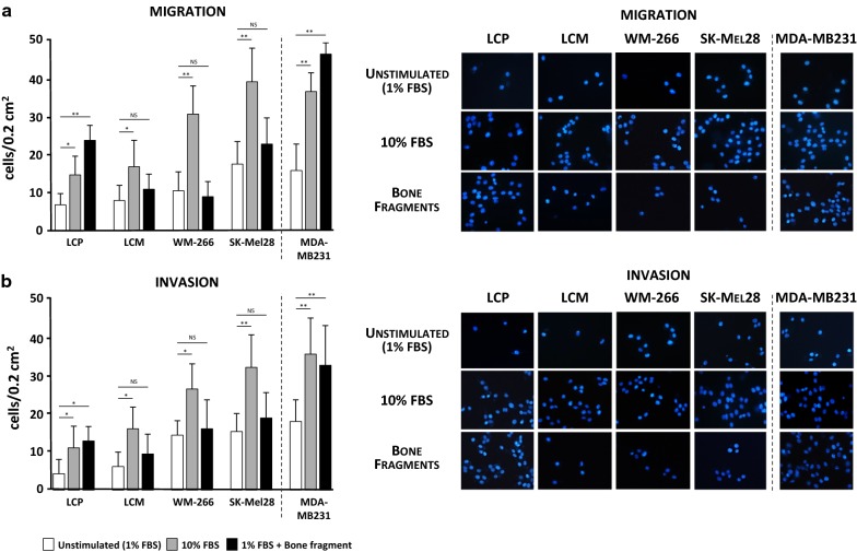
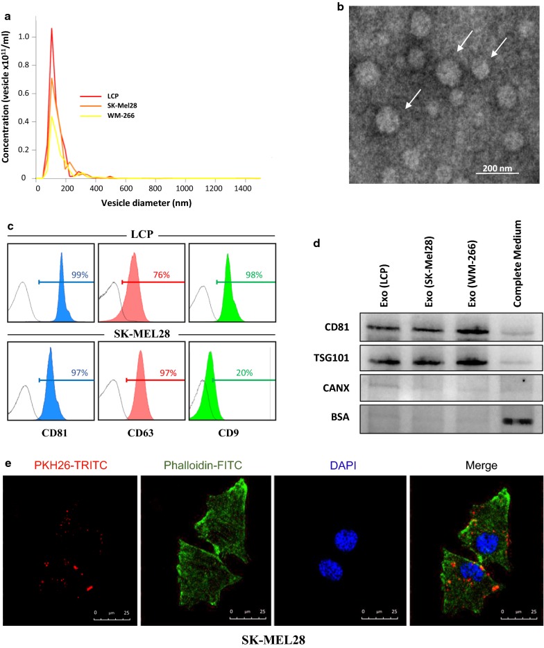
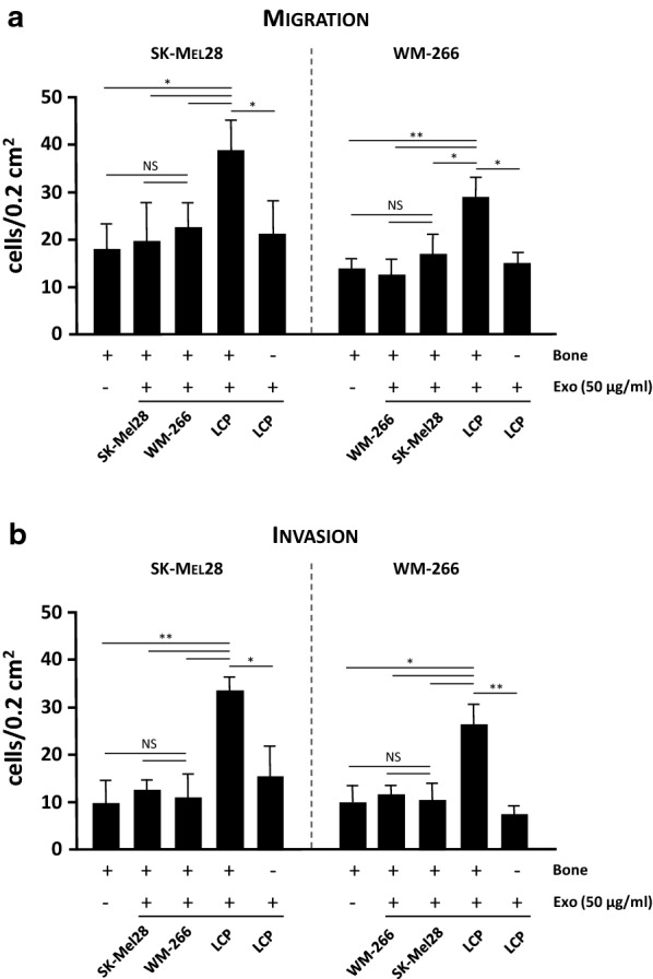
Methods: Trans-well assays explored the basal migratory and invasive potential of four melanoma cell lines and investigated their different propensity to be attracted toward the bone. Exosomes were purified from cell supernatants by ultracentrifugation and explored in their ability to influence the bone tropism of melanoma cells. The molecular machinery activated during this process was investigated by RT-PCR, droplet digital-PCR, flow-cytometry and Western blot, while loss of function studies with dedicated siRNAs defined the single contribute of CXCR4 and CXCR7 molecules.

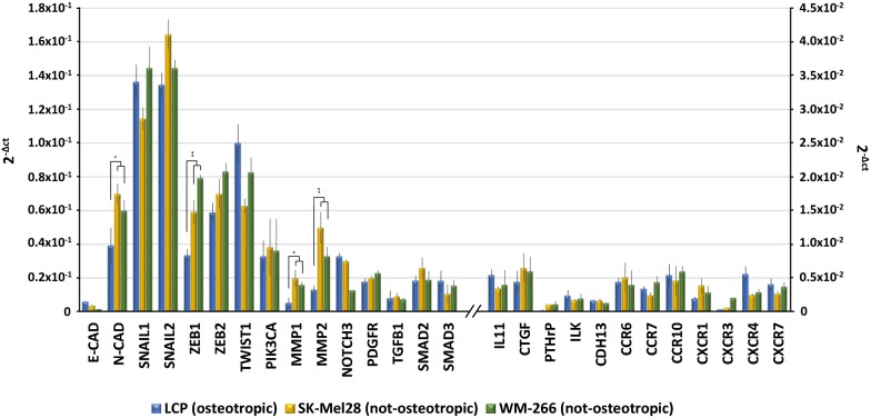
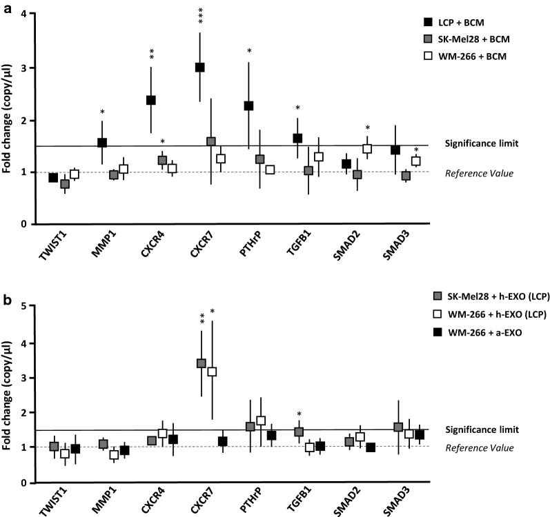
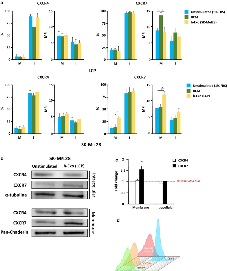
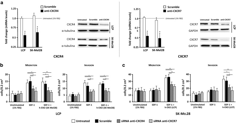
Results: Melanoma cells revealed a variable propensity to be attracted toward bone fragments. Gene profiling of both osteotropic and not-osteotropic cells did not show a different expression of those genes notoriously correlated to chemotaxis and bone metastasis. However, bone conditioned medium significantly increased CXCR4, CXCR7 and PTHrP expression solely to osteotropic cells, while their Exos were able to revert the original poor bone tropism of not-osteotropic cells through CXCR7 up-regulation. Silencing experiments also demonstrated that membrane expression of CXCR7 is required by melanoma cells to promote their chemotaxis toward SDF-1 gradients.

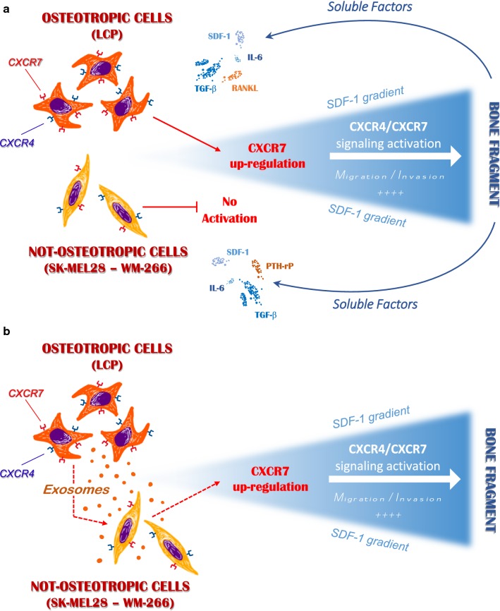
Conclusions: Our data correlated the osteotropism of melanoma cells to the activation of the SDF-1/CXCR4/CXCR7 axis following the exposition of tumor cells to bone-derived soluble factors. Also, we demonstrated in vitro that tumor-derived Exos can reprogram the innate osteotropism of melanoma cells by up-regulating membrane CXCR7. These results may have a potential translation to future identification of druggable targets for the treatment of skeletal metastases from malignant melanoma.

Keywords: Bone metastasis; CXCR4; CXCR7; Exosomes; Melanoma.


Conflict of interest statement

The authors declare that they have no competing interests.

Figures

Fig. 1
Fig. 1
Migration and invasion assays of melanoma cell lines. a The migration of melanoma cells was enhanced by 10% FBS as compared to unstimulated cells (average: 25.3 ± 5.5 vs 11.4 ± 2.8 cells/0.2 cm2). The stimulation with bone fragment significantly increased the migration of LCP with respect to unstimulated cells, while produced modest effects on LCM, WM-266 and SK-Mel28. MDA-MB231 breast cancer cells were the positive control. b The invasion assay also confirmed the general invasive attitude under the FBS stimulation (WM-266: 26.3 ± 7.8 cells/0.2 cm2; SK-Mel28: 38.9 ± 9.7 cells/0.2 cm2), although only LCP showed a significant increase of invasiveness (13.1 ± 3.4 vs 4.4 ± 2.9 cells/0.2 cm2) when stimulated with bone fragment as for MDA-MB231 cells (33.6 ± 10 vs 18.7 ± 4.8 cells/0.2 cm2). Bars are mean ± SEM. NS: not significant; *p < 0.05; **p < 0.01. Images on the right side are representative of fluorescence microscopy at ×40 magnification of cells trapped within the trans-well membranes as effect of different stimulations
Fig. 2
Fig. 2
Characterization of Exos from melanoma cells. a Exosome preparations were analyzed using NanoSight technology. Histogram represents the size distribution of nanovesicles purified from LCP, SK-Mel28 and WM-266 conditioned supernatants, and the analysis reveals more than 80% of vesicles with a diameter ranging from 30 to 150 nm. Results are the mean from three different measurements. b Representative panel showing Exos preparations by TEM reveals the presence of nanovesicles with typical cup-shaped morphology (arrows). c Flow-cytometry described the CD81, CD63 and CD9 tetraspanin expression in Exo preparations from representative LCP and SK-Mel28 cells whose values were higher than 95%. d Western blots for Exo markers (CD81 and TSG101) and potential contaminants (CANX, BSA) of Exo preparations isolated from supernatants of melanoma cells. Fresh Exo-free complete medium was used as control. e Representative confocal microscopy images at ×40 magnification showing the up-take of PKH26-labeled Exos (50 µg/ml) by melanoma cells after 4-h. Large Exo clusters (red dots) were found to be mostly distributed in the cytoplasm of SK-Mel28 cells (merge), whose actin filaments and nuclei were stained with phalloidin (green) and DAPI (blue), respectively
Fig. 3
Fig. 3
Exosomes influence the osteotropism of melanoma cells. The effects of Exos (50 µg/ml) on bone tropism of not-osteotropic SK-Mel28 and WM-266 cells were explored using bone fragments as chemoattractant. Both migration (a) and invasion (b) were significantly enhanced by h-Exos from osteotropic LCP, while they were not influenced by either a-Exos or h-Exos from not-osteotropic cells. The effect of LCP-derived Exos was abrogated in absence of bone fragments, thus suggesting their ability to sensitize not-osteotropic cells to the bone chemoattraction rather than stimulate their migration. Bars are mean ± SEM. NS not-significant; *p < 0.05; **p < 0.01
Fig. 4
Fig. 4
Basal gene expression of ‘osteotropic’ and ‘not-osteotropic’ melanoma cells. Real time-PCR analyzed osteotropic (LCP) and not-osteotropic cells (SK-Mel28 and WM-266) in their basal expression of 27 genes involved in the epithelial-to-mesenchymal transition (EMT), chemotaxis and bone metastasis development. Both osteotropic and not-osteotropic cells exhibit an EMT profile as evidenced by the over-expression of N-Cadherin (N-CAD), SNAIL, ZEB, TWIST and MMP. By contrast, genes notoriously described to be implicated in chemotaxis and bone metastasis were apparently down-regulated at baseline in each cell line. Results are expressed as 2−Δct and bars represent mean ± SEM. *p < 0.05; **p < 0.01
Fig. 5
Fig. 5
The exposition of melanoma cells to BCM or h-Exos induces transcriptomic modifications. a LCP, SK-Mel28 and WM-266 cells were analyzed by dd-PCR to identify potential gene expression variations induced by BCM stimulation. Dot plots refer to representative genes whose levels were influenced by BCM. Levels of MMP-1, CXCR4, CXCR7, PTH-rP and TGF-β were significantly up-regulated in LCP, while remained unchanged in SK-Mel28 and WM-266 cells. b Similar analyses explored the effects of LCP-derived Exos on not-osteotropic SK-Mel28 and WM-266 cells. A significant up-regulation of CXCR7 (> 3-fold increase) occurred in both cell lines, while stimulation of WM-266 cells with a-Exos failed to produce significant difference from baseline values. Results are expressed as fold change from basal mRNA concentration (copy/µl) and represent mean ± SEM. A threshold > 1.5-fold change (bold line) was arbitrarily identified as significant. *p < 0.05; **p < 0.01; ***p < 0.001
Fig. 6
Fig. 6
Expression of CXCR7 is enhanced by BCM in ‘osteotropic’ cells and is induced by h-Exos in ‘not-osteotropic’ cells. a Levels of CXCR4 and CXCR7 were measured by flow-cytometry in osteotropic LCP and not-osteotropic SK-Mel28 cells following the stimulation with BCM (green) or h-Exos (yellow). Unstimulated (1% FBS) cells were the controls (blue). Histograms are the percentage of positive cells and mean fluorescence intensity (MFI) ratio relative to membrane (M) and intracellular (I) expression of CXCR4 (left) and CXCR7 (right). Neither BCM, nor h-Exos influenced the expression of CXCR4 in LCP cells. However, the stimulation with BCM significantly improved the MFI ratio of membrane levels of CXCR7 in LCP, while no variation was induced by h-Exos. By contrast, the stimulation of SK-Mel28 with BCM failed to modify CXCR4 and CXCR7 levels, while both percentage of positive cells and MFI ratio relative to CXCR7 membrane expression were significantly up-regulated by LCP-derived h-Exos. b, c Western blots confirmed a significant up-regulation (≈ 1.5-fold increase; p < 0.05) of membrane CXCR7 induced by LCP-derived h-Exos, while intracellular expression was similar to baseline. d Image is representative of flow cytometry of LCP-derived Exos revealing the absence of CXCR4 and CXCR7 on Exo membranes. The same Exos stained for CD81 served as positive control. Results are expressed as fold change from basal mRNA concentration (copy/µl) and represent mean ± SEM. Bars represent mean ± SEM. *p < 0.05
Fig. 7
Fig. 7
Small interfering RNAs of CXCR4 and CXCR7 disables the osteotropism of melanoma cells. The contribution of each chemokine receptor in the osteotropism of LCP and SK-Mel28 cells was investigated by trans-well assays after silencing of either CXCR4 or CXCR7 with dedicated siRNAs. a The transfection efficiency of anti-CXCR4 and anti-CXCR7 siRNAs was evaluated by dd-PCR (left) and WB (right). Treatment with siRNAs restrained more than 50% of mRNA levels of CXCR4 and CXCR7 in both cell lines with respect to untreated cells, while were unchanged in scramble-treated cells. A similar decrease in terms of protein expression was demonstrated in the same cell lines. Results are expressed as fold change from basal mRNA concentration (copy/µl). b LCP silenced for CXCR4 or CXCR7 were studied in terms of migratory (left) and invasive (right) capacity using SDF-1 as chemoattractant, either in presence or absence of h-Exos, and compared to both scramble and untreated cells. Melanoma cells not stimulated by SDF-1 were the negative controls. Both the migratory and invasive capacity of LCP were increased by SDF-1 stimulation in scramble and untreated cells as compared to unstimulated populations, although they were impaired by silencing of either CXCR4 or CXCR7. Similar results were obtained using the h-Exos from SK-Mel28 cells. c Migration (left) and invasion (right) of untreated, scramble and silenced SK-Mel28 cells were not influenced by SDF-1 with respect to unstimulated cells. By contrast, the stimulation with h-Exos from LCP increased the migration and invasion of untreated and scramble cells that, conversely, were dampened by treatment with anti-CXCR4 and anti-CXCR7 siRNAs. Bars are mean ± SEM. *p < 0.05; **p < 0.01
Fig. 8
Fig. 8
Possible role of melanoma-derived Exos in regulating the in vitro osteotropism of melanoma cells. a Soluble factors released by the bone fragment up-regulate membrane-CXCR7 levels in osteotropic melanoma cells (LCP), thus stimulating their migration towards the SDF-1 through the CXCR4/CXCR7 signaling activation. By contrast, not-osteotropic cells (SK-Mel28 and WM-266) are refractory to the stimulation from bone-derived soluble factors and SDF-1-mediated chemoattraction cannot be activated. b Exosomes released by osteotropic melanoma cells stimulate not-osteotropic cells to increase membrane levels of CXCR7 resulting in a reprogramming effect on their original chemoattraction toward SDF-1

References

    1. Selby HM, Sherman RS, Pack GT. A roentgen study of bone metastases from melanoma. Radiology. 1956;67:224–228. doi: 10.1148/67.2.224. - DOI - PubMed
    1. Zekri J, Marples M, Taylor D, Kandukurti K, McParland L, Brown JE. Complications of bone metastases from malignant melanoma. J Bone Oncol. 2017;8:13–17. doi: 10.1016/j.jbo.2017.08.003. - DOI - PMC - PubMed
    1. Casimiro S, Ferreira AR, Mansinho A, Alho I, Costa L. Molecular mechanisms of bone metastasis: which targets came from the bench to the bedside? Int J Mol Sci. 2016;17:1415. doi: 10.3390/ijms17091415. - DOI - PMC - PubMed
    1. Sceneay J, Smyth MJ, Möller A. The pre-metastatic niche: finding common ground. Cancer Metastasis Rev. 2013;32:449–464. doi: 10.1007/s10555-013-9420-1. - DOI - PubMed
    1. Tucci M, Ciavarella S, Strippoli S, Brunetti O, Dammacco F, Silvestris F. Immature dendritic cells from patients with multiple myeloma are prone to osteoclast differentiation in vitro. Exp Hematol. 2011;39(773–83):e1. - PubMed

Publication types

MeSH terms