

Can the Injected Dose Be Reduced in 68Ga-PSMA-11 PET/CT While Maintaining High Image Quality for Lesion Detection?
- PMID: 31324710
- PMCID: PMC8801954
- DOI: 10.2967/jnumed.119.227207
Can the Injected Dose Be Reduced in 68Ga-PSMA-11 PET/CT While Maintaining High Image Quality for Lesion Detection?
Abstract
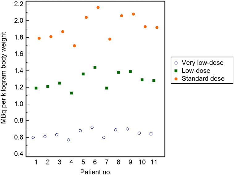
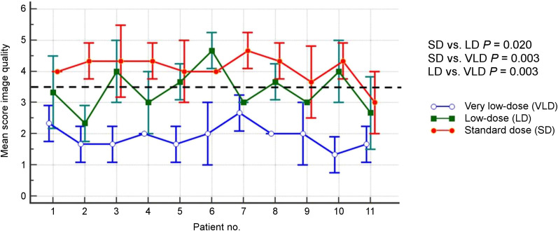
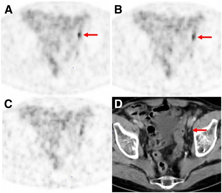
Our purpose was to define a clinically useful lower limit of injected dose for 68Ga-prostate-specific membrane antigen (PSMA)-11 PET/CT imaging of prostate cancer. Methods:68Ga-PSMA-11 PET/CT was performed on 11 patients. PET was acquired in list mode and reconstructed using a 3-min full acquisition, a 2-min acquisition, and a 1-min acquisition to generate images obtained with three thirds (standard dose), two thirds (low dose), and one third (very low dose) of the injected dose, respectively. Overall image quality (5-point scale) was assessed, and the detectability of PSMA-positive lesions was determined by 3 readers and compared with the reference standard. Results: Image quality declined with decreasing dose (mean score of 4.1 ± 0.4 for the standard dose, 3.4 ± 0.7 for the low dose, and 1.9 ± 0.4 for the very low dose; all P < 0.05). Readers 1, 2, and 3 correctly identified the lesions (n = 21) at a rate of 100%, 100%, and 95% with the standard dose; 95%, 81%, and 90% with the low dose; and 71%, 76%, and 59% with the very low dose, respectively. Conclusion:68Ga-PSMA-11 dose reduction is not feasible without a negative impact on image quality and lesion detectability.
Keywords: PSMA; dose reduction; image dose; prostate cancer.
© 2020 by the Society of Nuclear Medicine and Molecular Imaging.
Figures
Comment in
-
Lesion Detection and Administered Activity.J Nucl Med. 2020 Sep;61(9):1406-1410. doi: 10.2967/jnumed.120.244020. Epub 2020 Apr 3. J Nucl Med. 2020. PMID: 32245895 No abstract available.
References
-
- Rauscher I, Duwel C, Haller B, et al. Efficacy, predictive factors, and prediction nomograms for 68Ga-labeled prostate-specific membrane antigen-ligand positron-emission tomography/computed tomography in early biochemical recurrent prostate cancer after radical prostatectomy. Eur Urol. 2018;73:656–661. - PubMed
-
- Perera M, Papa N, Roberts M, et al. Gallium-68 prostate-specific membrane antigen positron emission tomography in advanced prostate cancer: updated diagnostic utility, sensitivity, specificity, and distribution of prostate-specific membrane antigen-avid lesions—a systematic review and meta-analysis. Eur Urol. February 14, 2019 [Epub ahead of print]. - PubMed
-
- Rauscher I, Maurer T, Beer AJ, et al. Value of 68Ga-PSMA HBED-CC PET for the assessment of lymph node metastases in prostate cancer patients with biochemical recurrence: comparison with histopathology after salvage lymphadenectomy. J Nucl Med. 2016;57:1713–1719. - PubMed
-
- Maurer T, Gschwend JE, Rauscher I, et al. Diagnostic efficacy of 68gallium-PSMA positron emission tomography compared to conventional imaging for lymph node staging of 130 consecutive patients with intermediate to high risk prostate cancer. J Urol. 2016;195:1436–1443. - PubMed
MeSH terms
Substances
LinkOut - more resources
Full Text Sources
Miscellaneous