. 2019 Jul 19;10(1):3251.
doi: 10.1038/s41467-019-11238-1.

NQO1 targeting prodrug triggers innate sensing to overcome checkpoint blockade resistance

Affiliations

NQO1 targeting prodrug triggers innate sensing to overcome checkpoint blockade resistance

Xiaoguang Li et al. Nat Commun. .

Abstract

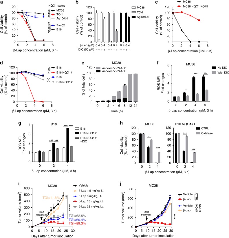
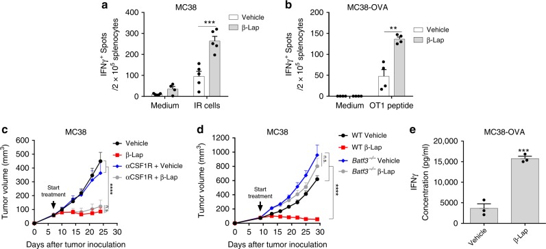
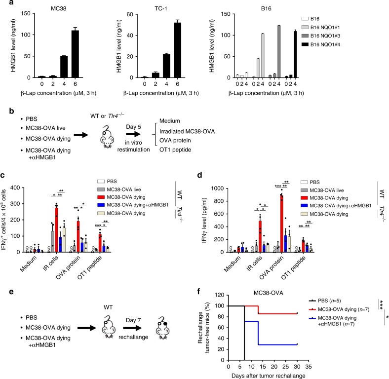
Lack of proper innate sensing inside tumor microenvironment (TME) limits T cell-targeted immunotherapy. NAD(P)H:quinone oxidoreductase 1 (NQO1) is highly enriched in multiple tumor types and has emerged as a promising target for direct tumor-killing. Here, we demonstrate that NQO1-targeting prodrug β-lapachone triggers tumor-selective innate sensing leading to T cell-dependent tumor control. β-Lapachone is catalyzed and bioactivated by NQO1 to generate ROS in NQO1high tumor cells triggering oxidative stress and release of the damage signals for innate sensing. β-Lapachone-induced high mobility group box 1 (HMGB1) release activates the host TLR4/MyD88/type I interferon pathway and Batf3 dendritic cell-dependent cross-priming to bridge innate and adaptive immune responses against the tumor. Furthermore, targeting NQO1 is very potent to trigger innate sensing for T cell re-activation to overcome checkpoint blockade resistance in well-established tumors. Our study reveals that targeting NQO1 potently triggers innate sensing within TME that synergizes with immunotherapy to overcome adaptive resistance.


Conflict of interest statement

The authors declare no competing interests.

Figures

Fig. 1
Fig. 1
β-Lap kills murine tumor cells in an NQO1-dependent manner. a NQO1 positive tumor cell lines MC38, TC-1, and Ag104Ld and NQO1 negative cell lines Panc02 and B16 grown in 96-well plates were treated with β-lap (0–6 μM) for a 3 h followed by washing and replacing medium. Cell viability was determined by Sulforhodamine B (SRB) Assay 48 h later. b MC38, TC-1 and Ag104Ld cells were exposed to 4 μM β-lap ± dicoumarol (DIC, 50 μM) for 3 h and cell survival was assessed 48 h later. c MC38 cells with CRISPR-based NQO1 knockout (MC38 NQO1KO #5) planted in 96-well plates were exposed to β-lap for 3 h and cell survival was assessed 48 h later. d B16 cells stalely harboring a pCMV-NQO1 expression vector (B16 NQO1#1) planted in 96-well plates were exposed to β-lap ± dicoumarol (DIC, 50 μM) for 3 h purse and survival assessed 48 h later. e MC38 cells were exposed to a lethal dose of β-lap (4 μM) for indicated times, then stained with 7AAD and Annexin V followed by flow cytometry analysis. f, g MC38 cells in f or B16 and B16 NQO1#1 cells in g were exposed to β-lap ± dicoumarol (DIC, 50 μM) for 3 h and then ROS level was determined by DCFDA cellular ROS assay. h MC38 and B16 NQO1#1 cells were exposed to β-lap for 3 h. Catalase (1000 U/ml) was added and cell survival was assessed 48 h later. i C57BL/6 mice (n = 5/group) were transplanted with 6 × 105 MC38 cells and treated with β-lap (1.5, 5, or 15 mg/kg, intratumorally; or 25 mg/kg, i.v.) every other day for four times. j C57BL/6 mice were transplanted with 6 × 105 MC38 cells (NQO1 WT or KO, n = 5/group) and treated with β-lap (15 mg/kg, i.t.) every other day for four times. Data are shown as mean ± SEM from three independent experiments. **p < 0.01, ***p < 0.001, ****p < 0.0001 determined by unpaired Student's t-test in fh or two-way ANOVA in i and j
Fig. 2
Fig. 2
β-Lap-mediated-antitumor effect depends on CD8+ T cells. a 6 × 105 MC38 cells were subcutaneously transplanted into C57BL/6 WT (n = 5 for vehicle group; n = 6 for β-lap treatment group) and Rag1−/− mice (n = 5/group), respectively. Tumor-bearing mice were treated with β-lap (15 mg/kg, i.t.) every other day for four times. Numbers of tumor-free mice after treatment were shown. b C57BL/6 mice (n = 5/group) were transplanted with 6 × 105 MC38 tumor and treated with β-lap (15 mg/kg, i.t.) every other day for four times. For CD8+ T cell depletion, 200 μg of anti-CD8 antibodies were intraperitoneally injected four times at three days interval during the treatment. c C57BL/6 mice were transplanted with 1.5 × 105 TC-1 tumor cells and were treated with β-lap (5 mg/kg, i.t.) for four times with or without anti-CD8 antibodies (n = 5 for β-lap treatment group; n = 4 for vehicle, vehicle + αCD8, and β-lap + αCD8 groups). d Naïve (n = 5/group) and β-lap cured MC38 tumor-free (n = 7/group) C57BL/6 mice were rechallenged subcutaneously with 3 × 106 MC38 cells on the opposite site from the primary tumor 30 days after complete rejection, and tumor growth curve was monitored. e 2 × 106 A549 cells were subcutaneously injected into C57BL/6 Rag1−/− mice (n = 5 for vehicle and β-lap groups; n = 6 for vehicle + OTI group; n = 7 for β-lap + OTI group). Thirty days later, the mice were i.v. adoptively transfected with 2 × 106 lymph node cells from OT-1 transgenic mice. On the following day, tumor-bearing mice were intratumorally treated with β-lap (10 mg/kg) every other day for four times. f 2 × 106 A549 cells were subcutaneously injected into NSG-SGM3 (n = 5/group) or NSG-SGM3 harboring human CD34+ hematopoietic stem cells (n = 5 for Hu-NSG vehicle group; n = 6 for Hu-NSG β-lap group). Tumor-bearing mice were treated with β-lap (10 mg/kg, i.t.) every other day for four times. Tumor growth was measured twice a week. Data are shown as mean ± SEM from three independent experiments. **p < 0.01, ****p < 0.0001 determined by unpaired Student's t-test in a and b, or two-way ANOVA in cf
Fig. 3
Fig. 3
β-Lap induces Batf3-dependent dendritic cell-mediated T cell cross-priming. a C57BL/6 mice (n = 5/group) were transplanted with 6 × 105 MC38 cells and treated with β-lap (15 mg/kg, i.t.) every other day for three times, and 10 days after the first treatment, lymphocytes from the spleens were isolated and stimulated with medium or MC38 cells irradiated with 60 Gy. b C57BL/6 mice (n = 4/group) were transplanted with 1 × 106 MC38-OVA cells and treated with β-lap (15 mg/kg, i.t.) every other day for three times, and 10 days after the first treatment, lymphocytes from the spleens were isolated and stimulated with 2.5 μg/ml of OTI peptide. IFNγ-producing cells were determined by ELISPOT assay. c C57BL/6 mice (n = 5/group) were transplanted with 6 × 105 MC38 cells and treated with β-lap (15 mg/kg, i.t.) every other day for four times. One hundred micrograms of anti-CSF1R Ab were intratumorally injected three times at three days interval during the treatment. d 6 × 105 MC38 cells were subcutaneously transplanted into C57BL/6 WT (n = 5/group) and Batf3−/− mice (n = 5 for vehicle treatment group; n = 6 for β-lap treatment group), respectively. Tumor-bearing mice were treated with β-lap (15 mg/kg, i.t.) every other day for four times. Tumor growth was monitored twice a week. e MC38-OVA-bearing mice (n = 3/group) were treated with β-lap (15 mg/kg, i.t.) for one dose, and 4 days later, CD11c+ dendritic cells were purified from the tumor drain lymph nodes, and co-cultured with CD8+ T cells isolated from the spleen of OT-1 transgenic mice. The activity of cross-priming of T cells was determined by the level of cell-secreted IFNγ via Cytometric Bead Array (CBA) mouse IFNγ assay. Data are shown as mean ± SEM from three independent experiments. **p < 0.01, ***p < 0.001, ****p < 0.0001 determined by unpaired Student's t-test in a, b and e or two-way ANOVA in c and d
Fig. 4
Fig. 4
Type I IFNs and TLR4/MyD88/ signaling are required for the antitumor effect of β-lap. a C57BL/6 mice (n = 5/group) were transplanted with MC38 cells and treated with β-lap (15 mg/kg, i.t.) every other day for four times. Anti-IFNAR blocking antibodies (150 μg, i.t.) were administrated every four days for three times during the treatment. b The MC38 tumor-bearing WT (n = 5/group) and Ifnαr1−/− (n = 4/group) C57BL/6 mice were treated with β-lap. c MC38 cells were treated with β-lap (4 μM) for 3 h followed by washing and replacing fresh medium. Twenty-four hours later, BMDCs from WT or Myd88−/− mice were co-cultured with β-lap-treated tumor cells for another 48 h. The level of IFNβ from the culture supernatant was detected by ELISA. d WT and Myd88−/− (n = 4/group) C57BL/6 mice were transplanted with MC38 cells and treated with β-lap. Four days later, tumor tissues were collected for protein extraction, and level of IFNβ was measured by ELISA. e WT (n = 5/group) and Myd88−/− (n = 3/group) C57BL/6 mice were transplanted with MC38 cells and treated with β-lap. f WT (n = 5/group) and Tlr4−/− (n = 4/group) C57BL/6 mice were transplanted with MC38 cells and treated with β-lap. g C57BL/6 mice (n = 5/group) were transplanted with MC38 cells and treated with β-lap (15 mg/kg, i.t.) every other day for four times. Anti-HMGB1 neutralized antibody (200 μg, i.p) was administrated every 3 days for three times during the treatment. h MC38 tumor-bearing WT (n = 4/group) or Tlr4−/− (n = 4/group) or Myd88−/− (n = 6/group) C57BL/6 mice were treated with β-lap. Anti-HMGB1 neutralized antibody (200 μg, i.p) was administrated every 3 days for three times during the treatment. Twelve days after the initial treatment, lymphocytes from the tumor drain lymph nodes were isolated and stimulated with MC38 tumor cells irradiated with 60 Gy. IFNγ-producing cells were determined by ELISPOTs assay. Data are shown as mean ± SEM from three independent experiments. *p < 0.05, **p < 0.01, ***p < 0.001, ****p < 0.0001 determined by two-way ANOVA test
Fig. 5
Fig. 5
β-Lap induces HMGB1-dependent immunogenic cell death. a MC38, TC-1, and B16 (NQO null and overexpression clones) were treated with β-lap for 3 h followed by washing and replacing medium. The level of HMGB1 released into the culture supernatant was determined by ELISA 24 h later. b The research schema for in vivo cross-presentation of tumor-specific antigen from β-lap-induced dying tumor cells in panels c and d. c, d Live or β-lap-induced dying MC38-OVA cells were subcutaneously inoculated into the flank of WT or Tlr4−/− C57BL/6 mice (n = 4 for MC38-OVA dying group; n = 3 for other groups) along with or without anti-HMGB1 antibody. Five days later, single-cell suspensions from tumor drain lymph nodes were collected and re-stimulated with OVA protein, OT-1 peptide, or irradiated MC38-OVA cells for 48 h. IFNγ-producing cells were determined by ELISPOTs assay in c, and IFNγ secretion level was quantified by CBA mouse IFNγ assay in d. e The research schema for the immunogenic vaccine assay in f. f MC38-OVA cells treated with β-lap in vitro were inoculated s.c. along with or without anti-HMGB1 antibody into the flank of C57BL/6 mice (n = 5 for PBS group; n = 7 for MC38-OVA dying and MC38-OVA dying + αHMGB1 groups). After 7 days, mice were rechallenged with live MC38-OVA cells by injection into the contralateral flank. The percentage of rechallenged tumor-free mice was shown. Data are shown as mean ± SEM from three independent experiments. *p < 0.05, **p < 0.01, and ***p < 0.001 determined by two-way ANOVA (c, d) or log-rank test in f
Fig. 6
Fig. 6
β-Lap overcomes therapeutic PD-L1/PD-1 blockade resistance. a Treatment schema for local β-lap treatment-based combinative therapy in panel b and panel c. b, c 6 × 105 MC38 tumor cells were s.c.inoculated into the flank of C57BL/6 mice (n = 5/group). Mice bearing small tumor (about 50 mm3, in b) or advanced tumor (about 150–200 mm3, in c) were locally treated with β-lap (15 mg/kg, i.t.) for four times with or without anti-PD-L1-based checkpoint blockage. Tumor growth was monitored twice a week, and numbers of tumor-free mice after treatment were shown. d Treatment schema for systemically β-lap treatment-based combinative therapy in panels e and f. e 6 × 105 MC38 tumor cells were s.c. inoculated into the flank of C57BL/6 mice (n = 5 for vehicle treatment group; n = 6 for β-lap and vehicle + αPD-L1 groups; n = 8 for β-lap + αPD-L1 treatment group). Tumor-bearing mice (about 50–100 mm3) were systemically treated with β-lap (30 mg/kg, i.p.) for six times with or without anti-PD-L1-based checkpoint blockade. Tumor growth was monitored twice a week, and numbers of tumor-free mice after treatment were shown. f Survival curve for MC38 tumor-bearing mice with combinative treatment in panel e. g, h C57BL/6 mice bearing MC38-OVA tumor (about 150 mm3, n = 4/group) were locally treated with β-lap (15 mg/kg, i.t.) every other day for four times or anti-PD-L1 (100 μg, i.p.) for three times, alone or combination. Twelve days after first treatment, tumor-infiltrating CD45+ cells and lymphocytes were analyzed by flow cytometry in g, and OT-1 antigen-specific T cells in the spleen were determined by IFNγ ELISPOT assay in h. Data are shown as mean ± SEM from two to three independent experiments. *p < 0.05, **p < 0.01, ***p < 0.001, ****p < 0.0001 determined by two-way ANOVA in b, c, e, g and h or log-rank test in f

References

    1. Topalian SL, Drake CG, Pardoll DM. Immune checkpoint blockade: a common denominator approach to cancer therapy. Cancer Cell. 2015;27:450–461. doi: 10.1016/j.ccell.2015.03.001. - DOI - PMC - PubMed
    1. Pauken KE, Wherry EJ. Overcoming T cell exhaustion in infection and cancer. Trends Immunol. 2015;36:265–276. doi: 10.1016/j.it.2015.02.008. - DOI - PMC - PubMed
    1. Sharma P, Hu-Lieskovan S, Wargo JA, Ribas A. Primary, adaptive, and acquired resistance to cancer immunotherapy. Cell. 2017;168:707–723. doi: 10.1016/j.cell.2017.01.017. - DOI - PMC - PubMed
    1. Jenkins RW, Barbie DA, Flaherty KT. Mechanisms of resistance to immune checkpoint inhibitors. Br. J. Cancer. 2018;118:9–16. doi: 10.1038/bjc.2017.434. - DOI - PMC - PubMed
    1. Garber K. A new cancer immunotherapy suffers a setback. Science. 2018;360:588. doi: 10.1126/science.360.6389.588. - DOI - PubMed

Publication types

MeSH terms