. 2019 Jul 19;10(1):3252.
doi: 10.1038/s41467-019-11202-z.

TurboID-based proximity labeling reveals that UBR7 is a regulator of N NLR immune receptor-mediated immunity

Affiliations

TurboID-based proximity labeling reveals that UBR7 is a regulator of N NLR immune receptor-mediated immunity

Yongliang Zhang et al. Nat Commun. .

Erratum in

Abstract

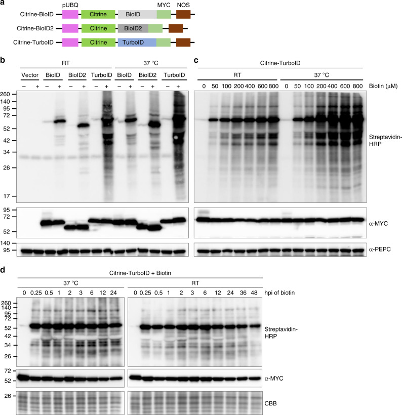
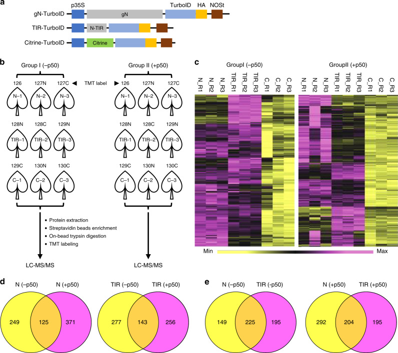
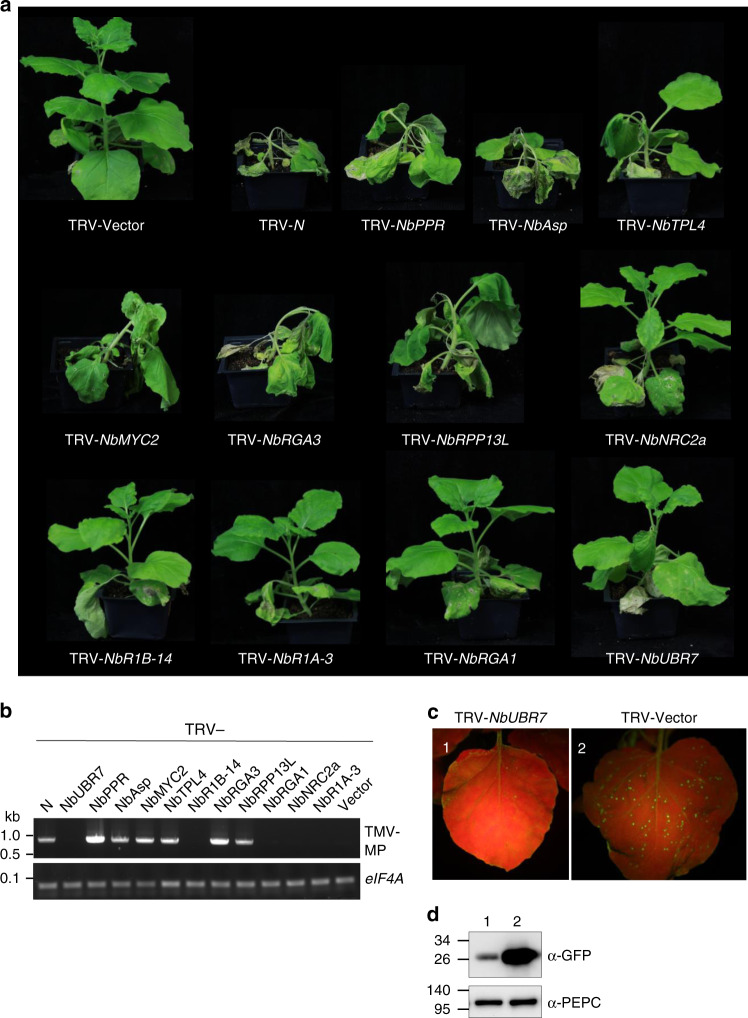
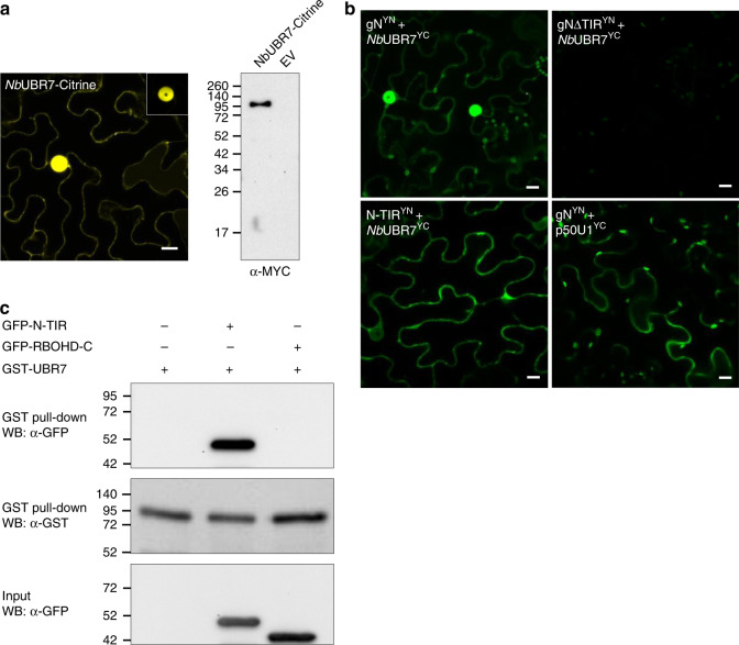
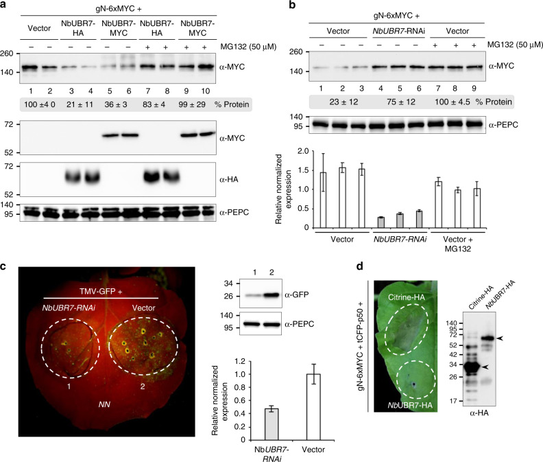
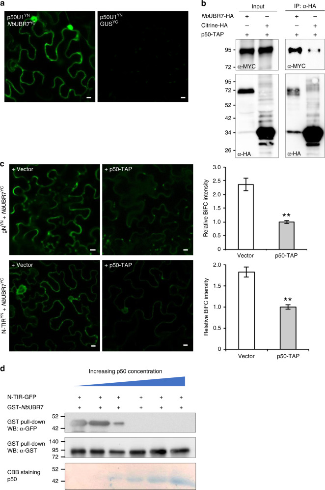
Nucleotide-binding leucine-rich repeat (NLR) immune receptors play a critical role in defence against pathogens in plants and animals. However, we know very little about NLR-interacting proteins and the mechanisms that regulate NLR levels. Here, we used proximity labeling (PL) to identify the proteome proximal to N, which is an NLR that confers resistance to Tobacco mosaic virus (TMV). Evaluation of different PL methods indicated that TurboID-based PL provides more efficient levels of biotinylation than BioID and BioID2 in plants. TurboID-based PL of N followed by quantitative proteomic analysis and genetic screening revealed multiple regulators of N-mediated immunity. Interestingly, a putative E3 ubiquitin ligase, UBR7, directly interacts with the TIR domain of N. UBR7 downregulation leads to an increased amount of N protein and enhanced TMV resistance. TMV-p50 effector disrupts the N-UBR7 interaction and relieves negative regulation of N. These findings demonstrate the utility of TurboID-based PL in plants and the N-interacting proteins we identified enhance our understanding of the mechanisms underlying NLR regulation.


Conflict of interest statement

The authors declare no competing interests.

Figures

Fig. 1
Fig. 1
Comparison and characterization of promiscuous protein biotinylation by different biotin ligases in plant. a Diagram of the expression cassettes used for the expression of three biotin ligases. Citrine was fused to the N-terminus while a MYC tag was added to the C-terminus of BioID, BioID2 and TurboID. Expression was under the control of Arabidopsis ubiquitin-10 promoter (pUBQ) and nopaline synthase terminator (NOSt). b The biotin ligases promiscuously biotinylate endogenous proteins in plant cells with varying efficiencies. N. benthamiana leaves were agroinfiltrated with the agrobacterium containing citrine-BioID-3xMYC (BioID), citrine-BioID2-3xMYC (BioID2) and citrine-TurboID-3xMYC (TurboID), or the empty vector control (vector), respectively. 36 h post-agroinfiltration (hpi), medium containing the buffer (−) or 200 µM biotin (+) were infiltrated into the previously agroinfiltrated leaves. Infiltrated N. benthamiana plants were directly incubated at room temperature (RT) or in a 37 °C chamber. Western blot analysis was performed on tissue collected 12 h after infiltration of biotin. Streptavidin-HRP and anti-MYC antibodies were used for detection of biotinylated proteins (top panel) and different biotin ligases (middle panel), respectively. PEPC served as loading control for equal protein loading (bottom panel). The molecular weight size markers in kDa are indicated at the left of each panel. An additional repeat of this experiment is shown in Supplementary Fig. 1b. c Determination of the optimal biotin concentrations required for TurboID-based proximity labeling in planta. Different concentrations of biotin, as indicated above the panels, were infiltrated into the N. benthamiana leaves expressing the citrine-TurboID. These plants were then incubated in the 37 °C chamber or at room temperature (RT) for 8 h followed by western blot analysis as described in (b). PEPC served as loading control for equal protein loading (bottom panel). The molecular weight size markers in kDa are indicated at the left of each panel. Additional repeat of this experiment is shown in Supplementary Fig. 1c. d Effect of incubation time on TurboID-based proximity labeling in plants. N. benthamiana leaves expressing citrine-TurboID were infiltrated with 200 µM biotin, plants were then incubated under 37 °C or RT followed by collection of leaves at different time points as indicated above the panels. Western blot analysis was then carried out as described in Fig. 1b. Due to the instability of rbcL protein levels at different time points after biotin treatment (Supplementary Fig. 2), the protein bands below that of the rbcL in the Coomassie Brilliant Blue (CBB)-stained gel is shown as a loading control (bottom panel). The molecular weight size markers in kDa are indicated at the left of each panel. Source data are provided as a Source Data file
Fig. 2
Fig. 2
Identification of proximal and interacting proteins of N NLR immune receptor. a Schematic representation of the constructs used for the identification of proximal and interacting proteins of N. b Diagram of the experimental design and labeling conditions. TurboID fusions without p50 (Group I) or with p50 that is under the control of the β-estradiol inducible promoter (Group II) were co-infiltrated into N. benthamiana leaves. At 36 hpi, 200 µM biotin and 30 µM 17-β-estradiol were infiltrated into both groups of pre-infiltrated leaves. 12 h later, leaf cells were lysed and biotinylated proteins were enriched with streptavidin beads, digested by trypsin, and labeled with tandem mass tags (TMT). Each treatment consisted of three independent biological replicates. All 9 samples in Group I or Group II, respectively, were then independently combined within group and analyzed by LC-MS/MS. c Hierarchical clustering of the Group I and Group II significantly enriched interacting proteins. Enriched interactors exhibited a >1.5 fold enrichment over the Citrine control and a q-value less than 0.05. d Venn diagrams depicting the proteins that interact with gN (left) or N-TIR (right) in the absence or presence of p50. e Overlap in the proteins that interact with gN and/or N-TIR in the absence (left) or presence (right) of p50
Fig. 3
Fig. 3
Genetic screening of the function of candidate N NLR immune receptor interacting proteins in N-mediated resistance to TMV. a Phenotypic observation of the TMV-U1-incoluted N-containing transgenic N. benthamiana plants after being silenced with various target genes as indicted. Various recombinant TRV vectors containing different gene fragments were inoculated onto the N-containing transgenic N. benthamiana plants. 10 days later, TMV-U1 was rub-inoculated onto the upper leaves. Photographs were taken at 7 days after TMV inoculation and representative results are shown. These silencing experiments were repeated 2–3 times. b Analysis of the TMV RNA corresponding to the movement protein coding region in the upper uninoculated leaves by RT-PCR (TMV-MP, top panel). eIF4A was used as an internal control to validate the equal amount of total RNA used for RT-PCR (bottom panel). c Silencing of NbUBR7 enhances N-mediated resistance to TMV. TMV-U1-GFP was rub-inoculated onto vector control or the NbUBR7-silenced N-containing transgenic N. benthamiana leaves. Photographs were taken under UV light at 5 days post inoculation and representative results are shown (upper panels). d The expression level of GFP in the inoculated leaves was examined by western blot analysis using antibodies against GFP (top panel). PEPC served as loading control for equal protein loading (bottom panel). The molecular weight size markers in kDa are indicated at the left of each panel. Source data are provided as a Source Data file
Fig. 4
Fig. 4
Analysis of the interaction between N and NbUBR7 in vivo and in vitro. a Subcellular localization of NbUBR7. NbUBR7 was fused to citrine (NbUBR7-citrine) followed by agroinfiltration. Confocal analysis was performed at 2 dpi. NbUBR7 is present in the cytoplasm and in the nucleus (left panel). The rectangular region on top right of the image indicated a lower gain value image to confirm the nuclear localization of NbUBR7. Scale bar represents 10 µm. NbUBR7 expression was confirmed by western blot analysis using antibodies against MYC tag (right panel). Empty vector served as the control. The molecular weight size markers in kDa are indicated at the left. b BiFC analysis of the interaction between NbUBR7 and N as well as its different domains. NbUBR7 fused to C-terminus of citrine (NbUBR7YC) was coexpressed with native promoter-driven full-length N (gN), TIR domain deleted N (gNΔTIR) or TIR domain alone (N-TIR) fused to N-terminus of citrine in the N. benthamiana leaves. Confocal analysis was performed at 2 dpi. gNYN and p50U1YC served as the positive control. Scale bars = 10 µm. c In vitro GST-pull down assay to examine the interaction between NbUBR7 and the TIR domain of N. The purified GFP-tagged TIR domain of N or C-terminal region of the respiratory burst oxidase homolog D (RBOHD) were incubated with GST-tagged NbUBR7. After being pulled-down with glutathione-sepharose beads, the proteins were detected by Western blot (WB) with anti-GFP or anti-GST antibodies. Source data are provided as a Source Data file
Fig. 5
Fig. 5
NbUBR7 modulates N protein level and functions as a negative regulator of N-mediated defense. a NbUBR7 overexpression reduced the stability of N in a proteasome-dependent manner. Agrobacterium containing expression sets consisting of an empty vector control (vector) or the agrobacterium containing the expression cassette of NbUBR7-HA or NbUBR7-MYC were infiltrated into N. benthamiana leaves followed by infiltration of MYC-tagged gN 24 later. 36 h after infiltration of gN, 50 µM proteasome inhibitor MG132 (+MG132) or the equal concentration of DMSO (−MG132) were further infiltrated into the pre-infiltrated leaves. Infiltrated leaf tissues were collected 12 h after DMSO or MG132 treatment and analyzed by western blot analysis. Antibodies used for the western blot analysis were indicated on the right of each panel. Equal protein loading is assessed by the similar amounts of PEPC protein. Molecular weight size markers in kDa are indicated on the left. Two independent samples (n = 2) for each treatment were analyzed in parallel. Band intensity was measured by Image J software and normalized to the PEPC protein control. Numbers below the top panel indicate the relative quantification of the corresponding band intensity, of which the empty vector control group was set to 100% [“±” indicates standard deviation (SD) of the mean]. b Double stranded hairpin-mediated silencing of NbUBR7 (NbUBR7-RNAi) enhances the stability of N. Empty vector control (vector) or the agrobacterium containing the hairpin NbUBR7 were infiltrated into the N. benthamiana leaves followed by infiltration of MYC-tagged N at 24 hpi. 36 h later, DMSO (-MG132) or MG132 (MG132) was then infiltrated into the pre-infiltrated leaves as described above. Infiltrated leaf tissues were collected 12 h after DMSO or MG132 treatment and analyzed by western blot. Three independent replicates (n = 3) were carried out for each treatment. Numbers below the top panel indicate the relative quantification of the corresponding band intensity, of which the MG132-treated empty vector control was set to 100% (“±” indicates SD). Equal protein loading is assessed by the similar amounts of PEPC protein (middle panel). Molecular weight size markers in kDa are indicated on the left. The downregulation of NbUBR7 in the infiltrated leaves were also confirmed by RT-qPCR (bottom panel). Data from three biological replicates were combined and values are shown as mean ± SD. c RNAi of NbUBR7 enhances the N-mediated resistance to TMV. Different regions of the leaf of N-containing transgenic N. benthamiana were first agroinfiltrated with hairpin NbUBR7 (1) and the control empty vector (2), respectively. 24 h later, TMV-U1-GFP was agroinfiltrated into the previously infiltrated regions. Photographs were taken under UV light at 5 dpi and representative results are shown (left panel). Leaf samples from the region 1 and 2 were then harvested and subjected to western blot analysis using the antibodies against GFP or PEPC (top right panels). RT-qPCR was performed to confirm the downregulation of NbUBR7 in the infiltrated region of the leaves (bottom right panel). Data from three biological replicates were combined and values are shown as mean ± SD. d Overexpression of NbUBR7 inhibits p50 effector-induced HR-PCD. Different leaf regions were infiltrated with MYC-tagged N (gN-6xMYC), TagCFP-tagged p50 (tCFP-p50) together with HA-tagged NbUBR7 (NbUBR7-HA) or citrine (Citrine-HA) control. Photographs were taken at 6 dpi and representative results are shown (left panel). The expression of citrine-HA or NbUBR7-HA was confirmed by Western blot analysis using anti-HA antibody (right panel). Arrowheads indicate the specific band of different HA fusions. Source data are provided as a Source Data file
Fig. 6
Fig. 6
TMV p50 effector interferes with the interaction between NbUBR7 and the TIR domain of N. a BiFC analysis of the interaction between p50U1 and NbUBR7. The constructs used for co-infiltration were indicated on the upper left of each panel. GUSYC served as a control for the specificity of associations involving p50U1YN. Scale bars = 10 µm. The expression of p50U1YN and NbUBR7YC were confirmed by western blot analysis (Supplementary Fig. 13b). b Co-IP analysis of the interaction between p50U1 and NbUBR7. HA-tagged UBR7 or citrine was coexpressed with TAP-tagged p50 in the N. benthamiana leaves. Protein extracts from the infiltrated leaf tissues were incubated with anti-HA antibody-conjugated agarose beads. Protein extracts (input) or immunoprecipitated (IP) complexes were separated by SDS-PAGE and probed with anti-MYC or anti-HA antibodies. p50-TAP was co-precipitated with NbUBR7-HA, but not with Citrine-HA. Molecular size markers in kDa are indicated on the left. c BiFC competition analysis showed the inhibitory effect of p50 on the interactions between NbUBR7 and N or NbUBR7 and TIR domain. BiFC constructs together with the empty vector control or the p50-TAP were coinfiltrated into the N. benthamiana leaves. Confocal analysis was performed at 2 dpi. Scale bars = 10 µm. Reconstituted citrine fluorescence intensity was quantified using the Image J software and p50-TAP-infitrated groups were used as the normalizer (=1). Error bars represent standard deviation from the mean (n = 3). Asterisk indicates statistically significant difference between vector and p50-TAP groups (Student’s t-test, **P= 0.004 for upper right panel and **P = 0.007 for bottom right panel). The expression of NbUBR7YC and p50-TAP were confirmed by western blot analysis (Supplementary Fig. 13c). d In vitro competitive GST pull-down assay showed the binding of NbUBR7 to TIR domain was inhibited with the addition of an increasing amount of p50. GST-tagged NbUBR7 or GFP-tagged TIR domain were expressed and purified from E. coli. These two proteins were pulled down with glutathione-agarose in the presence of an increasing concentration of p50 (0.1, 1, 2, 4, 8 µg). Pull-down samples were analyzed by western blot (WB) with anti-GST or anti-GFP antibodies. CBB staining of the increasing amounts of p50 protein is shown in the bottom panel. Source data are provided as a Source Data file
Fig. 7
Fig. 7
A model for the functional role of UBR7 in N-mediated resistance to TMV. a In uninfected naive condition, UBR7 interacts with the TIR domain of N resulting in the relatively low expression of N. b During TMV infection, the p50 effector disrupts the interaction between UBR7 and N by interacting with UBR7, thereby releasing the N from the N-UBR7 complex and leading to increased stability of N and activation of defense

References

    1. Baulcombe D. RNA silencing in plants. Nature. 2004;431:356–363. doi: 10.1038/nature02874. - DOI - PubMed
    1. Burgyan J, Havelda Z. Viral suppressors of RNA silencing. Trends Plant Sci. 2011;16:265–272. doi: 10.1016/j.tplants.2011.02.010. - DOI - PubMed
    1. Toruno TY, Stergiopoulos I, Coaker G. Plant-pathogen effectors: cellular probes interfering with plant defenses in spatial and temporal manners. Annu. Rev. Phytopathol. 2016;54:419–441. doi: 10.1146/annurev-phyto-080615-100204. - DOI - PMC - PubMed
    1. Cui H, Tsuda K, Parker JE. Effector-triggered immunity: from pathogen perception to robust defense. Annu. Rev. Plant Biol. 2015;66:487–511. doi: 10.1146/annurev-arplant-050213-040012. - DOI - PubMed
    1. Caplan JL, Padmanabhan M, Dinesh-Kumar SP. Plant NB-LRR immune receptors: from recognition to transcriptional reprogramming. Cell Host Microbe. 2008;3:126–135. doi: 10.1016/j.chom.2008.02.010. - DOI - PubMed

Publication types

MeSH terms